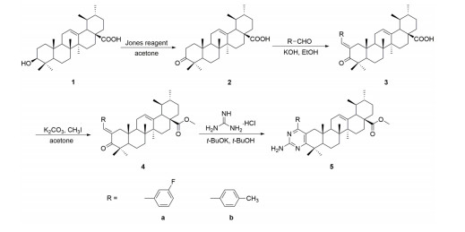
Scheme 1.
Synthetic route of the title compounds
Synthesis, Crystal Structures and Cytotoxic Activities of Two New Pyrimidine Derivatives of Ursolic Acid
Wen-Yan WANG , A-Liang LI , Qing-Song LIU , Yue SUN , Wen GU

As one of the leading causes of mortality all over the world, cancer poses a tremendous threat to human's health[1]. There were estimated 18.1 million new cancer cases and 9.6 million deaths from cancer worldwide in 2018[2]. Moreover, new cancer cases are expected to rise to almost 20 million by 2025[3]. Thus, the research and development of novel anticancer drugs with better efficacy have still been one of the most important issues for mankind.
Ursolic acid (UA), a natural pentacyclic triterpenic acid, is widely found in vegetables, fruits, and traditional herbal medicines[4]. As early as 1990, Japan listed ursolic acid as one of the most promising cancer chemopreventive drugs[5]. As a bioactive constituent in many traditional Chinese medicines, UA and its derivatives possess a variety of biological activities including antimicrobial, anticancer, antiviral, antioxidant, antiulcer, anti-osteoporosis, antiarrhy-thmic, anti-hyperlipidemic, and anti-neurodegenerative activities[6-12]. Therefore, ursolic acid and its synthetic derivatives have exhibited great potential in the discovery of new drugs treating some severe diseases such as cancer[6].
Pyrimidine, a kind of six-membered nitrogen-containing heterocycle, has been considered as a significant pharmacophore in drug discovery[13]. Pyrimidine derivatives exhibit various pharmacological properties such as anticancer, antimicrobial, antiviral, anti-tubercular, anti-inflammatory, antimalarial, anti-diabetic and antipyretic activities[14, 15]. Therefore, the introduction of pyrimidine moiety to the structure of UA may afford novel derivatives with promising anticancer activities. In this paper, we report the synthesis, characterization and crystal structures of two novel pyrimidine derivatives of UA. In addition, the results of their in vitro anticancer activities are also presented.
The melting point was determined by means of an XT-4 apparatus (Taike Corp., Beijing, China) and was uncorrected. The ESI-MS spectrum was measured on a Mariner System 5304 mass spectrometer. NMR spectra were accomplished in CDCl3 on a Bruker AV-400 and AV-600 spectrometer using TMS as the internal standard. Elemental analyses were performed on a CHN-O-Rapid instrument within ±0.4% of the theoretical values. Reactions were monitored by TLC which was carried out on silica gel IB-F flexible sheets (Mallinckrodt Baker Inc., Germany) and visualized in UV light (254 and 365 nm). Silica gel (300~400 mesh) for column chromatography was purchased from Qingdao Marine Chemical Factory, China. The reagents (chemicals) of A.R. grade were purchased from Shanghai Chemical Reagent Company (Shanghai, China). Ursolic acid (95%) was bought from Jingzhu Biological Technology Co., Ltd. (Nanjing, China).
The synthetic routes for compounds 5a and 5b are outlined in Scheme 1. Compound 2 was synthesized from UA (1) by oxidation with Jones reagent. To a solution of compound 2 (1.0 g, 2.3 mmol) in 100 mL of EtOH was added 3.0 mmol of corresponding benzaldehyde (3-fluorobenzaldehyde for 5a and 4-methylbenzaldehyde for 5b) and KOH (5.0 g, 89 mmol). The reaction mixture was stirred at room temperature for 4 h and monitored by TLC. At the end of the reaction, the mixture was extracted with ethyl acetate for three times (3 × 50 mL). The organic phase was combined, washed with water, saturated NaHCO3 solution and brine, dried over anhydrous Na2SO4, filtered and then concentrated in vacuo. The residue was purified by column chromatography on silica gel and eluted with petroleum ether-MeOH (500:1~300:1, v/v) to give compounds 3 (Yields: 67% for 3a and 58% for 3b).
To a solution of compound 3 (4.38 mmol) in 18 mL of acetone was added K2CO3 (0.6 g, 4.38 mmol) and CH3I (0.4 mL, 6.57 mmol). The reaction mixture was stirred at room temperature for 4 h and monitored by TLC. At the end of reaction, the mixture was extracted with ethyl acetate for three times (3 × 50 mL). The organic layer was combined, washed with water, saturated NaHCO3 solution and brine, dried over anhydrous Na2SO4, filtered and concentrated in vacuo to give a crude product of compound 4 which could be used directly for the next step.
To a solution of compound 4 (1.0 mmol) in 20 mL of t-butanol was added potassium t-butoxide (0.56 g, 5.0 mmol) and guanidine hydrochloride (0.4 g, 4.0 mmol). The reaction mixture was refluxed for 24 h and monitored by TLC. At the end of reaction, the mixture was extracted with ethyl acetate for three times (3 × 50 mL). The organic phase was combined, washed with water, saturated NaHCO3 solution and brine, dried over anhydrous Na2SO4, filtered and then concentrated in vacuo. Finally, the crude product was recrystallized repeatedly from methanol and filtered to give the title compound 5a or 5b (The purity was above 98%).
Compound 5a: yellow solid; 0.21 g, yield 81%; m.p. 243~247 ℃. 1H-NMR (600 MHz, CDCl3): δ 0.75 (s, 3H), 0.76 (s, 3H), 0.85 (d, J = 6.4 Hz, 3H), 0.92 (d, J = 6.3 Hz, 3H), 1.09 (s, 3H), 1.24 (s, 3H), 1.28 (s, 3H), 1.30~1.90 (m, 17H), 1.98 (td, J = 13.3, 5.7 Hz, 1H), 2.10 (d, J = 15.5 Hz, 1H), 2.21 (d, J = 11.3 Hz, 1H), 2.51 (d, J = 15.4 Hz, 1H), 3.57 (s, 3H), 5.08 (brs, 2H), 5.20 (t, J = 3.5 Hz, 1H), 7.09 (td, J = 8.4, 2.3 Hz, 1H), 7.15 (td, J = 9.3, 2.1 Hz, 1H), 7.21 (d, J = 7.7 Hz, 1H), 7.40 (td, J = 7.9, 5.8 Hz, 1H); 13C-NMR (150 MHz, CDCl3): δ 14.9, 16.8, 17.2, 20.3, 21.2, 23.4, 23.6, 23.9, 24.3, 28.1, 30.7, 31.6, 32.4, 36.2, 36.7, 38.9, 39.2, 39.5, 39.6, 41.8, 42.2, 45.7, 48.2, 51.5, 53.0, 53.1, 114.6, 115.6 (d, J = 18.0 Hz), 115.8 (d, J = 19.0 Hz), 124.4 (d, J = 2.9 Hz), 125.4, 129.9 (d, J = 8.2 Hz), 138.2, 141.2 (d, J = 7.3 Hz), 161.1, 162.6 (d, J = 245.1 Hz), 165.6, 175.0, 178.1; MS (ESI): m/z [M+H]+ 614.4. Anal. Calcd. for C39H52FN3O2: C, 76.31; H, 8.54; N, 6.85%. Found: C, 76.36; H, 8.51; N, 6.89%.
Compound 5b: yellow solid; 0.23 g, yield 87%; m.p. 195~198 ℃. 1H-NMR (400 MHz, CDCl3): δ 0.76 (s, 3H), 0.78 (s, 3H), 0.88 (d, J = 6.4 Hz, 3H), 0.94 (d, J = 6.1 Hz, 3H), 1.11 (s, 3H), 1.26 (s, 3H), 1.30 (s, 3H), 1.30~2.05 (m, 17H), 2.01 (td, J = 13.3, 4.4 Hz, 1H), 2.15 (d, J = 15.5 Hz, 1H), 2.23 (d, J = 11.4 Hz, 1H), 2.41 (s, 3H), 2.62 (d, J = 15.4 Hz, 1H), 3.60 (s, 3H), 4.98 (brs, 2H), 5.22 (t, J = 3.6 Hz, 1H), 7.26 (d, J = 8.0 Hz, 2H), 7.37 (d, J = 8.0 Hz, 2H). 13C-NMR (100 MHz, CDCl3): 14.9, 16.9, 17.2, 20.4, 21.3, 21.5, 23.5, 23.6, 23.9, 24.3, 28.1, 30.8, 31.7, 32.4, 36.2, 36.8, 39.0, 39.2, 39.5, 39.6, 42.1, 42.3, 45.8, 48.3, 51.6, 53.0, 53.1, 114.7, 125.6, 128.7 (2C), 129.0 (2C), 136.2, 138.2, 138.6, 161.1, 167.1, 174.5, 178.1; MS (ESI): m/z [M+H]+ 610.4. Anal. Calcd. for C40H55N3O2: C, 78.77; H, 9.09; N, 6.89%. Found: C, 78.71; H, 9.13; N, 6.92%.
The crystals suitable for X-ray diffraction were obtained by slow evaporation of the solutions of the title compounds 5a and 5b in EtOH at room temperature. The X-ray diffraction data were collected on an Enraf-Nonius CAD-4 diffractometer equipped with graphite-monochromated MoKα radiation (λ = 0.71073 Å) by using an ω-2θ scan mode at 293(2) K. For compound 5a, the intensity data were collected in the range of 1.37≤θ≤25.37° (0≤h≤14, 0≤k≤12, −18≤l≤17). A total of 3862 reflections were collected, of which 3685 were independent (Rint = 0.0668) and 1859 were observed with I > 2σ(I). For compound 5b, the intensity data were collected in the range of 1.37≤θ≤25.39° (0≤h≤14, 0≤k≤12, −18≤l≤17). A total of 3974 reflections were collected, of which 3794 were independent (Rint = 0.0237) and 2574 were observed with I > 2σ(I). The structures were solved by direct methods using SHELXS-97[16] and refined by full-matrix least-squares method based on F2 through SHELXL-97 software package[17]. Non-H atoms were refined anisotropically using all reflections with I > 2σ(I). All H atoms were generated geometrically and refined in terms of riding model (Uiso(H) = 1.5Ueq(C) for the atoms of methyl and Uiso(H) = 1.2Ueq(C) for others). Selected bond lengths and bond angles are given in Table 1.
 DownLoad:
CSV
DownLoad:
CSV
| Compound 5a | |||||||
| Bond | Dist. | Bond | Dist. | Bond | Dist. | ||
| N(1)–C(33) | 1.337(7) | O(2)–C(31) | 1.460(8) | C(11)–C(12) | 1.481(8) | ||
| N(1)–C(32) | 1.342(7) | O(3)–C(41) | 1.369(12) | C(12)–C(13) | 1.315(7) | ||
| N(2)–C(3) | 1.339(7) | C(1)–C(2) | 1.485(7) | C(17)–C(28) | 1.507(9) | ||
| N(2)–C(32) | 1.341(7) | C(2)–C(3) | 1.392(7) | C(33)–C(34) | 1.509(8) | ||
| N(3)–C(32) | 1.333(7) | C(2)–C(33) | 1.401(7) | C(36)–C(37) | 1.363(9) | ||
| O(1)–C(28) | 1.198(7) | C(3)–C(4) | 1.531(7) | C(40)–C(41) | 1.421(11) | ||
| O(2)–C(28) | 1.340(8) | C(4)–C(24) | 1.526(10) | F–C(36) | 1.342(8) | ||
| Angle | (°) | Angle | (°) | Angle | (°) | ||
| C(33)–N(1)–C(32) | 115.4(5) | C(1)–C(2)–C(3) | 119.8(5) | C(12)–C(13)–C(14) | 121.2(6) | ||
| C(3)–N(2)–C(32) | 117.5(5) | O(2)–C(28)–C(17) | 112.0(7) | C(34)–C(35)–C(36) | 116.8(6) | ||
| N(3)–C(32)–N(1) | 115.8(6) | O(1)–C(28)–O(2) | 122.2(7) | C(37)–C(36)–C(35) | 123.8(7) | ||
| F–C(36)–C(35) | 117.6(6) | O(3)–C(41)–C(40) | 110.5(11) | C(39)–C(34)–C(33) | 119.3(6) | ||
| C(1)–C(2)–C(3)–C(4) | 5.9(10) | F–C(36)–C(37)–C(38) | −179.5(7) | C(31)–O(2)–C(28)–O(1) | −1.4(11) | ||
| N(1)–C(33)–C(34)–C(39) | −36.5(8) | C(11)–C(12)–C(13)–C(14) | −1.0(11) | C(31)–O(2)–C(28)–C(17) | −178.3(6) | ||
| Compound 5b | |||||||
| Bond | Dist. | Bond | Dist. | Bond | Dist. | ||
| N(1)–C(33) | 1.329(6) | O(2)–C(31) | 1.449(10) | C(11)–C(12) | 1.486(7) | ||
| N(1)–C(32) | 1.333(7) | O(3)–C(41) | 1.374(15) | C(12)–C(13) | 1.318(7) | ||
| N(2)–C(3) | 1.355(7) | C(1)–C(2) | 1.492(6) | C(17)–C(28) | 1.514(9) | ||
| N(2)–C(32) | 1.322(7) | C(2)–C(3) | 1.387(7) | C(33)–C(34) | 1.491(7) | ||
| N(3)–C(32) | 1.365(7) | C(2)–C(33) | 1.405(7) | C(36)–C(37) | 1.386(8) | ||
| O(1)–C(28) | 1.195(7) | C(3)–C(4) | 1.531(7) | C(41)–C(42) | 1.429(9) | ||
| O(2)–C(28) | 1.343(8) | C(4)–C(24) | 1.528(9) | C(37)–C(40) | 1.490(8) | ||
| Angle | (°) | Angle | (°) | Angle | (°) | ||
| C(33)–N(1)–C(32) | 116.7(4) | C(1)–C(2)–C(3) | 121.3(4) | C(12)–C(13)–C(14) | 121.0(4) | ||
| C(3)–N(2)–C(32) | 116.3(4) | O(2)–C(28)–C(17) | 113.6(6) | C(34)–C(35)–C(36) | 121.4(5) | ||
| N(3)–C(32)–N(1) | 116.6(5) | O(1)–C(28)–O(2) | 121.9(6) | C(36)–C(37)–C(38) | 117.2(5) | ||
| C(36)–C(37)–C(40) | 121.2(5) | O(3)–C(41)–C(42) | 124.4(15) | C(39)–C(34)–C(33) | 123.5(5) | ||
| C(1)–C(2)–C(3)–C(4) | −2.9(8) | C(35)–C(36)–C(37)–C(40) | −177.1(6) | C(31)–O(2)–C(28)–O(1) | −2.1(9) | ||
| N(1)–C(33)–C(34)–C(39) | −138.8(5) | C(11)–C(12)–C(13)–C(14) | 1.7(10) | C(31)–O(2)–C(28)–C(17) | −179.6(6) | ||
Cytotoxic activities of compounds 5a and 5b were determined in vitro against human breast cancer cell line (MCF-7), human cervical cancer cell line (HeLa) and human hepatocarcinoma cell line (HepG2) via the MTT colorimetric method according to the literature method[18]. Briefly, different tumor cells were grown in DMEM medium supplemented with 10% fetal bovine serum, penicillin (100 U/mL), and streptomycin (50 μg/mL). Cells were harvested at log phase of growth and seeded in 96-well plates (100 μL/well at a density of 2 × 10⁵ cells/mL). After 24 h incubation at 37 ℃ and 5% CO2 to allow cell attachment, cultures were exposed to various concentrations of the isolated compounds for 48 h. Finally, MTT solution (2.5 mg/mL in PBS) was added (40 μL/well). Plates were further incubated for 4 h at 37 ℃, and the formazan crystals formed were dissolved by adding 150 μL/well of DMSO. Absorption at 570 nm was measured with an ELISA plate reader. The results were expressed as IC50 values with standard deviations, which was defined as the concentration at which 50% survival of cells was discerned. Etoposide was co-assayed as positive control.
The structures of the title compounds 5a and 5b were characterized on the basis of ESI-MS, NMR spectra and elemental analysis. For example, the molecular formula of compound 5a is determined to be C39H52FN3O2 by the ESI-MS spectrum (m/z 614.4 [M+H]+) and the data of elemental analysis. In its 1H-NMR spectrum, five singlets at δ 0.75, 0.76, 1.09, 1.24 and 1.28 ppm can be attributed to five methyl groups at C(23), C(24), C(25), C(26) and C(27), while two doublets at δ 0.85 and 0.92 ppm are due to the two adjacent methyl groups at C(29) and C(30). The singlet with three protons at δ 3.57 ppm can be attributed to the methyl ester group at C(31). In addition, the broad singlet at δ 5.08 ppm can be assigned to the protons of NH2 on the pyrimidine moiety, and the triplet with one proton at δ 5.20 ppm is the signal of the olefinic hydrogen at C(12). As for aromatic protons, four signals at δ 7.15, 7.09, 7.40 and 7.21 ppm can be assigned to H(35), H(37), H(38) and H(39), respectively through the chemical shifts and H–H, F–H coupling constants. In its 13C-NMR spectrum, there are 39 signals corresponding to the carbons of the molecule. Six doublets at δ 115.6, 115.8, 124.4, 129.9, 141.2 and 162.6 can be attributed to the carbons at C(34)~C(39). Among them, the doublet at δ 162.6 (d, J = 245.1 Hz) is the signal of C(36) due to its large JC–F. Moreover, the peak at δ 178.1 ppm could be assigned to the carbonyl carbon at C(28). All the 1H- and 13C-NMR data are in good agreement with the structures of the title compounds.
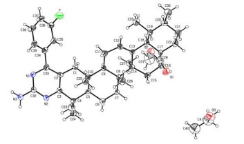
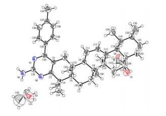
The single crystals of compounds 5a and 5b were obtained by slow evaporation of their ethanol solution. The crystal structures of compounds 5a and 5b with atomnumbering are shown in Figs. 1 and 2, respectively, and the selected bond lengths and bond angles are listed in Table 1. The two compounds have similar crystallographic results, so only compound 5a was selected to describe the crystal structure. It can be seen from Fig. 1 that each molecule of 5a is accompanied with one ethanol molecule, and possesses a pentacyclic triterpene skeleton with five six-membered rings (rings A, B, C, D and E) and a 2-amino-4-(3-fluorophenyl)-pyrimidine moiety fused to ring A. Rings A/B and B/C are fused via trans ring junction, while rings D/E adopt a cis junction. It can be observed that rings B (C(5)~C(10)), D (C(13)~C(18)) and E (C(17)~C(22)) possess chair conformations with three methyl groups (C(25), C(26) and C(27)) attached in axial positions, while rings A (C(1)~C(5), C(10)) and C (C(8), C(9), C(11)~C(14)) exhibit half-chair conformations with the torsion angles C(1)–C(2)–C(3)–C(4) and C(11)–C(12)–C(13)–C(14) of 5.9(10)° and −1.0(11)°, respectively. The dihedral angle between the pyrimidine and phenyl rings is 38.60°, indicating that the two aromatic rings are not coplanar. The bond length of C(12)–C(13) is 1.318(7) Å, within the normal range of C=C double bond. However, the bond length of N(3)–C(32) (1.365(7) Å) is shorter than the normal C–N bond length (1.47~1.50 Å), indicating p-π conjugation effect between the amino group and the pyrimidine ring.
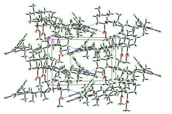
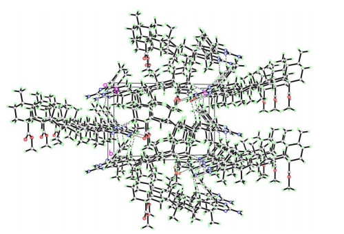
The molecular packing diagrams of compounds 5a and 5b are displayed in Figs. 3 and 4. For compound 5a, there are two orientations of molecules present in the crystal structure. Each title compound is connected with an ethanol molecule through two hydrogen bonds (N(3)–H(3A)⋅⋅⋅O(3) and O(3)–H(3C)⋅⋅⋅N(2)) (Table 2). The ethanol molecule also formed a hydrogen bond (C(1)–H(1C)⋅⋅⋅O(3)) with an adjacent molecule of 5a with different orientations. In addition, molecule 5a also formed two hydrogen bonds (N(3)–H(3B)⋅⋅⋅O(1) and C(22)–H(22B)⋅⋅⋅N(1)) with adjacent molecules in the same orientation, forming a chain along the c axis. The chains with different orientations pack alternately along the b axis, and those in the same orientation are packed along the a axis, thus forming the 3D crystal structure of compound 5a. The packing mode of compound 5b is very similar to 5a. There is an additional hydrogen bond (C(40)–H(40C)⋅⋅⋅N(1)) formed between the methyl proton at C(40) and the N(1) atom at the pyrimidine ring of its adjacent 5b molecule along the b axis.
 DownLoad:
CSV
DownLoad:
CSV
| Compound 5a | ||||
| D–H⋅⋅⋅A | d(D–H) | d(H⋅⋅⋅A) | d(D⋅⋅⋅A) | ∠DHA |
| N(3)–H(3A)⋅⋅⋅O(3)a | 0.86 | 2.34 | 3.071(8) | 143 |
| N(3)–H(3B)⋅⋅⋅O(1)a | 0.86 | 2.21 | 3.057(8) | 168 |
| O(3)–H(3C)⋅⋅⋅N(2)b | 0.82 | 2.23 | 2.837(7) | 131 |
| C(1)–H(1C)⋅⋅⋅O(3)c | 0.97 | 2.52 | 3.470(8) | 166 |
| C(22)–H(22B)⋅⋅⋅N(1)b | 0.97 | 2.55 | 3.492(8) | 163 |
| Compound 5b | ||||
| D–H⋅⋅⋅A | d(D–H) | d(H⋅⋅⋅A) | d(D⋅⋅⋅A) | ∠DHA |
| N(3)–H(3A)⋅⋅⋅O(3) | 0.86 | 2.38 | 3.091(8) | 141 |
| N(3)–H(3B)⋅⋅⋅O(1)b | 0.86 | 2.31 | 3.139(7) | 162 |
| O(3)–H(3C)⋅⋅⋅N(2) | 0.82 | 2.20 | 2.848(7) | 136 |
| C(1)–H(1C)⋅⋅⋅O(3)d | 0.97 | 2.56 | 3.514(8) | 167 |
| C(22)–H(22B)⋅⋅⋅N(1)a | 0.97 | 2.60 | 3.544(8) | 163 |
| C(40)–H(40C)⋅⋅⋅N(1)e | 0.96 | 2.57 | 3.409(9) | 146 |
| Symmetry codes: (a) x, y, −1 + z; (b) x, y, 1 + z; (c) 2 − x, −1/2 + y, 2 − z; (d) 1 − x, −1/2 + y, −z; (e) −x, −1/2 + y, −z | ||||
Compounds 5a and 5b were assayed for their in vitro cytotoxic activities against three human cancer cell lines (MCF-7, HeLa and HepG2) via the MTT colorimetric method. As a result, compound 5a showed moderate cytotoxic activities against three cancer cell lines (Table 3). Compound 5b exhibited stronger cytotoxicity against MCF-7 and HeLa cells than 5a with IC50 values of 10.71 ± 0.23 and 12.63 ± 0.31 μM, respectively, close to those of etoposide. These results indicated that this class of pyrimidine derivatives of ursolic acid could be a potential scaffold for the discovery of anticancer agents. Further researches on the in-depth structure-activity relationships (SAR) and their anticancer mechanisms will be carried out in the future.
DownLoad:
CSV
| Compound | IC50 value (µM) | ||
| MCF-7 | HeLa | HepG2 | |
| 5a | 33.07 ± 0.56 | 40.38 ± 0.62 | 26.50 ± 0.41 |
| 5b | 10.71 ± 0.23 | 12.63 ± 0.31 | > 50 |
| Etoposide | 6.51 ± 0.19 | 8.42 ± 0.16 | 8.46 ± 0.27 |
Tsai, C. J.; Nussinov, R. The molecular basis of targeting protein kinases in cancer therapeutics. Semin. Cancer Biol. 2013, 23, 235–42. doi: 10.1016/j.semcancer.2013.04.001
Bray, F.; Ferlay, J.; Soerjomataram, I.; Siegel, R. L.; Torre, L. A.; Jemal, A. Global cancer statistics 2108: GLOBOCAN estimates of incidence and mortality worldwide for 36 cancers in 185 countries. CA Cancer J. Clin. 2018, 68, 394–424. doi: 10.3322/caac.21492
Seo, D. Y.; Lee, S. R.; Heo, J. W.; No, M. H.; Rhee, B. D.; Ko, K. S.; Kwak, H. B.; Han, J. Ursolic acid in health and disease. Korean J. Physiol. Pharmacol. 2018, 22, 235–248. doi: 10.4196/kjpp.2018.22.3.235
Wen, J. Ursolic acid: pharmacokinetics process in vitro and in vivo, a mini review. Arch. Pharm. 2019, 352, e1800222. doi: 10.1002/ardp.201800222
Muto, Y.; Ninomiya, M.; Fujiki, H. Present status of research on cancer chemoprevention in Japan. Jpn. J. Clin. Oncol. 1990, 20, 219–224.
Hussain, H.; Green, I. R.; Ali, I.; Khan, I. A.; Ali, Z.; Al-Sadi, A. M.; Ahmed, I. Ursolic acid derivatives for pharmaceutical use: a patent review (2012–2016). Expert Opin. Ther. Pat. 2017, 27, 1061–1072. doi: 10.1080/13543776.2017.1344219
Jiang, W.; Huang, R. Z.; Zhang, J.; Guo, T.; Zhang, M. T.; Huang, X. C.; Zhang, B.; Liao, Z. X.; Sun, J.; Wang, H. S. Discovery of antitumor ursolic acid long-chain diamine derivatives as potent inhibitors of NF-kappa B. Bioorg. Chem. 2018, 79, 265–276. doi: 10.1016/j.bioorg.2018.05.005
Song, G.; Shen, X.; Li, S.; Li, Y.; Liu, Y.; Zheng, Y.; Lin, R.; Fan, J.; Ye, H.; Liu, S. Structure-activity relationships of 3-O-β-chacotriosyl ursolic acid derivatives as novel H5N1 entry inhibitors. Eur. J. Med. Chem. 2015, 93, 431–442. doi: 10.1016/j.ejmech.2015.02.029
Yang, H. M.; Yin, Z. Q.; Zhao, M. G.; Jiang, C. H.; Zhang, J.; Pan, K. Pentacyclic triterpenoids from Cyclocarya paliurus and their antioxidant activities in FFA-induced HepG2 steatosis cells. Phytochemistry 2018, 151, 119–127. doi: 10.1016/j.phytochem.2018.03.010
Ishikawa, T.; Donatini, R. S.; Diaz, I. E. C.; Yoshida, M.; Bacchi, E. M.; Kato, E. T. M. Evaluation of gastroprotective activity of Plinia edulis (Vell.) Sobral (Myrtaceae) leaves in rats. J. Ethnopharmacol 2008, 118, 527–529. doi: 10.1016/j.jep.2008.05.007
Fu, H. J.; Zhou, Y. R.; Bao, B. H.; Jia, M. X.; Zhao, Y.; Zhang, L.; Li, J. X.; He, H. L.; Zhou, X. M. Tryptophan hydroxylase 1 (Tph-1)-targeted bone anabolic agents for osteoporosis. J. Med. Chem. 2014, 57, 4692–4709. doi: 10.1021/jm5002293
Ramos-Hryb, A. B.; Pazini, F. L.; Kaster, M. P.; Rodrigues, A. L. S. Therapeutic potential of ursolic acid to manage neurodegenerative and psychiatric diseases. CNS Drugs 2017, 31, 1029–1041. doi: 10.1007/s40263-017-0474-4
Joshi, G.; Nayyar, H.; Alex, J. M.; Vishwakarma, G. S.; Mittal, S.; Kumar, R. Pyrimidine-fused derivatives: synthetic strategies and medicinal attributes. Curr. Top. Med. Chem. 2016, 16, 3175–3210. doi: 10.2174/1568026616666160506145046
Zhang, Y.; Wang, Y. Y.; Zhao, Y. X.; Gu, W.; Zhu, Y. Q.; Wang, S. F. Novel camphor-based pyrimidine derivatives induced cancer cell death through a ROS-mediated mitochondrial apoptosis pathway. RSC Adv. 2019, 9, 29711–29720. doi: 10.1039/C9RA05900H
El-Metwally, S. A.; Khalil, A. K.; El-Sayed, W. M. Design, molecular modeling and anticancer evaluation of thieno[2,3-d] pyrimidine derivatives as inhibitors of topoisomerase Ⅱ. Bioorg. Chem. 2020, 94, 103492. doi: 10.1016/j.bioorg.2019.103492
Sheldrick, G. M. SHELXS-97, Program for X-ray Crystal Structure Solution. University of Göttingen, Germany 1997.
Sheldrick, G. M. SHELXL-97, Program for X-ray Crystal Structure Refinement. University of Göttingen, Germany 1997.
Wang, C. J.; Delcros, J. G.; Biggerstaff, J.; Phanstiel, O. Synthesis and biological evaluation of N1-(anthracen-9-ylmethyl)triamines as molecular recognition elements for the polyamine transporter. J. Med. Chem. 2003, 46, 2663−2671. doi: 10.1021/jm030028w

Figure 1 Molecular diagram of compound 5a showing atomic labeling scheme. Displacement ellipsoids are drawn at the 30% probability level

Figure 2 Molecular diagram of compound 5b showing atom labeling scheme. Displacement ellipsoids are drawn at the 30% probability level
Table 1. Selected Bond Lengths (Å) and Bond Angles (°) of the Title Compounds
| Compound 5a | |||||||
| Bond | Dist. | Bond | Dist. | Bond | Dist. | ||
| N(1)–C(33) | 1.337(7) | O(2)–C(31) | 1.460(8) | C(11)–C(12) | 1.481(8) | ||
| N(1)–C(32) | 1.342(7) | O(3)–C(41) | 1.369(12) | C(12)–C(13) | 1.315(7) | ||
| N(2)–C(3) | 1.339(7) | C(1)–C(2) | 1.485(7) | C(17)–C(28) | 1.507(9) | ||
| N(2)–C(32) | 1.341(7) | C(2)–C(3) | 1.392(7) | C(33)–C(34) | 1.509(8) | ||
| N(3)–C(32) | 1.333(7) | C(2)–C(33) | 1.401(7) | C(36)–C(37) | 1.363(9) | ||
| O(1)–C(28) | 1.198(7) | C(3)–C(4) | 1.531(7) | C(40)–C(41) | 1.421(11) | ||
| O(2)–C(28) | 1.340(8) | C(4)–C(24) | 1.526(10) | F–C(36) | 1.342(8) | ||
| Angle | (°) | Angle | (°) | Angle | (°) | ||
| C(33)–N(1)–C(32) | 115.4(5) | C(1)–C(2)–C(3) | 119.8(5) | C(12)–C(13)–C(14) | 121.2(6) | ||
| C(3)–N(2)–C(32) | 117.5(5) | O(2)–C(28)–C(17) | 112.0(7) | C(34)–C(35)–C(36) | 116.8(6) | ||
| N(3)–C(32)–N(1) | 115.8(6) | O(1)–C(28)–O(2) | 122.2(7) | C(37)–C(36)–C(35) | 123.8(7) | ||
| F–C(36)–C(35) | 117.6(6) | O(3)–C(41)–C(40) | 110.5(11) | C(39)–C(34)–C(33) | 119.3(6) | ||
| C(1)–C(2)–C(3)–C(4) | 5.9(10) | F–C(36)–C(37)–C(38) | −179.5(7) | C(31)–O(2)–C(28)–O(1) | −1.4(11) | ||
| N(1)–C(33)–C(34)–C(39) | −36.5(8) | C(11)–C(12)–C(13)–C(14) | −1.0(11) | C(31)–O(2)–C(28)–C(17) | −178.3(6) | ||
| Compound 5b | |||||||
| Bond | Dist. | Bond | Dist. | Bond | Dist. | ||
| N(1)–C(33) | 1.329(6) | O(2)–C(31) | 1.449(10) | C(11)–C(12) | 1.486(7) | ||
| N(1)–C(32) | 1.333(7) | O(3)–C(41) | 1.374(15) | C(12)–C(13) | 1.318(7) | ||
| N(2)–C(3) | 1.355(7) | C(1)–C(2) | 1.492(6) | C(17)–C(28) | 1.514(9) | ||
| N(2)–C(32) | 1.322(7) | C(2)–C(3) | 1.387(7) | C(33)–C(34) | 1.491(7) | ||
| N(3)–C(32) | 1.365(7) | C(2)–C(33) | 1.405(7) | C(36)–C(37) | 1.386(8) | ||
| O(1)–C(28) | 1.195(7) | C(3)–C(4) | 1.531(7) | C(41)–C(42) | 1.429(9) | ||
| O(2)–C(28) | 1.343(8) | C(4)–C(24) | 1.528(9) | C(37)–C(40) | 1.490(8) | ||
| Angle | (°) | Angle | (°) | Angle | (°) | ||
| C(33)–N(1)–C(32) | 116.7(4) | C(1)–C(2)–C(3) | 121.3(4) | C(12)–C(13)–C(14) | 121.0(4) | ||
| C(3)–N(2)–C(32) | 116.3(4) | O(2)–C(28)–C(17) | 113.6(6) | C(34)–C(35)–C(36) | 121.4(5) | ||
| N(3)–C(32)–N(1) | 116.6(5) | O(1)–C(28)–O(2) | 121.9(6) | C(36)–C(37)–C(38) | 117.2(5) | ||
| C(36)–C(37)–C(40) | 121.2(5) | O(3)–C(41)–C(42) | 124.4(15) | C(39)–C(34)–C(33) | 123.5(5) | ||
| C(1)–C(2)–C(3)–C(4) | −2.9(8) | C(35)–C(36)–C(37)–C(40) | −177.1(6) | C(31)–O(2)–C(28)–O(1) | −2.1(9) | ||
| N(1)–C(33)–C(34)–C(39) | −138.8(5) | C(11)–C(12)–C(13)–C(14) | 1.7(10) | C(31)–O(2)–C(28)–C(17) | −179.6(6) | ||
 下载: 导出CSV
Table 2. Hydrogen Bond Lengths (Å) and Bond Angles (°)
| Compound 5a | ||||
| D–H⋅⋅⋅A | d(D–H) | d(H⋅⋅⋅A) | d(D⋅⋅⋅A) | ∠DHA |
| N(3)–H(3A)⋅⋅⋅O(3)a | 0.86 | 2.34 | 3.071(8) | 143 |
| N(3)–H(3B)⋅⋅⋅O(1)a | 0.86 | 2.21 | 3.057(8) | 168 |
| O(3)–H(3C)⋅⋅⋅N(2)b | 0.82 | 2.23 | 2.837(7) | 131 |
| C(1)–H(1C)⋅⋅⋅O(3)c | 0.97 | 2.52 | 3.470(8) | 166 |
| C(22)–H(22B)⋅⋅⋅N(1)b | 0.97 | 2.55 | 3.492(8) | 163 |
| Compound 5b | ||||
| D–H⋅⋅⋅A | d(D–H) | d(H⋅⋅⋅A) | d(D⋅⋅⋅A) | ∠DHA |
| N(3)–H(3A)⋅⋅⋅O(3) | 0.86 | 2.38 | 3.091(8) | 141 |
| N(3)–H(3B)⋅⋅⋅O(1)b | 0.86 | 2.31 | 3.139(7) | 162 |
| O(3)–H(3C)⋅⋅⋅N(2) | 0.82 | 2.20 | 2.848(7) | 136 |
| C(1)–H(1C)⋅⋅⋅O(3)d | 0.97 | 2.56 | 3.514(8) | 167 |
| C(22)–H(22B)⋅⋅⋅N(1)a | 0.97 | 2.60 | 3.544(8) | 163 |
| C(40)–H(40C)⋅⋅⋅N(1)e | 0.96 | 2.57 | 3.409(9) | 146 |
| Symmetry codes: (a) x, y, −1 + z; (b) x, y, 1 + z; (c) 2 − x, −1/2 + y, 2 − z; (d) 1 − x, −1/2 + y, −z; (e) −x, −1/2 + y, −z | ||||
下载: 导出CSV
Table 3. IC50 Values of Compounds 5a and 5b against Three Cancer Cells
| Compound | IC50 value (µM) | ||
| MCF-7 | HeLa | HepG2 | |
| 5a | 33.07 ± 0.56 | 40.38 ± 0.62 | 26.50 ± 0.41 |
| 5b | 10.71 ± 0.23 | 12.63 ± 0.31 | > 50 |
| Etoposide | 6.51 ± 0.19 | 8.42 ± 0.16 | 8.46 ± 0.27 |
下载: 导出CSV

 扫一扫看文章

扫一扫关注我们


 下载:
下载: