Login In
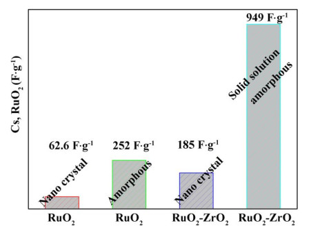
Microstructural Exploration of the High Capacitance in RuO2-ZrO2 Coating
- Corresponding author: Ji-Dong MA, majidong@xmut.edu.cn Jun-Qiu ZHU, junqiu@qztc.edu.cn
Figures(8)
Citation:
Ji-Dong MA, Yun-Miao WU, Chun-Hai JIANG, Hou-An ZHANG, Jun-Qiu ZHU. Microstructural Exploration of the High Capacitance in RuO2-ZrO2 Coating[J]. Chinese Journal of Structural Chemistry,
;2021, 40(1): 125-135.
doi:
10.14102/j.cnki.0254–5861.2011–2781

Figures(8)


Ferris, A.; Garbarino, S.; Guay, D.; Pech, D. 3D RuO2 microsupercapacitors with remarkable areal energy. Adv. Mater. 2015, 27, 6625–6629.
doi: 10.1002/adma.201503054
Wang, Y. L.; Gu, D. W.; Guo, J. R.; Xu, M. Y.; Sun, H. S.; Li, J. S.; Wang, L.; Shen, L. J. Maximized energy density of RuO2//RuO2 supercapacitors through potential dependence of specific capacitance. Chemelectrochem 2020, 7, 928–936.
doi: 10.1002/celc.201901898
Park, S.; Shin, D.; Yeo, T.; Seo, B.; Hwang, H.; Lee, J.; Choi, W. Combustion-driven synthesis route for tunable TiO2/RuO2 hybrid composites as high-performance electrode materials for supercapacitors. Chem. Eng. J. 2020, 384, 123269–5.
doi: 10.1016/j.cej.2019.123269
Jow, J. J.; Lai, H. H.; Chen, H. R.; Wang, C. C.; Wu, M. S.; Ling, T. R. Effect of hydrothermal treatment on the performance of RuO2-Ta2O5/Ti electrodes for use in supercapacitors. Electrochim. Acta 2010, 55, 2793–2798.
doi: 10.1016/j.electacta.2009.12.062
Hu, C. C.; Wang, C. W.; Wu, T. H.; Chang, K. H. Anodic composite deposition of hydrous RuO2-TiO2 nanocomposites for electrochemical capacitorss. Electrochim. Acta 2012, 85, 590–598.
Xiang, D.; Yin, L. W.; Wang, C. X.; Zhang, L. Y. High electrochemical performance of RuO2-Fe2O3 nanoparticles embedded ordered mesoporous carbon as a supercapacitor electrode material. Energy 2016, 106, 103–111.
doi: 10.1016/j.energy.2016.02.141
Gránásy, L.; James, P. F. Non-classical theory of crystal nucleation: application to oxide glasses: review. J. Non–Cryst. Solids 1999, 253, 210–230.
doi: 10.1016/S0022-3093(99)00354-3
Wong, W. Y.; Ho, C. L. Heavy metal organometallic electrophosphors derived from multi-component chromophores. Coord. Chem. Rev. 2009, 253, 1709–1758.
doi: 10.1016/j.ccr.2009.01.013
Simon, P.; Gogotsi, Y. Materials for electrochemical capacitors. Nat. Mater. 2008, 7, 845–854.
doi: 10.1038/nmat2297
Wang, G.; Zhang, L; Zhang, J. A review of electrode materials for electrochemical supercapacitors. Chem. Soc. Rev. 2012, 41, 797–828.
doi: 10.1039/C1CS15060J
González, A.; Goikolea, E.; Barrena, J. A.; Mysyk, R. Review on supercapacitors: technologies and materials. Sust. Energ. Rev. 2016, 58, 1189–1206.
doi: 10.1016/j.rser.2015.12.249
Shi, F.; Li, L.; Wang, X.; Gu, C.; Tu, J. Metal oxide/hydroxide-based materials for supercapacitors. RSC Adv. 2014, 4, 41910–41921.
doi: 10.1039/C4RA06136E
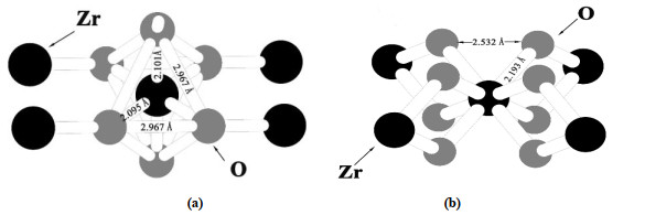
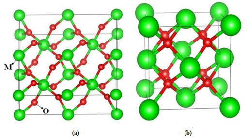
Zhu, J. Q.; Wang, X.; Yi, Z.; Tang, Z.; Wu, B.; Tang, D.; Lin, W. Stability of solid-solution phase and the nature of phase separation in Ru-Zr-O ternary oxide. J. Phys. Chem. C 2012, 116, 25832–25839.
doi: 10.1021/jp308310y
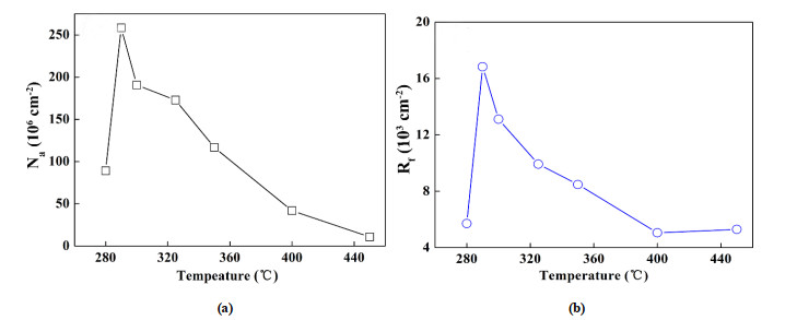
Ma, J. D.; Zuo, J.; Jiang, C. H.; Khan, D. F.; Zhang, H. A.; Zhu, J. Q. Effects of temperature on the capacitive performance of Ti/40%RuO2-60%ZrO2 electrodes prepared by thermal decomposition method. J. Electroanal. Chem. 2017, 789, 133–139.
doi: 10.1016/j.jelechem.2017.02.028
Siegrist, T.; Jost, P.; Volker, H.; Woda, M.; Merkelbach, P.; Schlockermann, C.; Wuttig, M. Disorder-induced localization in crystalline phase-change materials. Nat. Mater. 2011, 10, 202–208.
doi: 10.1038/nmat2934
Blakemore, J. D.; Mara, M. W.; Kushner-Lenhoff, M. N.; Schley, N. D.; Konezny, S. J.; Rivalta, I.; Negre, C. F. A.; Snoeberger, R. C.; Kokhan, O.; Huang, J.; Stickrath, A.; Tran, L. A.; Parr, M. L.; Chen, L. X.; Tiede, D. M.; Batista, V. S.; Crabtree, R. H.; Brudvig, G. W. Characterization of an amorphous iridium water-oxidation catalyst electrodeposited from organometallic precursors. Inorg. Chem. 2013, 52, 1860–1871.
doi: 10.1021/ic301968j
Tsuji, E.; Imanishi, A.; Fukui, K.; Nakato, Y. Electrocatalytic activity of amorphous RuO2 electrode for oxygen evolution in an aqueous solution. Electrochim. Acta 2011, 56, 2009–2016.
doi: 10.1016/j.electacta.2010.11.062
Patake, V. D.; Pawar, S. M.; Shinde, V. R.; Gujar, T. P.; Lokhande, C. D. The growth mechanism and supercapacitor study of anodically deposited amorphous ruthenium oxide films. Curr. Appl. Phys. 2010, 10, 99–103.
doi: 10.1016/j.cap.2009.05.003
Ishimaru, M.; Hirata, A.; Naito M. Electron diffraction study on chemical short-range order in covalent amorphous solids. Meth. Phy. Res. Sect. B 2012, 277, 70–76.
Playford, H.; Keen, D.; Tucker, M. Local structure of crystalline and amorphous materials using reverse monte carlo methods. Neutron News 2016, 27, 17–21.
Hrovat, M.; Holc, J.; Kolar, D. Thick film ruthenium oxide/yttria-stabilized zirconia-based cathode material for solid oxide fuel cells. Solid State Ionics 1994, 68, 99–103.
doi: 10.1016/0167-2738(94)90241-0
Altwasser, S.; Glaser, R.; Lo, A.; Liu, P.; Chao, K.; Weitkamp, J. Incorporation of RuO2 nanoparticles into MFI-type zeolites. Micropor. Mesopor. Mat. 2006, 89, 109–122.
doi: 10.1016/j.micromeso.2005.10.017
Chang, C. J.; Chu, Y. C.; Yan, H. Y.; Liao, Y. F.; Chen, H. M. Revealing the structural transformation of rutile RuO2 via in situ X-ray absorption spectroscopy during the oxygen evolution reaction. Dalton T. 2019, 48, 7122–7129.
doi: 10.1039/C9DT00138G
Nagai, Y.; Yamamoto, T.; Tanaka, T.; Yoshida, S.; Nonaka, T.; Okamoto, T.; Suda, A.; Sugiura, M. X-ray absorption fine structure analysis of local structure of CeO2-ZrO2 mixed oxides with the same composition ratio (Ce/Zr = 1). Catal. Today 2002, 74, 225–234.
doi: 10.1016/S0920-5861(02)00025-1
Biskupek, J.; Kaiser, U.; Falk, F. Heat and electron-beam-induced transport of gold particles into silicon oxide and silicon studied by in situ high-resolution transmission electron microscopy. Microsc. 2008, 57, 83–89.
Colomer, M. T.; Jurado, J. R. Preparation and characterization of gels of the ZrO2-Y2O3-RuO2 system. J. Non-Cryst. Solids 1997, 217, 48–54.
doi: 10.1016/S0022-3093(97)00125-7
Djurado, E.; Roux, C.; Hammou, A. Synthesis and structural characterization of a new system: ZrO2-Y2O3-RuO2. J. Eur. Ceram. Soc. 1996, 16, 767–771.
doi: 10.1016/0955-2219(95)00202-2
Kimura, T.; Goto, T. Preparation of RuO2-YSZ nano-composite films by MOCVD. Surf. Coat. Tech. 2003, 167, 240–244.
doi: 10.1016/S0257-8972(02)00913-1
Haines, J.; Ger, J. M.; Schulte O. Pa3 modified fluorite-type structures in metal dioxides at high pressure. Science 1996, 271, 629–631.
doi: 10.1126/science.271.5249.629
Trasatti, S. Electrocatalysis: understanding the success of DSA®. Electrochim. Acta 2000, 45, 2377–2385.
doi: 10.1016/S0013-4686(00)00338-8
Sugimoto, W.; Yokoshima, K.; Murakami, Y.; Takasu, Y. Charge storage mechanism of nanostructured anhydrous and hydrous ruthenium-based oxides. Electrochim. Acta 2006, 52, 1742–1748.
doi: 10.1016/j.electacta.2006.02.054
Pico, F.; Morales, E.; Fernandez, J. A.; Centeno, T. A.; Ibañez, J.; Rojas, R. M.; Amarilla, J. M.; Rojo, J. M. Ruthenium oxide/carbon composites with microporous or mesoporous carbon as support and prepared by two procedures. a comparative study as supercapacitor electrodes. Electrochim. Acta 2009, 54, 2239–2245.
doi: 10.1016/j.electacta.2008.10.028
Kim, H.; Popov, B. N. Characterization of hydrous ruthenium oxide/carbon nanocomposite supercapacitors prepared by a colloidal method. J. Power Sources 2002, 104, 52–61.
doi: 10.1016/S0378-7753(01)00903-X
Nanni, L.; Polizzi, S.; Benedetti, A.; De Battisti, A. Morphology, microstructure, and electrocatalytic properties of RuO2-SnO2 thin films. J. Electrochem. Soc. 1999, 146, 220–225.
doi: 10.1149/1.1391590
Chabanier, C.; Irissou, E.; Guay, D.; Pelletier, J.; Sutton, M.; Lurio, L. Hydrogen absorption in thermally prepared RuO2 electrode. Electrochem. Solid-State Lett. 2002, 5, E40–E42.
doi: 10.1149/1.1485806
Ziang Shang , Heyu Sui , Zeyi Huang , Xueting Feng , Guanzhen Chen , Jiena Weng , Yu Xiong , Yaqiong Su , Yunhu Han . Mo, B-induced local structure and electron redistribution of RuO2 for efficient acidic oxygen evolution. Chinese Chemical Letters, 2026, 37(6): 111016-. doi: 10.1016/j.cclet.2025.111016
Min LUO , Xiaonan WANG , Yaqin ZHANG , Tian PANG , Fuzhi LI , Pu SHI . Porous spherical MnCo2S4 as high-performance electrode material for hybrid supercapacitors. Chinese Journal of Inorganic Chemistry, 2025, 41(2): 413-424. doi: 10.11862/CJIC.20240205
Wen LUO , Lin JIN , Palanisamy Kannan , Jinle HOU , Peng HUO , Jinzhong YAO , Peng WANG . Preparation of high-performance supercapacitor based on bimetallic high nuclearity titanium-oxo-cluster based electrodes. Chinese Journal of Inorganic Chemistry, 2024, 40(4): 782-790. doi: 10.11862/CJIC.20230418
Hongren RONG , Gexiang GAO , Zhiwei LIU , Ke ZHOU , Lixin SU , Hao HUANG , Wenlong LIU , Qi LIU . High-performance supercapacitor based on 1D cobalt-based coordination polymer. Chinese Journal of Inorganic Chemistry, 2025, 41(6): 1183-1195. doi: 10.11862/CJIC.20250034
Mengying XU , Wen LI , Junzhong MEI , Cheng ZHANG , Kannan Palanisamy , Lei LU , Lianpeng ZHANG , Peng WANG . Manganese-doped poly(1,5-diaminonaphthalene) based high-performance supercapacitors. Chinese Journal of Inorganic Chemistry, 2026, 42(2): 387-397. doi: 10.11862/CJIC.20250211
Shiqi Peng , Yongfang Rao , Tan Li , Yufei Zhang , Jun-ji Cao , Shuncheng Lee , Yu Huang . Regulating the electronic structure of Ir single atoms by ZrO2 nanoparticles for enhanced catalytic oxidation of formaldehyde at room temperature. Chinese Chemical Letters, 2024, 35(7): 109219-. doi: 10.1016/j.cclet.2023.109219
Jiahong ZHENG , Jingyun YANG . Preparation and electrochemical properties of hollow dodecahedral CoNi2S4 supported by MnO2 nanowires. Chinese Journal of Inorganic Chemistry, 2024, 40(10): 1881-1891. doi: 10.11862/CJIC.20240170
Ao XIA , Botao YU , Jun CHEN , Guoqiang TAN . Preparation and electrochemical property of Ce-doped MnO2. Chinese Journal of Inorganic Chemistry, 2025, 41(12): 2514-2526. doi: 10.11862/CJIC.20250163
Jin CHANG . Supercapacitor performance and first-principles calculation study of Co-doping Ni(OH)2. Chinese Journal of Inorganic Chemistry, 2024, 40(9): 1697-1707. doi: 10.11862/CJIC.20240108
Qing Li , Yumei Feng , Yuhua Xie , Qi Xu , Yifei Li , Yingjie Yu , Fang Luo , Zehui Yang . MOF derived RuO2/V2O5 nanoneedles for robust and stable water oxidation in acid. Chinese Chemical Letters, 2025, 36(7): 111074-. doi: 10.1016/j.cclet.2025.111074
Qing Li , Yumei Feng , Yingjie Yu , Yazhou Chen , Yuhua Xie , Fang Luo , Zehui Yang . Engineering eg filling of RuO2 enables a robust and stable acidic water oxidation. Chinese Chemical Letters, 2025, 36(3): 110612-. doi: 10.1016/j.cclet.2024.110612
Juan Zhu , Jingxiang Xia , Wei Luo . Oxophilic support mediated interfacial water reconstruction on RuO2 for high-efficiency proton exchange membrane electrolysis. Chinese Journal of Structural Chemistry, 2026, 45(4): 100857-100857. doi: 10.1016/j.cjsc.2025.100857
Jing Cao , Dezheng Zhang , Bianqing Ren , Ping Song , Weilin Xu . Mn incorporated RuO2 nanocrystals as an efficient and stable bifunctional electrocatalyst for oxygen evolution reaction and hydrogen evolution reaction in acid and alkaline. Chinese Chemical Letters, 2024, 35(10): 109863-. doi: 10.1016/j.cclet.2024.109863
Xiaoyu Zhao , Kai Gao , Sen Xue , Wei Ran , Rui Liu . Synergistic effects of oxygen vacancies and Pd single atoms on Pd@TiO2−x for efficient HER catalysis. Chinese Chemical Letters, 2025, 36(6): 110309-. doi: 10.1016/j.cclet.2024.110309
Qiangqiang SUN , Pengcheng ZHAO , Ruoyu WU , Baoyue CAO . Multistage microporous bifunctional catalyst constructed by P-doped nickel-based sulfide ultra-thin nanosheets for energy-efficient hydrogen production from water electrolysis. Chinese Journal of Inorganic Chemistry, 2024, 40(6): 1151-1161. doi: 10.11862/CJIC.20230454
Anqun LAI , Qiaoyu WU , Qingqing LIANG , Qiyong LI , Guowen DONG , Yongjie DING , Jia′nan CHEN , Qing YAN , Zhonghua PAN , Wangchuan XIAO . Electrocatalytic water oxidation properties of Nd-Co polynuclear complexes. Chinese Journal of Inorganic Chemistry, 2025, 41(12): 2527-2535. doi: 10.11862/CJIC.20250151
Doudou Liu , Weiwei Guo , Guoliang Mei , Youpeng Dan , Rong Yang , Chao Huang , Yanling Zhai , Xiaoquan Lu . Application of catalyst Cu-t-ZrO2 based on the electronic metal-support interaction in electrocatalytic nitrate reduction. Chinese Chemical Letters, 2025, 36(8): 110578-. doi: 10.1016/j.cclet.2024.110578
Zhaomei LIU , Wenshi ZHONG , Jiaxin LI , Gengshen HU . Preparation of nitrogen-doped porous carbons with ultra-high surface areas for high-performance supercapacitors. Chinese Journal of Inorganic Chemistry, 2024, 40(4): 677-685. doi: 10.11862/CJIC.20230404
Yanhui XUE , Shaofei CHAO , Man XU , Qiong WU , Fufa WU , Sufyan Javed Muhammad . Construction of high energy density hexagonal hole MXene aqueous supercapacitor by vacancy defect control strategy. Chinese Journal of Inorganic Chemistry, 2024, 40(9): 1640-1652. doi: 10.11862/CJIC.20240183
Huirong BAO , Jun YANG , Xiaomiao FENG . Preparation and electrochemical properties of NiCoP/polypyrrole/carbon cloth by electrodeposition. Chinese Journal of Inorganic Chemistry, 2025, 41(6): 1083-1093. doi: 10.11862/CJIC.20250008
 DownLoad: