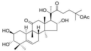
Figure 1.
Structure of cucurbitacin IIa
Biological Activities and Crystal Structure of the Natural Anti-cancer Drug: Cucurbitacin IIa
Kun YU , Ying LI , Hai-Jiao CHEN , Bo LIU , Qing-Qiang YAO

Cucurbitacin IIa mainly exists in the rhizome of Hemsleya, which is a cucurbitaceous plant. It has the functions of anticancer, bacteriostasis, anti-gastric ulcer, antipyretic, detoxification, pain relief and anti-HIV. It is widely used in the treatment of hepatitis, coronary heart disease and tracheitis[1-3]. Cucurbitacin IIa was investigated for in vitro anti-cancer activities in different cancer cell lines such as HepG2 (hepatic carcinoma), MCF-7 (breast carcinoma), SKOV3 (ovarian carcinoma), HT-29 and LOVO (colon carcinoma) using SRB assay[4].
In these experiments, cisplatin, a known anti-cancer drug, was used as a positive control[5, 6]. In this paper, cucurbitacin IIa was evaluated for the inhibition of CDK1/cyclinB, AMPKα2, EGFR, GSK3α, JAK2, MAPK1, mTOR, PKBα, and PI3K p110α(E542K)/p85α via a KinaseProfiler radiometric protein kinase assay, and the structural characteristic and anti-cancer activity of cucurbitacin IIa were introduced in order to provide a reference for further study of this excellent natural medicine.
DMSO, methanol, Hexane, dichloromethane, sulforhodamine B, fetal bovine serum, sodium bicarbonate and other materials were obtained from commercial sources and used without further purification.
High-resolution mass spectroscopy (HRMS) was performed on an AB SCIEX X500R Accurate Mass Q-TOF by using electrospray ionization (ESI). The NMR spectra were recorded on a Bruker AM-600 spectrometer (Billerica, MA).
White solid, HRMS (ESI) calcd. for C32H50O8Na (M+Na)+ 585.3403, found 585.3388.
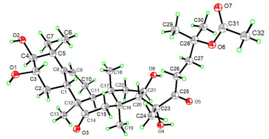
A single crystal of cucurbitacin IIa suitable for X-ray diffraction analysis was grown by slow evaporation of CH2Cl2/hexane solutions of cucurbitacin IIa at 4 ℃. X-ray diffraction was performed on a Bruker D8 Venture diffractometer[9]. Data were collected at 173 K by using a graphite monochromator with CuKα radiation (1.54178 Å) in the ω-ϕ scanning mode. Using ShelXL-2014/7, the structure was solved with the ShelXS17 structure solution program by direct methods and refined with the ShelXL-2014/7 refinement package using the least-squares minimization. Hydrogen and oxygen atoms were located using the geometric method. Details of crystal data, data collections, and structure refinement are summarized in Table 1.
 DownLoad:
CSV
DownLoad:
CSV
| Bond | Dist. | Bond | Dist. | Bond | Dist. | ||
| C(31)–O(7) | 1.214(4) | C(23)–O(4) | 1.421(3) | C(3)–O(1) | 1.417(4) | ||
| C(31)–O(6) | 1.317(4) | C(14)–O(3) | 1.210(4) | C(4)–O(2) | 1.432(3) | ||
| C(25)–O(5) | 1.216(4) | C(3)–C(4) | 1.517(5) | C(21)–O(8) | 1.325(4) | ||
| Angle | (°) | Angle | (°) | Angle | (°) | ||
| O(4)–C(23)–C(24) | 108.7(3) | C(23)–C(20)–C(21) | 113.3(2) | O(3)–C(14)–C(15) | 119.9(3) | ||
| O(4)–C(23)–C(25) | 107.1(2) | C(23)–C(20)–C(16) | 120.7(2) | O(9)–Ca(1)–O(3) | 138.40(5) | ||
| O(4)–C(23)–C(20) | 111.3(2) | C(21)–C(20)–C(16) | 103.1(2) | O(3)–Ca(1)–O(4) | 139.17(4) | ||
| O(3)–C(14)–C(12) | 122.1(2) | C(13)–C(12)–C(11) | 107.8(2) | C(24)–C(23)–C(25) | 106.8(3) | ||
| O(8)–C(21)–H(21) | 109.7 | C(3)–C(2)–C(1) | 111.2(2) | C(4)–C(3)–C(2) | 108.7(2) | ||
| C(3)–C(4)–C(5) | 113.0(2) | C(3)–C(4)–C(5) | 113.0(2) | C(8)–C(5)–C(4) | 110.5(2) | ||
| C(1)–C(8)–C(5) | 115.7(2) | O(8)–C(21)–C(22) | 113.6(2) | C(13)–C(12)–C(1) | 110.6(2) | ||
| O(1)–C(3)–C(4) | 110.5(2) | O(1)–C(3)–C(2) | 113.8(2) | O(2)–C(4)–C(3) | 111.7(3) |
LOVO (colon carcinoma), SKOV3 (ovarian carcinoma), HT-29 (colon carcinoma), MCF-7 (breast carcinoma), and HepG2 (hepatic carcinoma) cell lines (purchased from the Cell Bank of the Chinese Academy of Sciences, Shanghai, China) were cultured in minimum essential medium (modified) with 1.5 mM L-glutamine adjusted to contain 2.2 g/L sodium bicarbonate (90%) and fetal bovine serum (10%). All cells were cultured in a humidified atmosphere containing 5% CO2 at 37 ℃.
The antiproliferative activities of cucurbitacin IIa against LOVO, SKOV3, HT-29, MCF-7 and HepG2 cell lines were measured using the sulforhodamine B (SRB) assay with cisplatin as reference. Cells were seeded in 96-well plates and then treated with different drug concentrations. After incubation for 72 h, cells were fixed with 10% trichloroacetic acid for 1 h at 4 ℃, washed five times with tap water, and air-dried. Cells that survived were stained with 0.4% (w/v) sulforhodamine B (SRB) for 20 min at room temperature and washed five times with 1% acetic acid. Bound SRB was solubilized with 10 mM Tris and absorbance was measured at 540 nm[4].
Kinases CDK1/cyclinB, AMPKα2, EGFR, GSK3α, JAK2, and PKBα were diluted with a buffer comprising 20 mM MOPS, 1 mM EDTA, 0.01% Brij-35, 5% glycerol, 0.1% β-mercaptoethanol, and 1 mg/mL BSA before adding to the reaction mixture. Kinase mTOR was diluted with a buffer comprising 500 mM HEPES, 10 mM EGTA, and 0.1% Tween 20 before adding to the reaction mixture. Kinase MAPK1 was diluted with a buffer comprising 50 mM TRIS, 0.1 mM EGTA, 0.1 mM Na3VO4, 0.1% β-mercaptoethanol, and 1 mg/mL BSA before adding to the reaction mixture. CDK1/cyclinB (h) was incubated with 8 mM MOPS (pH 7.0), 0.2 mM EDTA, 0.1 mg/mL histone H1, 10 mM magnesium acetate, and [γ-33P]-ATP (specific activity and concentration as required). The reaction was initiated by adding the Mg/ATP mix. After incubating for 40 min at room temperature, the reaction was stopped by adding phosphoric acid to a concentration of 0.5%. A 10 μL sample of the reaction was then spotted onto a P30 filtermat and washed with 0.425% phosphoric acid four times for 4 min, and once in methanol, prior to drying and scintillation counting.
Cucurbitacin IIa was characterized by IR and NMR spectroscopy. For instance, the IR spectra showed three absorption bands in the range of 3566~3408 cm-1 for hydroxyl groups and the 1691 cm-1 peak indicates that the compound has carbonyl group. The 13C-NMR spectra exhibited characteristic peaks at δ 215.1 and 213.1 ppm for carbonyls[10], and at δ 142.4 and 118.7 ppm for the double bond of cucurbitacin IIa.
The single-crystal X-ray diffraction study of cucurbitacin IIa was performed to confirm the structure of the obtained compound. The crystallographic data with refinement parameters, selected bond lengths and bond angles are given in the Supporting Information. The X-ray crystal structure of cucurbitacin IIa is shown in Fig. 2. The selected bond lengths and bond angles are given in Table 1, and the C–C bond angles of the six-membered ring including C(1) to C(5) and C(8) fall in the 108.7~115.7° range, which is consistent with chair conformation[11]. The bond angles of O(1)–C(3)–C(2) and O(2)–C(4)–C(5) are 113.8° and 108.8°, indicating these two hydroxyl groups are in para to each other. The bond angle 111.3° of O(4)–C(23)–C(20) proves the chirality of branched chains. The bond lengths of C(31)–O(7) and C(14)–O(3) of two carbonyl functionalities in the compound are 1.214 and 1.210 Å. The bond length of C(8)–C(9) is 1.325 Å, which proves the existence of double bonds in the ring of cucurbitacin IIa[12].
The in vitro cell proliferation assays were performed as previously described. Briefly, the antiproliferative effects of cucurbitacin IIa against HepG2 (hepatic carcinoma), MCF-7 (breast carcinoma), SKOV3 (ovarian carcinoma), HT-29 and LOVO (colon carcinoma) cell lines were measured by the SRB assay, and cucurbitacin IIa has better activity than cisplatin against cancer cell lines.
DownLoad:
CSV
| Compounds | IC50 (μM)a | ||||
| HepG2 | MCF-7 | SKOV3 | HT-29 | LOVO | |
| Cucurbitacin IIa | 0.45 ± 0.07 | 0.22 ± 0.01 | 0.15 ± 0.07 | 0.25 ± 0.07 | 0.10 ± 0.01 |
| Cisplatinb | 2.00 ± 0.14 | 1.65 ± 0.01 | 0.50 ± 0.14 | 2.15 ± 0.07 | 0.90 ± 0.14 |
| a IC50 values were calculated by using the SRB assay after 72 h treatment. The values were reported as the Mean ± SD. b Cisplatin is a positive control drug. |
|||||
To explore the mechanism of cucurbitacin IIa, we tested the biochemical activities of cucurbitacin IIa against CDK1/cyclinB, AMPKα2, EGFR, GSK3α, JAK2, MAPK1, mTOR, PKBα, and PI3K p110α(E542K)/p85α via a KinaseProfiler radiometric protein kinase assay[13-15]. As shown in Tables 3, cucurbitacin IIa has a high selectivity because it has a good inhibitory activity aganist the kinase CDK1/cyclin B, but no inhibitory effects against other kinases.
DownLoad:
CSV
| Compound | Activity (% control) | ||||||||
| CDK1/cyclinB | AMPKα2 | EGFR | GSK3α | JAK2 | MAPK1 | mTOR | PKBα | PI3K p110α(E542K)/p85α | |
| Cu IIa | 26 | 81 | 89 | 104 | 114 | 103 | 71 | 90 | 87 |
In summary, the structure of cucurbitacin IIa from the rhizomes of Hemsleya pengxianensis was characterized by IR, HRMS, NMR spectroscopy, and its structure was confirmed by X-ray crystallography. The antiproliferative effects of cucurbitacin IIa against HepG2, MCF-7, SKOV3, HT-29 and LOVO cell lines were better than the cisplatin against cancer cell lines. Cucurbitacin IIa was evaluated for inhibition of CDK1/cyclinB, AMPKα2, EGFR, GSK3α, JAK2, MAPK1, mTOR, PKBα, and PI3K p110α(E542K)/p85α via a Kinase-Profiler radiometric protein kinase assay. It was found that cucurbitacin IIa has high inhibitory activity and good selectivity to CDK1/cyclinB. Structural characterization and activity determination of cucurbitacin IIa provide meaningful references for the future pharmacological applications and structural modification.
Boykin, C.; Zhang, G.; Chen, Y. H.; Zhang, R. W.; Fan, X. E.; Yang, W. M.; Lu, Q. Cucurbitacin IIa: a novel class of anti-cancer drug inducing non-reversible actin aggregation and inhibiting survivin independent of JAK2/STAT3 phosphorylation. Brit. J. Cancer 2011, 104, 781−789. doi: 10.1038/bjc.2011.10
Tian, R. R.; Chen, J. C.; Zhang, G. H.; Qiu, M. H.; Wang, Y. H.; Du, L.; Shen, X.; Liu, N. F.; Zheng, Y. T. Anti-HIV-1 activities of hemslecins A and B. Chin. J. Nat. Med. 2008, 6, 214−218. doi: 10.3724/SP.J.1009.2008.00214
Zhi, B. Y.; Yan, Y.; Yi, Z. L. Investigation of antimicrobial model of Hemsleya pengxianensis W. J. Chang and its main active component by metabolomics technique. J. Ethnopharmacol. 2008, 116, 89−95. doi: 10.1016/j.jep.2007.11.008
Quan, H. T.; Liu, H. F.; Li, C.; Lou, L. G. 1,4-Diamino-2,3-dicyano-1,4-bis(methylthio)butadiene (U0126) enhances the cytotoxicity of combretastatin A4 independently of mitogen-activated protein kinase kinase. J. Pharmacol. Exp. Ther. 2009, 330, 326−333. doi: 10.1124/jpet.109.153320
Wang, W. W.; Yang, H. R.; Li, Y.; Zheng, Z. F.; Liu, Y. J.; Wang, H. Y.; Mu, Y. L.; Yao, Q. Q. Identification of 16,25-O-diacetyl-cucurbitane F and 25-O-acetyl-23,24-dihydrocucurbitacin F as novel anti-cancer chemicals. R. Soc. Pen. Sci. 2018, 5, 1−16.
Li, Y.; Wang, W. X.; Zheng, Z. F.; Mu, Y. L.; Liu, Y. J.; Wang, H. Y.; Li, L.; Yao, Q. Q. Eight new cucurbitane triterpenoids from "Xue Dan", the roots of Hemsleya pengxianensis. J. Asian. Nat. Prod. Res. 2017, 20, 36−48.
Reguero, M. T.; Mata, R.; Bye, R.; Linares, E.; Delgado, G. Chemical studies on Mexican plants used in traditional medicine, Ⅱ: cucurbitacins from Hintonia latiflora. J. Nat. Pro. 1987, 50, 315−316. doi: 10.1021/np50050a050
Maloney, K. N.; Fujita, M.; Eggert, U. S.; Schroeder, F. C.; Field, C. M.; Mitchison, T. J.; Clardy, J. Actin-aggregating cucurbitacins from physocarpus capitatus. J. Nat. Pro. 2008, 71, 1927−1929. doi: 10.1021/np8005259
Zhang, X.; Ma, X. Y.; Zhang, T. Y.; Li, B.; Jiang, S.; Zhang, G. H.; Hai, L.; Wang J. C.; Shao, X. The influence of phosphine ligand substituted [2Fe2S] model complexes as electro-catalyst on proton reduction. RSC Adv. 2018, 8, 42262−42268. doi: 10.1039/C8RA08016J
Dang, G. V.; Rode, B. M.; Stuppner, H. Quantitative electronic structure-activity relationship (QESAR) of natural cytotoxic compounds: maytansinoids, quassinoids and cucurbitacins. Eur. J. Pharm. Sci. 1994, 2, 331−350. doi: 10.1016/0928-0987(94)00061-1
Gorobets, E.; Stepanenko, V.; Wicha, J. Enantioselective total synthesis of a trans-hydrindane rings/side-chain building-block of Vitamin D-asymmetric induction in an acid-catalyzed conjugate-addition reaction. J. Nat. Pro. 2004, 2004, 783−799.
Ren, J.; Shi, X.; Li, X. N.; Li, L. W.; Su, J.; Shao, L. D.; Zhao, Q. S. Synthesis of a small-molecule library with skeletal diversity from hemslecin A via the reaction-discovery strategy. Org. Lett. 2016, 18, 3948−3951. doi: 10.1021/acs.orglett.6b01654
Bengoechea-Alonso, M. T.; Ericsson, J. Cdk1/cyclin B-mediated phosphorylation stabilizes SREBP1 during mitosis. Cell Cycle (Georgetown, Tex. ) 2006, 5, 1708−1718. doi: 10.4161/cc.5.15.3131
Chu, R.; Terrano, D. T.; Chambers, T. C. Cdk1/cyclin B plays a key role in mitotic arrest-induced apoptosis by phosphorylation of Mcl-1, promoting its degradation and freeing Bak from sequestration. Biochem. Pharmacol. 2012, 83, 199−206. doi: 10.1016/j.bcp.2011.10.008
Kundu, J.; Kim, D. H.; Kundu, J. K.; Chun, K. S. Thymoquinone induces heme oxygenase-1 expression in HaCaT cells via Nrf2/ARE activation: Akt and AMPKα as upstream targets. Food Chem. Toxicol. 2014, 65, 18−26. doi: 10.1016/j.fct.2013.12.015
Table 1. Selected Bond Lengths (Å) and Bond Angles (°)
| Bond | Dist. | Bond | Dist. | Bond | Dist. | ||
| C(31)–O(7) | 1.214(4) | C(23)–O(4) | 1.421(3) | C(3)–O(1) | 1.417(4) | ||
| C(31)–O(6) | 1.317(4) | C(14)–O(3) | 1.210(4) | C(4)–O(2) | 1.432(3) | ||
| C(25)–O(5) | 1.216(4) | C(3)–C(4) | 1.517(5) | C(21)–O(8) | 1.325(4) | ||
| Angle | (°) | Angle | (°) | Angle | (°) | ||
| O(4)–C(23)–C(24) | 108.7(3) | C(23)–C(20)–C(21) | 113.3(2) | O(3)–C(14)–C(15) | 119.9(3) | ||
| O(4)–C(23)–C(25) | 107.1(2) | C(23)–C(20)–C(16) | 120.7(2) | O(9)–Ca(1)–O(3) | 138.40(5) | ||
| O(4)–C(23)–C(20) | 111.3(2) | C(21)–C(20)–C(16) | 103.1(2) | O(3)–Ca(1)–O(4) | 139.17(4) | ||
| O(3)–C(14)–C(12) | 122.1(2) | C(13)–C(12)–C(11) | 107.8(2) | C(24)–C(23)–C(25) | 106.8(3) | ||
| O(8)–C(21)–H(21) | 109.7 | C(3)–C(2)–C(1) | 111.2(2) | C(4)–C(3)–C(2) | 108.7(2) | ||
| C(3)–C(4)–C(5) | 113.0(2) | C(3)–C(4)–C(5) | 113.0(2) | C(8)–C(5)–C(4) | 110.5(2) | ||
| C(1)–C(8)–C(5) | 115.7(2) | O(8)–C(21)–C(22) | 113.6(2) | C(13)–C(12)–C(1) | 110.6(2) | ||
| O(1)–C(3)–C(4) | 110.5(2) | O(1)–C(3)–C(2) | 113.8(2) | O(2)–C(4)–C(3) | 111.7(3) |
 下载: 导出CSV
Table 2. IC50 (μM) Values of Cucurbitacin IIa and Cisplatin against Cancer Cell Lines
| Compounds | IC50 (μM)a | ||||
| HepG2 | MCF-7 | SKOV3 | HT-29 | LOVO | |
| Cucurbitacin IIa | 0.45 ± 0.07 | 0.22 ± 0.01 | 0.15 ± 0.07 | 0.25 ± 0.07 | 0.10 ± 0.01 |
| Cisplatinb | 2.00 ± 0.14 | 1.65 ± 0.01 | 0.50 ± 0.14 | 2.15 ± 0.07 | 0.90 ± 0.14 |
| a IC50 values were calculated by using the SRB assay after 72 h treatment. The values were reported as the Mean ± SD. b Cisplatin is a positive control drug. |
|||||
下载: 导出CSV
Table 3. Inhibition of Kinases for Cucurbitacin IIa (1 µM)
| Compound | Activity (% control) | ||||||||
| CDK1/cyclinB | AMPKα2 | EGFR | GSK3α | JAK2 | MAPK1 | mTOR | PKBα | PI3K p110α(E542K)/p85α | |
| Cu IIa | 26 | 81 | 89 | 104 | 114 | 103 | 71 | 90 | 87 |
下载: 导出CSV

 扫一扫看文章

扫一扫关注我们


 下载:
下载: