Table 1.
Selected Bond Lengths (Å) and Bond Angles (°) for 1
Citation:
Li-Ping XUE, Ke-Xin DING. Synthesis, Structure and Luminescence Property of a New Two-dimensional Znic(Ⅱ) Coordination Polymer[J]. Chinese Journal of Structural Chemistry,
2020, 39(7): 1295-1300.
doi:
10.14102/j.cnki.0254–5861.2011–2561

Synthesis, Structure and Luminescence Property of a New Two-dimensional Znic(Ⅱ) Coordination Polymer
English
Synthesis, Structure and Luminescence Property of a New Two-dimensional Znic(Ⅱ) Coordination Polymer
Abstract:
Based on 5-dimethylamino-isophthalic acid (H2dia) and 1,2-bis(4-pyridyl)ethylene (bpe) mixed linkers, a new d10-configuration Zn(Ⅱ) coordination polymer material, [Zn(dia)(bpe)(H2O)]n (1), was synthesized. In the asymmetric unit, there are one Zn2+ ion, two dia2− anions, one neutral bpe linker and one free H2O molecule. In the complex, adjacent Zn2+ ions are alternately bridged by the dia2− dianions to generate chains which are further connected by flexible bpe ligands to form a two-dimensional (2D) layer structure. In topology, the structure of 1 represents a non-interpenetrating 4-connected sql topology. Moreover, infrared spectroscopy, elemental analyses, thermogravimetric analysis, powder X-ray diffraction, solid UV-Vis absorption spectra and photoluminescent property of 1 are also investigated.
-
1. INTRODUCTION
Coordination polymers (CPs), which can be readily self-assembled from metal centers and organic ligands, are attractive crystalline materials with real-world applications in ion exchange, sensors, luminescence, molecular magnetism and heterogeneous catalysis[1-4]. Generally speaking, many factors, for instance, organic ligands, central metals, solvent, counter anion, pH, concentration, the ratio of metal-to-ligand and temperature are associated with its synthesis[5-12]. Among the variety of organic ligands thus far used for the assembly of CPs, polycarboxylic acids are the most extensively explored owing to the diverse coordination modes and strong binding ability to metal centers.
As an isophthalate derivative, the rigid 5-dimethylamino-isophthalic acid (H2dia) possesses potential coordination sites involving four oxygen atoms of two carboxylate groups thereby paving a pathway for the construction of new CPs. Moreover, the rigid dimethylamino group of H2dia may exhibit additional structural constraint in controlling the assembly of CPs. In this regard, we hope to reveal some structural factors of the H2dia for dominating the self-assembly of CPs, and this will provide more useful information of the substituent effect. Though as a good polycarboxylic ligand, H2dia remains largely unexplored, and only few CPs have been reported[13, 14].
Recently, we have reported a zinc(Ⅱ) CP material with Lewis basic pyridyl sites by adjusting the ratio of metal-to-ligand[14]. In order to further enrich CPs from H2dia ligand, herein we report a new CP, [Zn(dia)(bpe)(H2O)]n, constructed from H2dia and 1,2-bis(4-pyridyl)ethylene (bpe) mixed linkers by adjusting the ratio of metal-to-ligand. The complex is a 2D layer structure with sql topology. What is more, the solid UV-Vis absorption spectra and photoluminescent property were investigated.
2. EXPERIMENTAL
All chemicals used in this work were obtained from commercial sources and required no further purification. Elemental analysis (C, H, N) was carried out on a Flash 2000 elemental analyzer. The IR spectra were measured on a Nicolet Avatar-360 spectrometer in the range of 4000~400 cm−1 range. Powder X-ray diffraction (PXRD) measurements were performed on a Bruker D8-ADVANCE X-ray diffractometer with CuKα radiation (λ = 1.5418 Å). Thermogravimetric analyses (TGA) were carried out on a SDTQ600 thermogravimetric analyzer. A platinum pan was used for heating the sample with a heating rate of 10 ℃/min under a dry air atmosphere. The solid-state UV-Vis diffuse-reflectance spectra were recorded with a TU-1901 spectrophotometer equipped with IS19-1 integrating sphere. Fluorescence measurements were recorded on a Hitachi F4500 fluorescence spectrophotometer.
2.1 Synthesis of [Zn(dia)(bpe)(H2O)]n
A mixture of H2dia (20.9 mg, 0.1 mmol), Zn(NO3)2·6H2O (29.8 mg, 0.1 mmol), bpe (9.1 mg, 0.05 mmol), 0.10 M NaOH (0.25 mL) and 5 mL deionized water was sealed in a 25 mL glass bottle and heated at 60 ℃ for 2 days, followed by cooling to room temperature at a rate of 5 ℃·h-1. Red block crystals of 1 were collected (yield: 35% based on bpe). Elemental analysis calcd. (%) for C22H21N3O5Zn: C, 55.84; H, 4.44; N, 8.88. Found (%): C, 55.73; H, 4.49; N, 8.73. IR data (KBr, cm-1): 3453(w), 3072(w), 2937(w), 1602(s), 1541(s), 1560(m), 1412(m), 1373(s), 1270(s), 1226(m), 1201(m), 1149(w), 1123(w), 1066(s), 1025(m), 1013(m), 999(m), 951(s), 829(s), 765(s), 719(s), 678(m), 659(m).
2.2 X-ray structure determination
The structure of 1 was determined by single-crystal X-ray diffraction technique. Diffraction data were collected on an Oxford Diffraction Gemini with MoKα radiation (λ = 0.71073 Å) at 293 K. The structure was solved by direct methods using the Olex2 program as an interface together with the SHELXT and SHELXL programs in order to solve and refine the structure, respectively[15-17]. All non-hydrogen atoms were refined anisotropically. The hydrogen atoms on water molecules were located from difference Fourier maps and refined using a riding model. Other hydrogen atoms were placed at the calculation positions. Selected bond distances and bond angles of 1 are summarized in Table 1. Crystal data for 1: triclinic, space group P
$ \overline 1 $ , a = 9.6641(4), b = 11.0116(5), c = 11.2348(4) Å, α = 110.762(4)°, β = 109.708(4)°, γ = 92.548(3)°, V = 1034.03(8) Å3, Z = 2, Mr = 472.79, Dc = 1.518 Mg/m3, μ = 1.228 mm-1, F(000) = 488, the final R = 0.0480 and wR = 0.1318 for 3630 observed reflections with I > 2σ(I) and R = 0.0534 and wR = 0.1355 for all data. The goodness-of-fit indicator (S) is 1.077.Table 1

 DownLoad:
CSV
DownLoad:
CSV
Bond Dist. Bond Dist. Bond Dist. Zn(1)–O(1) 2.005(3) Zn(1)–N(2) 2.189(3) Zn(1)–N(1) 2.072(3) Zn(1)–O(5) 2.180(3) Zn(1)–O(4)#3 2.061(3) Angle (°) Angle (°) Angle (°) N(1)–Zn(1)–N(2) 92.84(12) O(5)–Zn(1)–N(1) 90.80(13) O(4)#3–Zn(1)–O(5) 86.82(13) O(5)–Zn(1)–N(2) 176.31(12) O(5)–Zn(1)–O(3)#3 84.06(12) O(4)#3–Zn(1)–N(1) 153.69(12) O(4)#3–Zn(1)–N(2) 89.64(11) Symmetry transformation: #1: 1 – x, –y, –z; #2: 2 – x, –y, 2 – z; #3: 1 + x, y, z 3. RESULTS AND DISCUSSION
Single-crystal X-ray diffraction analysis reveals that 1 crystallizes in triclinic system with space group P
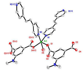
$ \overline 1 $ . In the asymmetric unit, there are one crystallographically independent Zn2+ ion, one dia2− anion, two halves of neutral bpe linkers, and one free H2O molecule. As shown in Fig. 1, each central zinc atom is coordinated by two oxygen atoms from two individual dia2− anions, two pyridyl nitrogen atoms from two bpe linkers, and one oxygen atom from one free water molecule. The Addison parameter τ for the Zn(1) atom is 0.38, indicating that the coordination environment of Zn(1) atom is closer to the square-pyramidal geometry than a trigonal-bipyramidal configuration. The Zn−O/N bond lengths falling in the range of 2.005(3)~2.189(3) Å are in a normal range (Table 1)[18, 19].Figure 1
 Figure 1. Coordination environment of the Zn2+ ions in 1, showing the atom numbering scheme. Displacement ellipsoids are drawn at the 30% probability level. All hydrogen atoms are omitted. Symmetry codes: (#1) 1 – x, –y, –z; (#2) 2 – x, –y, 2 – z; (#3) 1 + x, y, z
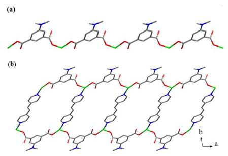
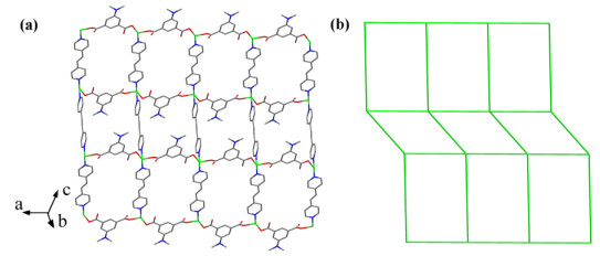
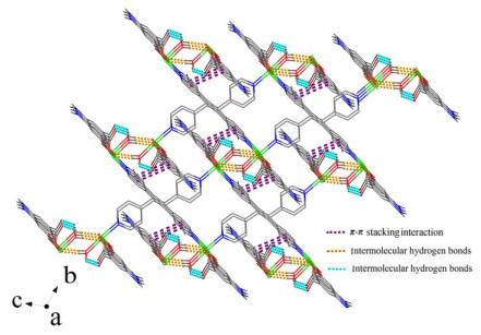
In 1, adjacent Zn2+ ions are interlinked by bridging dia2− anions to form a one-dimensional (1D) [ZnL]n chain extending along the ac plane (Fig. 2a), in which the dia2− anion adopts a bis-monodentate coordination fashion. Noted, bpe linkers with anti conformation could be classified into two types according to their contributions to the structure formation. The bpe1 takes as a bridging linker to extend 1D chains into a double-chain structure (Fig. 2b). Such arrays are further interlinked via bpe2 to finally produce a 2D coordination network (Fig. 3a). From the view of topology, the Zn2+ ions can act as four-connecting nodes that are jointed to each other via dia2− anions and bpe linkers, affording a non-interpenetrating 44-sql network (Fig. 3b). Compared with the literature, many Znic(Ⅱ) CPs synthesized based on bpe and functionalized isophthalate ligands with flexible substituents such as 5-ethoxy, 5-n-propoxy, 5-n-butoxy, 5-n-pentyloxy, 5-azido and 5-(hydroxymethyl) groups are more likely to exhibit interpenetrating or polycatenating structures[20-22]. In addition, such 2D layers of 1 are held together via π-π stacking interaction involving the phenyl rings of dia2− anions and pyridyl rings of bpe linkers (centroid-to-centroid distance of 3.767(3) Å and dihedral angle of 19.658°), as well as intermolecular hydrogen bonds between the carboxylate oxygen atoms and water molecules, generating a three-dimensional (3D) supramolecular structure (Table 2, Fig. 4).
Figure 2
 Figure 2. (a) 1D chain of complex 1. (b) View of the double chain of complex 1
Figure 3
 Figure 3. (a) 2D coordination layer of 1. (b) Schematic representation of the 4-connected sql topology
Table 2
Table 2. Hydrogen Bond Lengths (Å) and Bond Angles (°) in 1DownLoad:
CSV
D–H…A d(D–H) d(H…A) d(D…A) ∠DHA O(5)–H(1W)···O(2) 0.86 2.04 2.577(6) 120 O(5)–H(2W)···O(3)#4 0.86 1.88 2.702(5) 161 Symmetry code: #4: 1 – x, 1 – y, 1 – z Figure 4
 Figure 4. View of the 3D supramolecular structure of 1
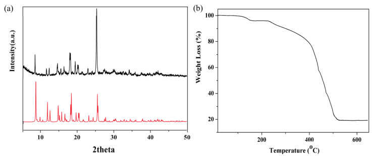
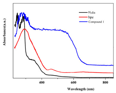
As seen in Fig. 5a, the PXRD pattern of the obtained crystalline complex 1 agreed well with that simulated from the single crystal data, proving the high phase purity of the synthesized sample. The TGA was also investigated under dry air atmosphere at a heating rate of 10 ℃/min (Fig. 5b). The weight loss of 3.85% from 141 to 150 ℃ is attributed to the loss of free H2O molecules (calcd. 3.81%). And the resulting water-free structure is stable up to 249 ℃. Beyond 249 ℃, the abrupt weight loss corresponds to the release of organic ligands. In order to further characterize complex 1, the solid UV-Vis absorption spectra of complex 1 together with the H2L and bpe ligands are investigated at room temperature (Fig. 6). The H2L ligand exhibits two peaks at 255 and 289 nm, and the bpe ligand itself displays absorption bands at 297 nm with a weak shoulder band at 449 nm. The absorption bands of free organic ligands could be attributed to the intraligand transition. In contrast, complex 1 with red color displays an absorption band at 269 nm and a wide band in the range of 372~564 nm, which may be derived from the charge-transfer transition between aromatic moieties of electron rich dia2- unit and electron-poor pyridyl unit[23].
Figure 5
 Figure 5. (a) PXRD patterns simulated from X-ray single-crystal diffraction data and experimental data. (b) TGA curve
Figure 6
 Figure 6. UV-vis absorption spectra for free organic ligands and 1
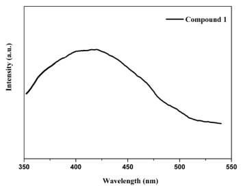
The solid photoluminescence spectra of complex 1 are displayed in Fig. 7. Upon excitation at 307 nm, complex 1 exhibits the emission at ca. 420 nm. The free H2dia ligand shows the emission at 434 nm (λex = 306 nm), and the free bpe ligand shows the luminescence emission bands at ca. 354 and 370 nm (λex = 334 nm), respectively[14]. Generally, Zn(Ⅱ) CPs with d10 configuration ions more commonly exhibit ligand-based emissions. The emission peak of complex 1 should be assigned to the intraligand cooperative effect of the bpe and the dia mixed ligands[24-26].
Figure 7
 Figure 7. Solid-state emission spectra for complex 1
4. CONCLUSION
In summary, a Zn(Ⅱ) coordination polymer has been synthesized based on 5-dimethylamino-isophthalic acid and 1,2-bis(4-pyridyl)ethylene mixed organic linkers. The structure and corresponding weak interactions (hydrogen bonding and π-π stacking interactions) of the complex are identified. Moreover, the solid UV-Vis absorption spectra and luminescence property of the complex have also been studied.
-
-
[1]
Li, Y.; Lun, H.; Xiao, C.; Xu, Y.; Wu, L.; Yang, J.; Niu, J.; Xiang, S. A bilayer triangular lattice with crown-like Co7 spin cluster SBUs exhibiting high spin frustration. Chem. Commun. 2014, 50, 8558–8560. doi: 10.1039/C4CC02910K
-
[2]
Wang, Z. P.; Hu, B.; Qi, X. H.; Shen, N. N.; Huang, X. Y. Microwave-assisted ionothermal synthesis of a water-stable Eu-coordination polymer: Ba2+ ion detector and fluorescence thermometer. Dalton Trans. 2016, 45, 8745–8752. doi: 10.1039/C6DT00641H
-
[3]
Miao, S. B.; Li, Z. H.; Xu, C. Y.; Ji, B. M. Syntheses, characterization, and luminescence properties of three novel Ag(Ⅰ) coordination polymers based on polycarboxylic acid ligands and 1,3-di-(1,2,4-triazole-4-yl)benzene. CrystEngComm. 2016, 18, 4636–4642. doi: 10.1039/C6CE00625F
-
[4]
Deng, D. S.; Guo, H.; Ji, B. M.; Wang, W. Z.; Ma, L. F.; Luo, F. Size-selective catalysts in five functionalized porous coordination polymers with unsaturated zinc centers. New J. Chem. 2017, 41, 12611–12616. doi: 10.1039/C7NJ02021J
-
[5]
Li, Z. H.; Xue, L. P.; Miao, S. B.; Zhao, B. T. Assembly of 4-, 6- and 8-znnnected Cd(Ⅱ) pseudo-polymorphic coordination polymers: synthesis, solvent-dependent structural variation and properties. J. Solid State Chem. 2016, 240, 9–15. doi: 10.1016/j.jssc.2016.05.005
-
[6]
Wang, X. L.; Luan, J.; Lin, H. Y.; Le, M.; Liu, G. C. The various architectures and properties of a series of coordination polymers tuned by the central metals. Dalton Trans. 2014, 43, 8072–8082. doi: 10.1039/c4dt00064a
-
[7]
Li, Z. H.; He, S. J.; Xue, L. P.; Wang, X. N.; Zhang, D. D.; Zhao, B. T. Exploring methyl-3-hydroxy-5-carboxy-2-thiophenecarboxylate and varying flexible bis(imidazole)-based synthons as building blocks for the construction of diverse cadmium coordination polymers. Dyes and Pigments 2018, 149, 498–504. doi: 10.1016/j.dyepig.2017.10.036
-
[8]
Drabent, K.; Ciunik, Z. Counter anion dependent symmetry of CuⅡ-4-amino-1,2,4-triazole polymeric chains. Chem. Commun. 2001, 1254–1255.
-
[9]
Yang, J. X.; Zhang, X.; Cheng, J. K.; Zhang, J.; Yao, Y. G. pH influence on the structural variations of 4,4′-oxydiphthalate coordination polymers. Cryst. Growth Des. 2011, 12, 333–345.
-
[10]
Beheshti, A.; Clegg, W.; Nobakht, V.; Harrington, R. W. Metal-to-ligand ratio as a design factor in the one-pot synthesis of coordination polymers with [MS4Cun] (M = W or Mo, n = 3 or 5) cluster nodes and a flexible pyrazole-based bridging ligand. Cryst. Growth Des. 2013, 13, 1023–1032. doi: 10.1021/cg301106g
-
[11]
Tong, M. L.; Kitagawa, S.; Chang, H. C.; Ohba, M. Temperature-controlled hydrothermal synthesis of a 2D ferromagnetic coordination bilayered polymer and a novel 3D network with inorganic Co3(OH)2 ferrimagnetic chains. Chem. Commun. 2004, 418–419.
-
[12]
Hao, Z. M.; Zhang, X. M. Ligand concentration controlled supramolecular isomerism in two CuSCN based coordination polymers with in situ synthesized 4,4'-dipyridylsulfide as a co-ligand. Cryst. Growth Des. 2007, 7, 64–68. doi: 10.1021/cg060371c
-
[13]
Du, X. G.; Mi, G.; Liu, J. C.; Zhang, J. A two-dimensional Zn(Ⅱ) coordination polymer with a three-dimensional supramolecular architecture comprising 5-dimethylamino-isophthalic acid and 1,3-bis(4-pyridyl)propane. Mol. Cryst. Liq. Cryst. 2016, 624, 44−50. doi: 10.1080/15421406.2015.1044155
-
[14]
Li, Z. H.; Xue, L. P.; Qin, Q. P.; Zhang, J.; Wang, J. M.; Zhang, X. Y.; Zhao, B. Tun. A zinc(Ⅱ) coordination polymer material with Lewis basic pyridyl sites: structure, photoluminescence, and heterogeneous catalysis. J. Solid State Chem. 2019, 274, 81−85. doi: 10.1016/j.jssc.2019.03.020
-
[15]
Dolomanov, O. V.; Bourhis, L. J.; Gildea, R. J.; Howard, J. A. K.; Puschmann, H. OLEX2: a complete structure solution, refinement and analysis program. J. Appl. Cryst. 2009, 42, 339–341. doi: 10.1107/S0021889808042726
-
[16]
Sheldrick, G. M. SHELXT-integrated space-group and crystal-structure determination. Acta Cryst. 2015, A71, 3–8.
-
[17]
Sheldrick, G. M. Crystal structure refinement with SHELXL. Acta Cryst. 2015, C71, 3–8.
-
[18]
Xin, L. Y.; Liu, G. Z.; Ma, L. F.; Zhang, X.; Wang, L. Y. Structural diversity and fluorescence regulation of three ZnⅡ coordination polymers assembled from mixed ligands tectons. Aust. J. Chem. 2015, 68, 758–765. doi: 10.1071/CH14347
-
[19]
Xue, L. P.; Chang, X. H.; Ma, L. F.; Wang, L. Y. Four d10 metal coordination polymers based on bis(2-methyl imidazole) spacers: syntheses, interpenetrating structures and photoluminescence properties. RSC Adv. 2014, 4, 60883–60890. doi: 10.1039/C4RA10331A
-
[20]
Wang, T.; Zhu, R. R.; Zhang, X. F.; Yan, T.; Wang, Q.; Feng, J.; Zhou, J.; Du, L.; Zhao, Q. H. Assembly of a series of zinc coordination polymers based on 5-functionalized isophthalic acids and dipyridyl. RSC Adv. 2018, 8, 7428–7437. doi: 10.1039/C7RA12874F
-
[21]
Mukherjee, S.; Ganguly, S.; Manna, K.; Mondal, S.; Mahapatra, S.; Das, D. Green approach to synthesize crystalline nanoscale ZnⅡ-coordination polymers: cell growth inhibition and immunofluorescence study. Inorg. Chem. 2018, 57, 4050–4060.
-
[22]
Xu, Z. X.; Ma, Y. L. Helical coordination polymer with a 3-fold interpenetration structure based on 5-(hydroxymethyl)isophthalic acid. Chin. J. Struct. Chem. 2017, 36, 1193–1198.
-
[23]
Das, S.; Bharadwaj, P. K. Self-assembly of a luminescent zinc(Ⅱ) complex: a supramolecular host-Guest fluorescence signaling system for selective nitrobenzene inclusion. Inorg. Chem. 2006, 45, 5257–5259. doi: 10.1021/ic060518p
-
[24]
Xin, L. Y.; Liu, G. Z.; Ma, L. F.; Wang, L. Y. Coligand-regulated assembly, fluorescence, and magnetic properties of Co(Ⅱ) and Cd(Ⅱ) complexes with a non-coplanar dicarboxylate. J. Solid State Chem. 2013, 206, 233–241. doi: 10.1016/j.jssc.2013.08.010
-
[25]
Xue, L. P. A dinuclear Cd(Ⅱ) cluster-based coordination polymer: synthesis, structure and luminescence property. Chin. J. Struct. Chem. 2018, 37, 119–124.
-
[26]
Zhi, S. C.; Wang, Y. L.; Sun, L.; Cheng, J. W.; Yang, G. Y. Linking 1D transition-metal coordination polymers and different inorganic boron oxides to construct a series of 3D inorganic-organic hybrid borates. Inorg. Chem. 2018, 57, 1350−1355. doi: 10.1021/acs.inorgchem.7b02765
-
[1]
-
Figure 1 Coordination environment of the Zn2+ ions in 1, showing the atom numbering scheme. Displacement ellipsoids are drawn at the 30% probability level. All hydrogen atoms are omitted. Symmetry codes: (#1) 1 – x, –y, –z; (#2) 2 – x, –y, 2 – z; (#3) 1 + x, y, z
Figure 3 (a) 2D coordination layer of 1. (b) Schematic representation of the 4-connected sql topology
Figure 5 (a) PXRD patterns simulated from X-ray single-crystal diffraction data and experimental data. (b) TGA curve
Table 1. Selected Bond Lengths (Å) and Bond Angles (°) for 1
Bond Dist. Bond Dist. Bond Dist. Zn(1)–O(1) 2.005(3) Zn(1)–N(2) 2.189(3) Zn(1)–N(1) 2.072(3) Zn(1)–O(5) 2.180(3) Zn(1)–O(4)#3 2.061(3) Angle (°) Angle (°) Angle (°) N(1)–Zn(1)–N(2) 92.84(12) O(5)–Zn(1)–N(1) 90.80(13) O(4)#3–Zn(1)–O(5) 86.82(13) O(5)–Zn(1)–N(2) 176.31(12) O(5)–Zn(1)–O(3)#3 84.06(12) O(4)#3–Zn(1)–N(1) 153.69(12) O(4)#3–Zn(1)–N(2) 89.64(11) Symmetry transformation: #1: 1 – x, –y, –z; #2: 2 – x, –y, 2 – z; #3: 1 + x, y, z  下载: 导出CSV
Table 2. Hydrogen Bond Lengths (Å) and Bond Angles (°) in 1
D–H…A d(D–H) d(H…A) d(D…A) ∠DHA O(5)–H(1W)···O(2) 0.86 2.04 2.577(6) 120 O(5)–H(2W)···O(3)#4 0.86 1.88 2.702(5) 161 Symmetry code: #4: 1 – x, 1 – y, 1 – z
下载: 导出CSV
-


 扫一扫看文章
计量
- PDF下载量: 2
- 文章访问数: 675
- HTML全文浏览量: 1
 下载:
下载:
