Lung cancer is one of the most common malignant tumors with the fastest increase in the incidence rate and mortality. Even after maximum tumor resection assistance with a radiotherapy and chemotherapy combination, the recurrence of non-small cell lung cancer is still inevitable. In addition, low targeting efficiency and poor permeability of drug delivery systems strongly affect the therapeutic efficiency of anti-cancer drugs on non-small cell lung cancer. Here we designed a gemcitabine (GEM) loaded arginine-glycine-aspartic acid-cysteine (RGDc)-modified gold mineralization "hybrid nanozyme bomb" (RGTG) to overcome those obstacles. RGDc modification improved the active targeting of liposomes to the tumor tissues with the second near-infrared (NIR-II)-triggered gold-shell disruption and GEM release. The collapsed gold-shell particles with a smaller size could penetrate the tumor solid barrier and act as photothermal therapy (PTT) agents to improve PTT therapy and starvation therapy via generating gluconic acid and reactive oxygen species (ROS). Moreover, the resting reversal effect of gold particles on tumor fibroblasts can achieve accelerating tumor penetration of gold particles and GEM. Compared to monotherapy, RGTG showed significant improvement in tumor inhibition, with a tumor volume reduction of 83% compared to the control group, which provides a promising tumor treatment platform for non-small cell lung cancer (NSCLC).
Bladder cancer is the 9th most prevalent cancer type and ranks fourth globally among diseases affecting males [1,2]. Conventional chemotherapy drugs, particularly doxorubicin (DOX), may be effective but targeting is poor, and significant side effects occur, including impaired bone marrow hematopoietic function and cardiotoxicity. Nanotechnology may improve drug accumulation and targeting of bladder cancer cells by increasing the efficiency of drug delivery. Thus, novel nanomedicine treatments are urgently required to facilitate chemotherapy [3,4].
Delivering drugs to the tumor region is a challenge in cancer therapy. Lipid-based organelles, such as liposomes, which are spherical vesicles with a phospholipid bilayer may be used to encapsulate chemotherapeutic agents and show promise for drug delivery [5]. Liposomes have low toxicity and good biocompatibility and prevent drug leakage in the circulation, controlling drug release at tumor areas and increasing intratumor concentration. Enhanced permeability and retention (EPR) effects of solid tumor tissues damages microvessels and lymphatic vessels and aids the accumulation of liposomes or macromolecules in tumors [6]. Drug resistance and bladder cancer progression limit the therapeutic effect of conventional chemotherapy and off-target drug distribution leads to adverse reactions and low intratumor drug accumulation. Encouragingly, liposomes may overcome some of these difficulties and aid chemotherapeutic drug delivery [7].
Advanced photothermal materials, such as gold nanoparticles (GNPs), have indicated the potential of photothermal therapy (PTT) [8–10]. GNPs are laser-activated photothermal therapeutic agents [11] and their physicochemical, optical, and electronic properties make them suitable for biomedical applications [12–14]. The small and spherical nanoparticles produce fluorescent/thermal/reactive oxygen species under near-infrared (NIR) light. Adriamycin-loaded GNPs have been shown to have slower drug release and prolonged systemic circulation compared with free adriamycin-loaded PLZ4-nanoporphyrin (PNP) [15], showing them to be multipurpose agents for thermal ablation and drug delivery [16]. Intravenous injection of NIR radiation-absorbing gold-silicon nano shells resulted in the clearance of the majority from the blood and retention in the bladder for 30–50 min, where particles were distributed throughout the bladder wall in the tumor-associated stroma [17,18]. PTT under NIR light excitation thus has considerable specificity [19]. Poly(N-isopropyl acrylamide) (PNIPAM) is a novel temperature-sensitive polymer and is used for the integration of photothermal transition particles, including GNPs [20–22]. The resulting nanoparticles allow the enhancement of liposome-delivered intratumor drugs and have the potential to reduce side effects.
However, chemotherapeutic drugs have been widely used clinically for various cancers. Chemotherapy is not effective, Shi et al. found that pH-sensitive nanomaterials for anti-tumor drug delivery are stronger than chemotherapy alone [23]. Li et al. found that synergistic effects of chemotherapy and PTT applied to nanomaterials produced favorable therapeutic effects [24]. Therefore, combining chemotherapy and PTT for synergistic treatment of bladder cancer is highly promising.
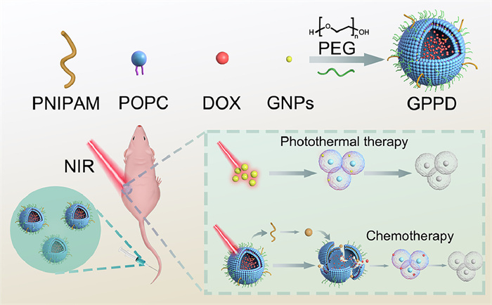
Liposomes containing DOX and embedded with GNPs and PNIPAM to form a functional GPPD structure were constructed during the current study. After NIR light irradiated the cells in the tumor region, the gold nanoparticles locally generated heat and produced a photothermal therapeutic effect, which killed the tumor cells. In addition, contraction denaturation of the PNIPAM structure led to structural disintegration, releasing gold nanoparticles and chemotherapeutic agents, further facilitating PTT and chemotherapy (Fig. 1). GPPD was injected into mice through the tail vein and accumulation in tumor regions and tumor apoptosis promoted under 808 nm NIR light irradiation. High cytotoxicity was produced from the photothermal conversion of the GNPs with NIR light irradiation. Thermal stimulation caused PNIPAM to undergo a coil-like to spherical transition which ruptured the liposomes, allowing DOX to be released [25]. The GNPs embedded in the liposomes enhanced the PNIPAM phase transition by increasing the local temperature when excited by NIR light. As a result, GNPs and DOX had a synergistic effect on bladder cancer.
Figure 1
Figure 1.
The system model was assembled using a rotary steam bottle, and the protective shell was made of PEG. The system was embedded with GNPs and PNIPAM to form a functional GPPD structure for delivering chemotherapy drugs. The figure is a schematic diagram of the mechanism of killing tumor cells by GPPD in mice. GPPD was injected into mice through the tail vein, and GNPs exerted photothermal therapeutic effects under NIR light and disintegration of the system enabled the synergistic release of chemotherapeutic agents to kill tumor cells.
1-Palmitoyl-2-oleoyl-sn-glycerol-3-phosphatecholine (POPC) was used for liposome construction [26] and GNPs and PNIPAM dendritic molecules were embedded in the membrane. GNPs were adsorbed onto polymers modified with polyethylene glycol (PEG) and PNIPAM by positive and negative charges for liposome insertion (Table S1 in Supporting information). The physical and chemical properties of GPPD were adjusted during self-assembly by fine-tuning the composition of GNPs, POPC, PEG, and PNIPAM to form nanoparticles with a defined structure.
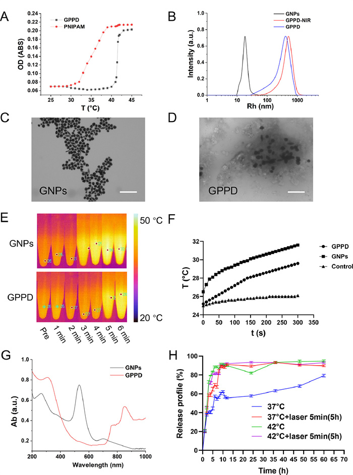
The PNIPAM low critical solution temperature (LCST) was approximately 32 ℃ but could be modified and that of GPPD was approximately 42 ℃ (Fig. 2A). GNP- and DOX-loaded polymers thus had temperature-controlled release. GNP and GPPD size distributions were evaluated by dynamic light scattering (DLS). The particle size increased after GNPs embedding on the liposome membrane with a size of 400–500 nm. The particle size of GPPD was also increased after irradiation by NIR light (Table S1). GPPD doped with PNIPAM showed an increase in particle size after NIR irradiation and became spherical due to self-polymerization (Fig. 2B). Good photothermal conversion in vitro was shown (Figs. 2A and B). The photothermal conversion efficiency (η) of GPPD was 16.3% (Fig. S1 in Supporting information). GPPD had a volume phase transition temperature of approximately 42 ℃. POPC delivery of DOX and NIR irradiation of GNP, which led to the breakage of hydrogen bonds as the temperature increased, gave the system reduced toxicity. PNIPAM underwent a chain to globular shape transition, enlarging the liposome void and releasing the drug. The photothermal PNIPAM contraction, which allowed controlled drug release, indicated successful system construction and precipitated tumor cell death. PNIPAM contraction produced thermosensitive changes which mediated passive targeting [27]. Therapeutic nanomedicine applications of the photothermal properties of gold have previously been demonstrated by Zhang et al. [28]. The combination of GNP and PNIPAM has great potential to improve past research results. Transmission electron microscopy (TEM) was used to examine the GNPs (Fig. 2C) and the size of the GPPD was found to be approximately 400–500 nm (Fig. 2D). A similar value was derived by DLS. The DLS (Table S1) and TEM (Figs. 2C and D) data demonstrated the successful synthesis of the system. The photothermal conduction efficiency of 100 µg/mL GNPs and GPPD with continuous NIR light (808 nm, 1 W/cm2, 6 min) at different time points allowed the temperature to increase with illumination duration (Fig. 2E). The favorable photothermal conversion efficiency would facilitate the PNIPAM structural transformation which enables drug release. The photothermal effect of 100 µg/mL GNP was greater than that of 100 µg/mL GPPD and both were good relative to ddH2O (Fig. 2F). Small GNPs were readily embedded into the liposome membrane for tissue permeability and retention. The GPPD encapsulation efficiency was 48.21% and drug loading was 17.84% (Table S1). The percentage of GNPs and PNIPAM in GPPD nanoparticles analyzed by thermal gravimetric analyzer (TGA) was 9.37% and 26.58% (Fig. S2 in Supporting information). Ultraviolet–visible spectroscopy (UV–vis) measured GNP absorbance at 400–600 nm and GPPD at 800–1000 nm (Fig. 2G). The 808 nm laser was chosen for subsequent experiments. Drug release was stimulated by laser irradiation for 5 min at 37 ℃ and also by increased temperature so that the drug could be measured in the solution (Fig. 2H). However, liposomes failed to reach a stable drug release plateau quickly at 37 ℃ in vitro. Laser irradiation caused the tumor microenvironment to achieve a temperature that exceeded the GPPD LCST of 42 ℃ with the result that PNIPAM released the chemotherapeutic drugs. In summary, GPPD with temperature-controlled release and photothermal synergy was successfully constructed.
Figure 2
Figure 2.
(A) Discriminant volume phase transition temperature (VPTT) by UV‒vis spectrophotometer determination of optical density (OD) value, testing the same concentration of PNIPAM in anhydrous ethanol solution and the temperature dependence of the GPPD radius. (B) Particle size distribution of GNPs, GPPD, and GPPD-NIR. (C, D) TEM images of GNPs and GPPD system (scale bar: 100 nm). (E) Photothermal images of GNPs and GPPD were captured using an infrared camera. (F) Photothermal effect of GPPD, GNPs and the control group (ddH2O) in vitro. (G) DLS. Light absorption curve of GNPs (100 µg/mL) and GPPD (100 µg/mL). (H) Release curve of in vitro GPPD at 37 and 42 ℃ and GPPD at 37 and 42 ℃ with a laser (808 nm), n = 3. Data are mean ± standard deviation (SD).
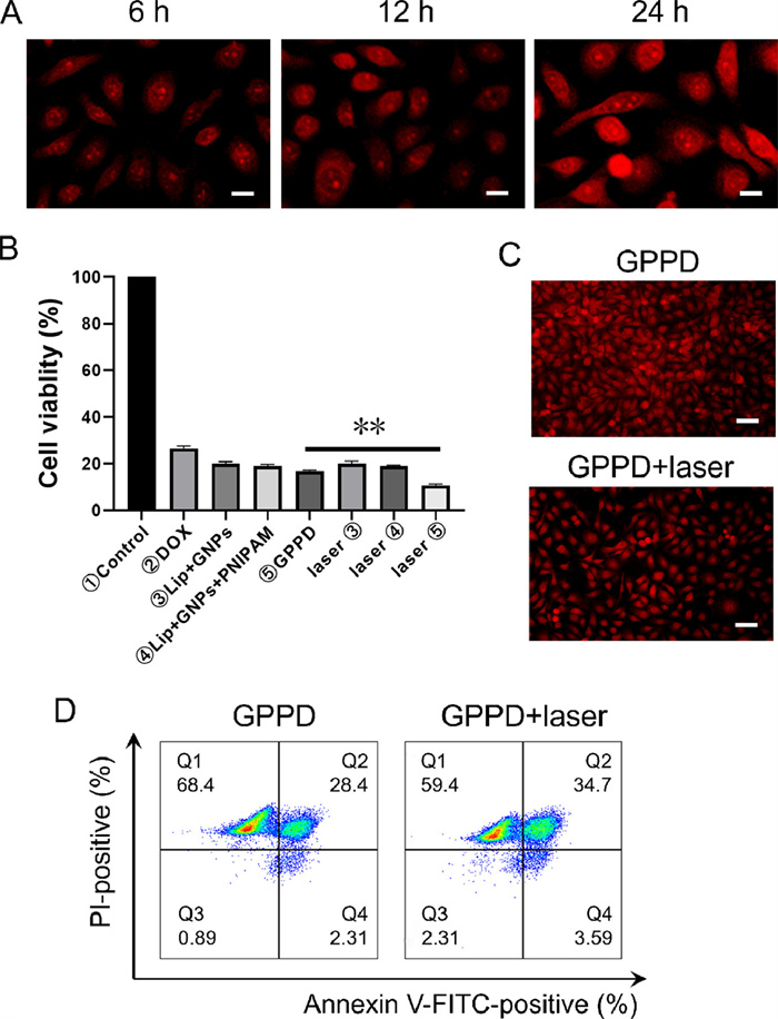
Inverted fluorescence microscopy was used to observe the endocytosis of GPPD, 5637 human bladder cancer cells were treated with 10 mg/mL GPPD. Subsequently, dead cells were washed away with PBS, and drug accumulation was observed from 6 h to 24 h in the nucleus (Fig. 3A). GNPs interacted with organelles and lysosomes induced apoptosis [29,30]. The nuclear binding of DOX is shown in Fig. 3A. Cell counting kit-8 (CCK-8) assays showed the half maximal inhibitory concentration (IC50) to be 10.8 mg/mL (GPPD) and 2.5 mg/mL (DOX) for bladder cancer cells and these concentrations were used with a 2 h incubation for subsequent experiments (Fig. S3 in Supporting information). Laser irradiation enhanced cytotoxicity, reducing the number of bladder cancer cells, and GPPD produced higher cytotoxicity than GNPs. The photothermal effect was monitored with an inverted fluorescence microscope after NIR light exposure (Figs. 3B and C). The combination index (CI) value is 0.22 according to the cell viability measured by single DOX and combined drugs, which indicated a strong synergistic effect of GPPD (Fig. S3). Flow cytometry showed that irradiation increased apoptosis in bladder cancer cells (Fig. 3D). Cancer cell apoptosis has been reported to be driven by mitochondrial signals [31,32].
Figure 3
Figure 3.
(A) 5637 cells absorb assessment GPPD at 6, 12, and 24 h (scale bar: 10 µm). (B) CCK-8 assay of 5637 cells. (C) 5637 cells condition of GPPD and GPPD with laser. After 5 min laser irradiation for GPPD, 5637 cells continued to develop 24 h (scale bar: 100 µm). (D) Scatter plots of apoptosis in 5637 cells measured by flow cytometry for GPPD and GPPD with laser, n = 3. Data are mean ± SD. **P < 0.01.
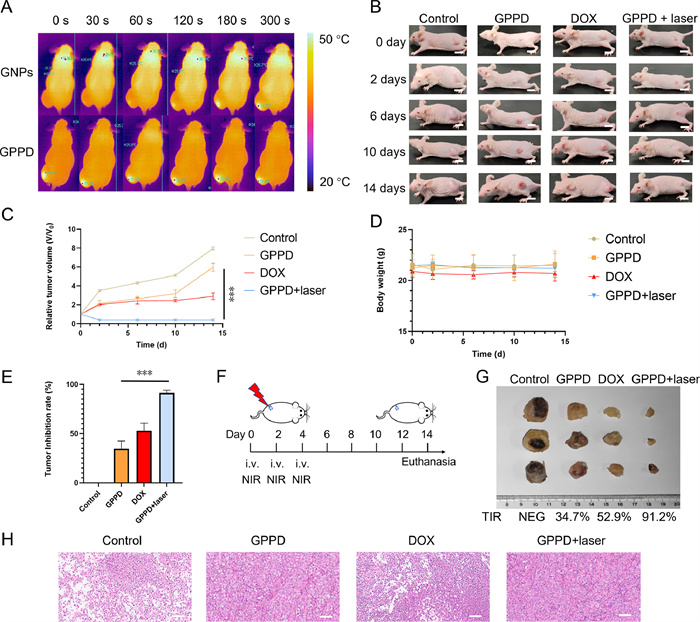
Animal welfare and experimental procedures were reviewed and approved by the Animal Ethics Committee of Shanghai General Hospital (No. 2021KY046). Photothermal effects of GPPD and GNPs were tested in vivo and intratumor injection gave a weaker effect of GPPD than of GNPs (Fig. 4A). Tail vein administration of GPPD to tumor-bearing mice followed by NIR light irradiation showed the highest tumor volume for GPPD with laser treatment, followed by DOX and GPPD (Figs. 4B and C). No change was seen for mouse body weight (Fig. 4D). Tumor inhibition rates (TIR) were 34.7% for GPPD, 52.9% for DOX, and 91.2% for GPPD with the laser treatment (Fig. 4E). Mice were injected and irradiated on 0, 2 and 4 d and sacrificed on 14 d (Fig. 4F) for harvesting of tumors (Fig. 4G) and hematoxylin-eosin (HE) staining (Fig. 4H). The photothermal effect and tumor suppression were higher for GPPD with laser than for GPPD alone, perhaps due to the EPR effect. Thus, GPPD with laser treatment appeared to promote bladder cancer cell death.
Figure 4
Figure 4.
There were four groups in the anti-tumor effect experiment: the PBS group, DOX group, GPPD group, and GPPD with NIR light irradiation group. (A) The photothermal effect of 808 nm excitation light in tumor-bearing mice was observed by an infrared camera. (B) A tumor suppression test was performed in the control group, GPPD group, DOX group, and GPPD group with laser, and tumor growth was observed (0, 2, 6, 10 and 14 d) (scale bar: 5 cm). (C) Changes in relative tumor volume (V/V0) in each group, n = 3. (D) Body weight changes in each group, n = 3. (E) Tumor inhibition rates of different groups were calculated, n = 3. (F) The experimental schedule in vivo, from 0 d to 14 d. (G) The collected tumors in the different group treatments with the measurement scale. (H) HE staining results of tumors (scale bar: 100 µm). Data are mean ± SD. ***P < 0.001.
GPPD was stable (Fig. S4 in Supporting information) with no sedimentation or variation in particle size (Table S2 in Supporting information), and biocompatibility tests, involving injection of 200 µL GPPD (4 mg/kg) every two days for a total of 3 times, in ICR mice showed no significant effect of the drugs on mouse survival. The mice were in good condition with no significant abnormalities after one month. HE staining of the liver, heart, spleen, lung and kidney indicated favorable GPPD biocompatibility (Fig. S5 in Supporting information). Therefore, GPPD appeared to be suitable for human use.
Administration of fluorescein isothiocyanate (FITC)-loaded but DOX-free liposomes to tumor-bearing mice showed insignificant tumor site enrichment of FITC but significant enrichment was observed after laser irradiation. Accumulation was seen in the liver, heart, and lung after 2 h but minimal tissue content after 24 h, indicating good biocompatibility (Figs. S6A–J in Supporting information). Drug release and accumulation were promoted by the use of the laser after both 2 and 24 h, demonstrating the passive targeting property of GPPD (Figs. S6K–N in Supporting information). Temperature-responsive structures have previously been shown to improve cellular uptake and drug release [33]. Although the current drug system had a good targeting capacity, its tissue retention capacity requires further improvement. Targeting and retention were improved by increasing the administration frequency but this represents a shortcoming of the system. Nonetheless, NIR light improved GPPD enrichment in tumor tissues.
In conclusion, a thermo- and chemotherapy-sensitive dual-function GPPD liposome structure was constructed. The EPR effect of GPPD, with stable mosaic structure and excellent biocompatibility, led to accumulation at tumor sites. Drugs were released into the tumor region by the PNIPAM-regulated photothermal effect of GNPs, leading to the accumulation of high concentrations of DOX. Co-therapy with NIR light-induced chemotherapy and PTT has an anti-tumor effect in vivo and in vitro. Moreover, GPPD has the advantage over the previous tissue ablation strategies since normal tissue is preserved. We introduce the NIR light-dependent nanodrug delivery system of GPPD with therapeutic potential for drug delivery in bladder cancer.
Declaration of competing interest
The authors declare that they have no known competing financial interests or personal relationships that could have appeared to influence the work reported in this paper.
Acknowledgments
This work was funded by the National Natural Science Foundation of China (Nos. 92059112, 82072821 and 31470964), the Shanghai Songjiang Municipal Science and Technology Commission Natural Science Foundation (No. 20SJKJGG250), University of Shanghai for Science and Technology (No. 10–21–302–405) and sponsored by the Program of Shanghai Academic Research Leader (No. 22XD1404700). EditSprings provided the linguistic services.
Supplementary materials
Supplementary material associated with this article can be found, in the online version, at doi:10.1016/j.cclet.2023.109199.
References
[1]
H. Sung, J. Ferlay, R.L. Siegel, et al., CA Cancer J. Clin. 71 (2021) 209–249.
doi: 10.3322/caac.21660
[2]
S.K. Alam, L. Wang, Z. Zhu, L.H. Hoeppner, NPJ Precis. Oncol. 7 (2023) 33.
doi: 10.1038/s41698-023-00370-3
[3]
S. Garinet, P. Wang, A. Mansuet-Lupo, et al., Cancers 14 (2022) 1400.
doi: 10.3390/cancers14061400
[4]
J. Vansteenkiste, E. Wauters, B. Reymen, et al., Annals Oncol. 30 (2019) 1244–1253.
doi: 10.1093/annonc/mdz175
[5]
M. You, Z. Xie, N. Zhang, et al., Signal Transduc. Target. Ther. 8 (2023) 196.
doi: 10.1038/s41392-023-01442-3
Jialiang XU
, Jiabin CUI
. Recent biological applications of corroles: From diagnosis to therapy. Chinese Journal of Inorganic Chemistry,
2024, 40(12): 2303-2317.
doi: 10.11862/CJIC.20240245
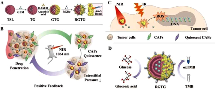
Scheme 1. Illustration of preparation steps of RGTG and its antitumor mechanism. (A) The preparation of targeting RGTG. (B) Penetration promoting mechanism. (C, D) Starvation therapy and cocktail therapy of RGTG.
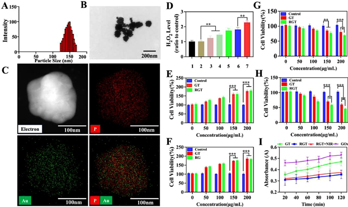
Figure 1. The particle size (A) and transmission electron microscope (TEM) images (B) of RGTG (n=3). Scale bar: 200 nm. (C) The EDS mapping of RGTG. Scale bar: 100 nm. (D) H2O2 level was catalyzed by different formulations. 1: Control; 2: TSL; 3: GTG; 4: RGTG; 5: RGTG + NIR; 6: RGTG + IR; 7: RGTG + NIR + IR. (E–H) Cell viability without glucose at 24 h (E) and 48 h (F). Cell viability containing glucose at 24 h (G) and 48 h (H). (I) Results of catalytic activity of glucose like oxidase. Each bar represents the mean ± standard deviation (SD), n=3. *P < 0.05, **P < 0.01, ***P < 0.001.
Figure 2. (A) The confocal laser microscope of different C6 labeled formulations into LLC tumor spheroids to different depths for 6 h. Scale bar: 100 µm. 1: free C6; 2: TSL-C6; 3: GT-C6; 4: GT-C6 + NIR; 5: RGT-C6 + NIR; 6: RGT-C6 + NIR + IR. (B) Penetration efficiency of fluorescence of different preparations in mixture tumor spheroids. (C) Inhibition of LLC mixture tumor spheroid growth by GEM preparations at 5 µg/mL for 5 days. Scale bar: 100 µm. (D) Statistical results of the inhibition of tumor spheroid growth by GEM preparations at 5 µg/mL for 5 days. *P < 0.05, **P < 0.01, ***P < 0.001. (E) Western blot of the expression of related proteins in tumor globules after different treatment groups. (F) Representative immunofluorescence images of collage I, fibronectin, and HIF-1α expression after various therapeutic strategies. Scale bar: 200 µm. 1: PBS; 2: Free; 3: GTG; 4: GTG + NIR; 5: RGTG + NIR; 6: RGTG + NIR + IR. GAPDH: glyceraldehyde-3-phosphate dehydrogenase. Each bar represents the mean ± SD, n=3.
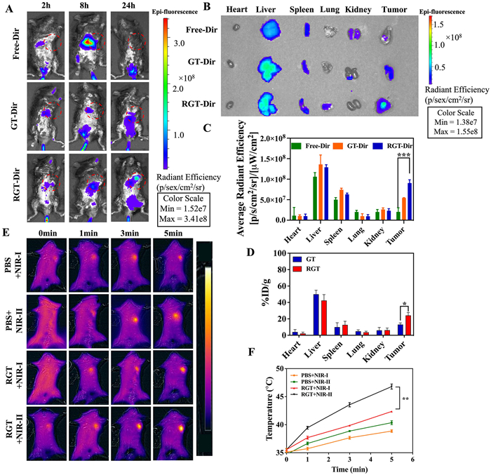
Figure 3. (A) The in vivo imaging of LLC tumor mice after intravenous injection of free Dir, GT-Dir, and RGT-Dir. Red circles represent the tumor site. (B) The images of ex vivo organs of mice at 24 h after injection free Dir, GT-Dir, and RGT-Dir. (C) The average fluorescence intensity of ex vivo organs and tumors of mice at 24 h after injection of different groups (n = 3). (D) The average Au amount of ex vivo organs and tumors of mice at 24 h after injection of different groups (n = 3). (E) In vivo photothermal imaging with a NIR laser at 0, 1, 3 and 5 min of LLC tumor mice 24 h after intravenous injection of PBS and RGT. (F) Temperature changes of the LLC tumor mice during NIR-I and NIR-II irradiation at different points of different groups (n = 3). *P < 0.05, **P < 0.01, ***P < 0.001. %ID/g: the percentage injected dose per gram of tissue. Each bar represents the mean ± SD.
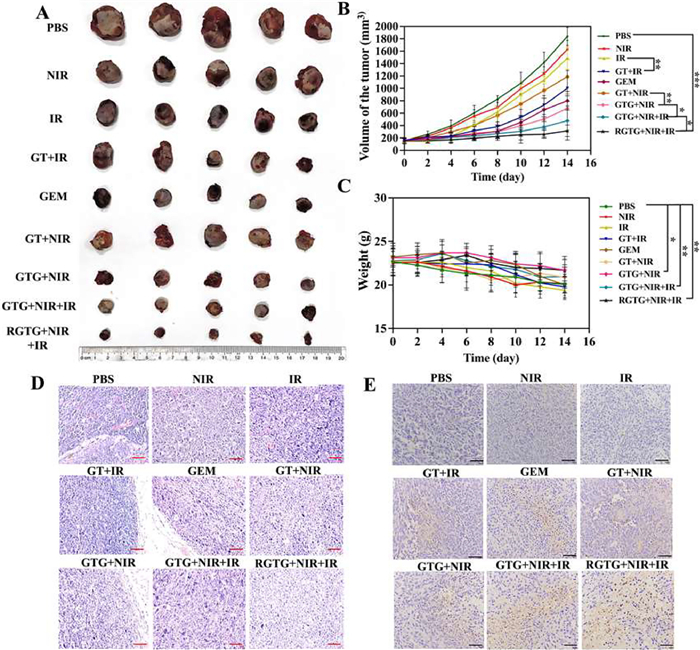
Figure 4. (A) The tumor images of different treatment groups after 14-day treatment. (B) Tumor volume changes in different treatment groups within 14 days (n = 5). (C) The weight changes of mice in all at 14 days. (D) Representative H&E staining images of tumor tissues after different treatments. (E) Representative TUNEL staining images of tumor tissues after different treatments. Scale bar: 100 µm. mean ± SD. *P < 0.05, **P < 0.01, ***P < 0.001.

 Login In


 DownLoad:

 DownLoad: