Department of Critical Care Medicine, Frontiers Science Center for Disease-related Molecular Network, State Key Laboratory of Biotherapy, West China Hospital, Sichuan University, Chengdu 610041, China
b.
Medical Research Center, The First Affiliated Hospital of Zhengzhou University, Zhengzhou University, Zhengzhou 450000, China
* Corresponding authors. E-mail addresses: yqwei@scu.edu.cn (Y. Wei), songxr@scu.edu.cn (X. Song). 1 These authors contributed equally to this work.
Received Date:
19 April 2023 Revised Date:
21 June 2023 Accepted Date:
30 June 2023 Available Online:
3 July 2023
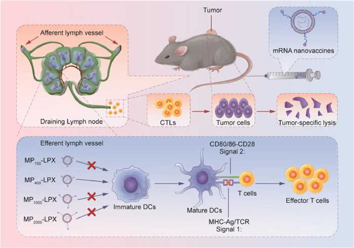
Dendritic cell (DC)-targeted delivery of mRNA is a prominent method to boost the efficacy of mRNA tumor vaccines. The targeting ligands are often modified on nanocarriers by polyethylene glycol (PEG) linker in mRNA delivery systems. Whether the PEG linker length influences the targeting delivery efficiency of mRNA nanocarrier in vivo remains unclear. Here, we designed and constructed DC-targeted mRNA delivery systems modified by mannose via different PEG linker lengths (100/400/1000/2000) (MPn-LPX). The top candidate MP400-LPX (the linker was PEG400) showed the optimal mRNA expression and antigen presentation owing to the highly efficient uptake by DCs. Furthermore, MP400-LPX could better inhibited tumor growth and extended survival in the E.G7-OVA lymphoma and TC-1 cervical tumor mouse model. Collectively, these results demonstrated that PEG400 was the optimal linker for the PEGylated DC-targeted mRNA vaccines. Our findings provided a new platform for the rational design of targeted mRNA nanovaccines with shorter-length PEG.
Chiral separation has become one of the main disciplines in analytical chemistry, and will continue to be prevalent in pharmaceutical and agrochemical industry as well as food science and technology, due to the different biological interactions, pharmacology, and toxicity of pure antipodes [1–3]. Researchers engrossed in enantioseparation keep seeking for versatile chiral separation selectors for a long time. However, there is always a tradeoff between versatility and selectivity due to negative influence among the recognition domains and the relatively low surface concentrations in a limited support surface area.
Developments in the past decade include the expansion of traditional chiral resolution materials such as cyclodextrin (CD)-based selectors, polysaccharide-based selectors and natural alkaloids-based selectors [4–6]. These materials have been evaluated and commercialized [7], which greatly contribute to the rapid development of modern-day biomedical industry. Besides, a variety of new recognition and separation materials such as chiral metal organic frameworks [8], chiral covalent organic frameworks [9,10], chiral pillar[n]arenes [11] and chiral organic polymers [12] have been expanded. However, the current chiral selectors cannot exhibit good versatility in screening various categories of racemates on a single column due to the intrinsic recognition domain of a single selector is generally designed for separating specific analytes.
Dual chiral selectors system may provide an alternative to improve the separation and some successful applications have been performed by incorporating two chiral selectors on same silica support in high performance liquid chromatography (HPLC). A series of novel bridged bis(β-CD) were constructed and covalently immobilized on a support to extend the enantioselectivity profile of chiral stationary phase (CSP), while steric hindrance in the anchoring step can affect loading of selectors [13–15]. Wei group prepared a biselector bonded-type CSP using two brush-type selectors but its enantioseparation ability was relatively lower than that of single selector CSP [16]. According to previous studies, it could hardly improve the enantioselectiviy compared to the CSPs with individual selector due to pseudo-enantiomerical behaviors and negative effects from non-enantioselective interactions [17–19]. Therefore, the key point is how to realize the dual functions without interfering while maintaining a high surface loading of selectors. A good approach is to incorporate more types of interaction sites and chiral centers on one support surface via surface-up construction. Our group reported an ion-exchange type chiral selector quinine (QN) bridged functional CD selector and the complementarity between QN and CD significantly broadens the separation profile and enhances the enantioselectivity [20]. Dual selectors with more types of action sites can avoid functional interference and the surface-up strategy ensures the maximum loading on a limited support area.
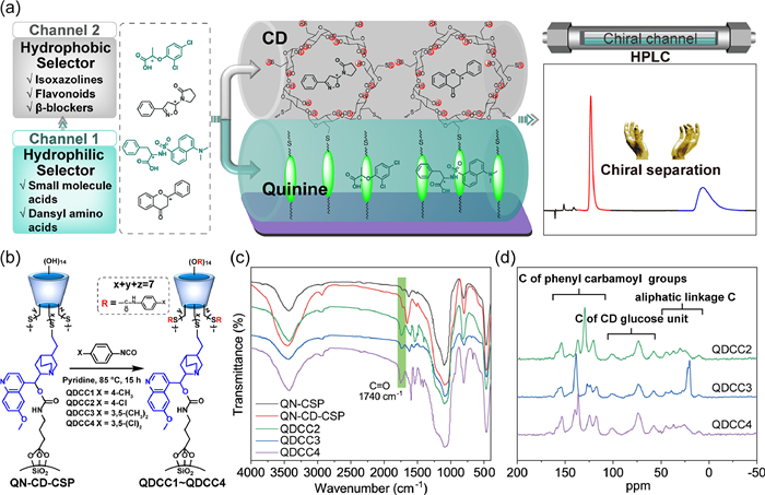
Based on the previous research, we herein propose a concept of quasi-dual-chiral-channel (QDCC) enantioseparation platform, where three novel chiral selectors were prepared by tandemly linking QN and different functional CDs on silica surfaces (Fig. 1a). The function-separated well-defined dual channels were constructed by hydrophilic layer (QN) and hydrophobic layer (functional CD). The QDCC structure can imitate two independent chiral channels accommodating different racemates to achieve wide spectrum chiral resolution. The powerful chiral resolving capabilities of the bilayer selectors were evaluated and affirmed in HPLC by screening model analytes such as isoxazolines, flavonoids, organic acids and β-blockers. Furthermore, the separation mechanism of the QDCC structure was investigated combined with molecular docking. It is expected that this work can inspire the development of versatile enantioseparation strategies.
Figure 1
Figure 1.
Construction and characterization of the dual chiral selectors system. (a) The design of QDCC. (b) Synthetic pathway of the QDCCs. (c) FTIR and (d) solid state 13C NMR of QDCCs.
The synthetic details of QN combined native CD selector (QN-CD-CSP) are exhibited in Fig. S1 (Supporting information) according to previously reported procedure [20]. QN combined per(4-methyl)phenylcarbamoylated-β-CD (QDCC1) was obtained from previously reported selectors by our group [20]. Three novel QDCCs including QN combined per(4-chloro)phenylcarbamoylated-β-CD CSP (QDCC2), QN combined per(3,5-dimethyl)phenylcarbamoylated-β-CD CSP (QDCC3) and QN combined per(3,5-dichloro)phenylcarbamoylated-β-CD CSP (QDCC4) were prepared as shown in Fig. 1b. The synthetic details and the chiral column fabrication processes are described in the Supporting Information. QDCC2~QDCC4 were characterized by Fourier-transform infrared (FTIR), solid state 13C NMR, thermal gravimetric analysis (TGA) and elemental analysis. As shown in Fig. 1c, after thiol-ene click reaction, the FTIR spectrum reveal the enhancement of -OH (3450 cm−1) and C-H (2960 cm−1) absorptions on QN-CD-CSP due to the introduction of CD. The absorption of carbonyl groups (1729 cm−1) was significantly enhanced due to the introduction of phenylcarbamoyl moieties after functionalization reaction. The selectors were further characterized with 13C NMR spectroscopy (Fig. 1d). QDCC2~QDCC4 show obvious CD carbon signal peaks at 60-110 ppm, the peaks between 110 and 170 ppm belong to the carbon atoms of phenylcarbamoyl and the other peaks between 0 and 50 ppm can be assigned to the aliphatic linkage carbon atoms. The TGA weight loses of QN-CSP, QN-CD-CSP and QDCC2~QDCC4 were shown in Fig. S2 (Supporting information), indicating the successful immobilization of CDs and post-derivatization. The surface loadings of chiral selectors were determined by elemental analysis (Table S1). The loading of QN was calculated to be 0.73 µmol/m2 and CD was 0.93 µmol/m2 according to the reported equation [21].
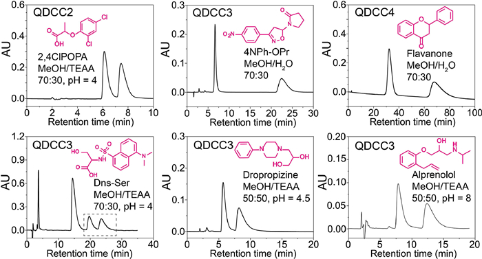
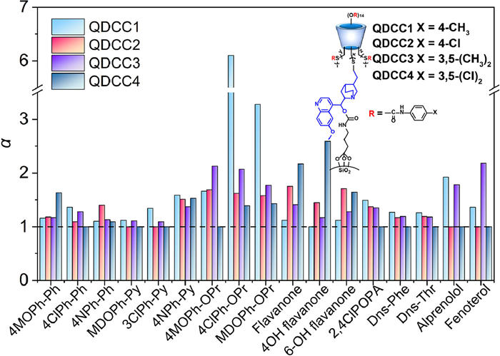
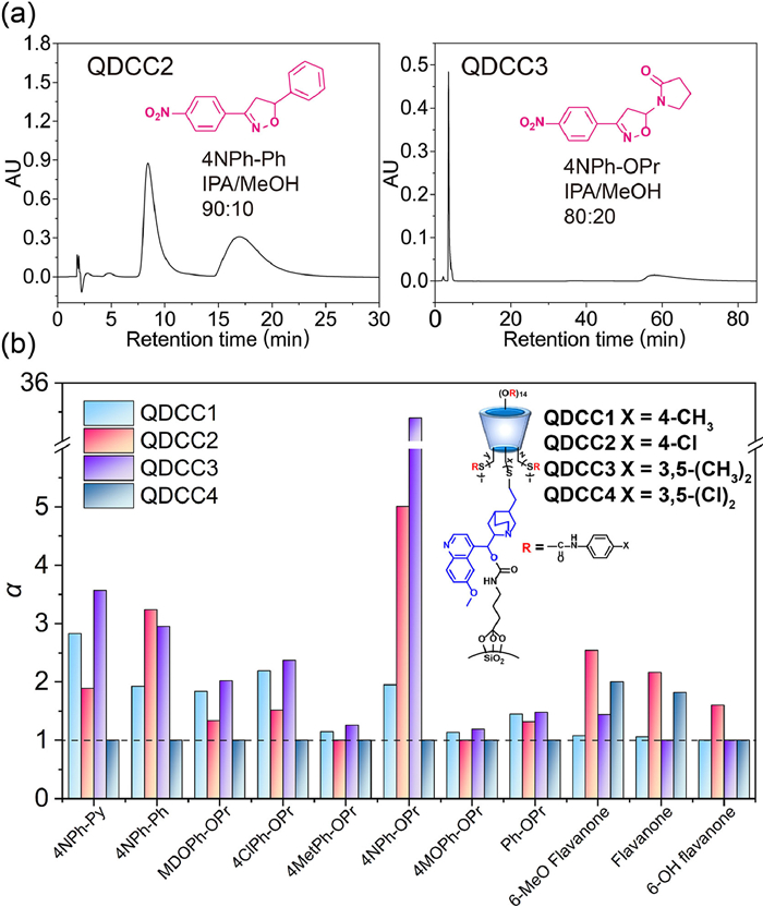
In order to investigate the versatile enantioselectivity of QDCC selectors and evaluate the independent separation ability of the dual channel, dansyl amino acids, small molecule acids, isoxazolines, flavonoids, β-blockers and some basic racemates were selected for evaluation in reversed-phase (RP) mode (Structures see Fig. S3 in Supporting information). All the separations were performed with a flow rate of 1 mL/min at 30 ℃. The triethyl ammonium acetate buffer (TEAA) was prepared by adding triethylamine (v/v, 1%) into ultrapure water and adjusted to required pH using acetic acid. Methanol (MeOH) or acetonitrile (ACN) mixed with ultrapure water or TEAA were used as mobile phases. Tables S2-S4 (Supporting information) list the separation results and some representative chromatograms are shown in Fig. 2. The comparison of selectivity (α) of QDCC1~QDCC4 in the RP mode for the separation of different types of analytes under the same condition is shown in Fig. 3. As far as we know, functional CD have the ability to separate neutral and basic racemates but cannot separate organic acids [20,22]. However, QN is regarded as a superior selector for organic acids racemates owing to it has a bulky quinuclidine moiety with an easily protonated tertiary amino group under acidic condition, which can provide anion-exchange sites together with other interactions [23]. QDCC1 and QDCC3 exhibited chiral resolution ability for all types of analytes used in current work as shown in Figs. 2 and 3, which reflects the complementary functions of dual channel. QDCC2 and QDCC4 exhibits specific enantioseparation abilities towards the analytes. Besides, comparison of QDCC3 with the commonly used commercial chiral columns CHIRALPAK®IA, IB and IC with chemically bonded polysaccharide was shown in Fig. S4 (Supporting information). QDCC3 exhibits comparable or better chiral selectivity over the three commercial chiral columns for the representative racemates. We also found that the retention time and α increased with the decrease of column temperature (Fig. S5 in Supporting information), indicating that the separation ability of QDCCs could be improved by adjusting the chromatographic conditions.
Figure 2
Figure 2.
Representative chromatograms on QDCCs of each type of analyte in RP mode.
Figure 3.
Comparison of QDCC1~QDCC4 in the RP mode. Conditions: MeOH/H2O (v/v, 70:30) for isoxazolines and flavonoids. MeOH/TEAA (v/v, 70:30), pH 4.0 for organic acids. MeOH/TEAA (v/v, 50:50), pH 8.0 for β-blockers.
Meanwhile, the functionality of the dual channel is influenced by the property of the derived groups on CD. For most isoxazolines, QDCC1/QDCC3 with electron-donating methyl exhibit better separation. All the flavonoids had better enantioseparations on QDCC2/QDCC4 with chlorine moieties, which may be due to the electron-withdrawing groups on the CD derivative QDCCs strengthening the π-π interaction. The basic enantiomers were not effectively resolved on QDCC2 and QDCC4 (data not shown), indicating that the chloro substitution on the CD rim phenyl ring is unfavorable for the resolution of analytes with amine moieties. It is worth mentioning that QDCC4 has no separation ability for organic acids, implying that the QN channel loses its functionality. The phenylcarbamates bear strong electron-withdrawing substituents on QDCC4 (3,5-dichloro). These substituents appear to perturb the polarity of the carbamate group through an inductive effect and affect the interaction mode between the dual channel and the organic acids. When the excessive electron-withdrawing substituents are introduced on the phenyl moiety, the acidity of the -NH proton of the carbamate groups increased [24,25]. Therefore, the acidity of the electron-withdrawing substituents interferes with the interaction between organic acids and the QN channel. Then we further prove our explanation using molecular docking simulation.
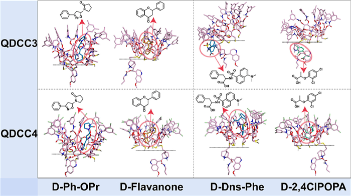
Based on the chromatographic separation ability of the dual channel, AutoDock was used to simulate the binding conformation between the QDCC selectors and different kinds of racemates. QDCC3 and QDCC4 with different derivative groups on CD were selected to probe the mechanism of action of the dual channel. Flavanone, Ph-OPr, Dns-Phe and 2, 4ClPOPA were selected as representative analytes. Chemical structures and AutoDock-optimized geometries of the QDCC structure and racemates were shown in Fig. S6 (Supporting information). The binding conformations were shown in Fig. 4 and Fig. S7 (Supporting information). As shown in Fig. 4, for the isoxazoline and flavanone, they tend to enter the CD channel during the separation process on QDCC3/QDCC4. For the organic acids (Dns-Phe and 2, 4ClPOPA), they tend to enter QN channel due to the strong electrostatic effect when they interact with QDCC3. However, the organic acids tend to enter CD channel when it interacts with QDCC4, which does not reflect the function of QN channel. This phenomenon is consistent with the results for the chromatographic separation. Therefore, the dual channel enantioseparation is achievable, but its implementation requires a rational design. Dual selectors with electron-donating derived groups on CD rims are more favorable for the functional realization of the dual channel in this work.
Figure 4
Figure 4.
The binding conformations analyzed via molecular docking.
To verify the versatile separation of the QDCCs in the polar-organic (PO) mode, isopropanol (IPA) and MeOH solvent was selected as the mobile phase to separate isoxazolines and flavonoids (Fig. 5 and Tables S5-S7 in Supporting information). Representative chromatograms are shown in Fig. 5a. It was noteworthy that pure solvent IPA enabled the enantioseparation of racemates due to a single solvent can be facilely recycled. Most of the isoxazolines and some of the flavonoids used in the current work were effectively resolved on these QDCCs. QDCC3 separated all the isoxazolines used in the current work with high enantioselectivity (α), in which the α of 4NPh-OPr reach 35.40. Similar to the RP-mode results, flavonoids were better separated on QDCC2/QDCC4 with chlorine substituents (Fig. 5b).
Figure 5
Figure 5.
Enantioseparation performance in PO mode. (a) Representative chromatograms of selected analytes. (b) Comparison of α between QDCC1~QDCC4. Conditions: IPA 100% for isoxazolines and flavonoids except for 4NPh-Ph and 4NPh-OPr.
Finally, Table S8 (Supporting information) provides a quick reference guidance for the researchers to choose suitable chiral dual selectors for specific enantioseparation. Dual selectors with electron-donating moieties on CD rims (QDCC1 and QDCC3) have the broadest separation profile, allowing for the separation of all types of analytes used in the current work. Although QDCC4 has the poorest separation profile, some specific analytes like flavanones have the highest resolution according to the study in the previous sections. We hope that this table can provide a reference for researchers to quickly choose appropriate QDCCs for enantioseparation of their targeting racemates.
In conclusion, this work proposes a novel QDCC chiral selector design principle. A series of dual chiral selectors with functional complementarity including QN and CD were well-prepared to achieve versatile enantioseparation. Neutral and basic racemates enter the functional CD channel but organic acids enter the QN channel for efficient separation. Dual selectors with electron-donating derived group on CD rims exhibits the most powerful chiral resolution for most analytes own to the dual function implementation, while dual selectors with strong electron-withdrawing group perturb dual functions. Our work gives guidance to avoid functional interference between the two chiral selectors thus obtaining the QDCCs constructed rationally.
Declaration of competing interest
The authors declare that they have no known competing financial interests or personal relationships that could have appeared to influence the work reported in this paper.
Acknowledgments
This work was financially funded by the National Key R&D Program of China (No. 2019YFC1905500), National Natural Science Foundation of China (No. 21922409) and National Natural Science Foundation of China (No. 22274109).
Supplementary materials
Supplementary material associated with this article can be found, in the online version, at doi:10.1016/j.cclet.2023.108342.
Figure 1. Schematic diagram of MPn-LPX targeting to DCs for potent cancer immunotherapy. LPX decorated with MP400-CH had optimal target ability to DCs owing to the maximum exposure of mannose. MP400-LPX exerted the best antigen presentation efficiency and therapeutic cancer immune protection efficacy among MP100-LPX, MP1000-LPX and MP2000-LPX in vivo.
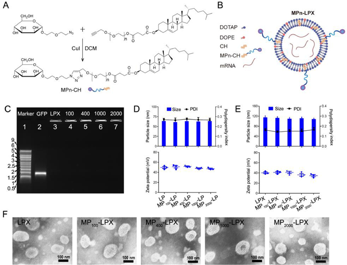
Figure 2. Preparation and characterization of MPn-LPX. (A) Schematic of the synthetic steps for MPn-CH. The number of ethylene glycol unit (n) in MP100-CH, MP400-CH, MP1000-CH, and MP2000-CH are 0, 9, 22, and 45, respectively. (B) Schematic structure of MPn-LPX. (C) Gel electrophoresis retardation assay of MPn-LPX. Marker (Marker, lane 1), free GFP-mRNA (GFP, lane 2), LPX (LPX, lane 3), MP100-LPX (100, lane 4), MP400-LPX (400, lane 5), MP1000-LPX (1000, lane 6) and MP2000-LPX (2000, lane 7). (D, E) Size, polydispersity index and zeta potential of MPn-LP (D) and MPn-LPX (E) (n = 3). (F) TEM images of MPn-LPX with different PEG linker lengths. Scale bar: 100 nm. Error bars indicate mean ± standard error of mean (SEM).
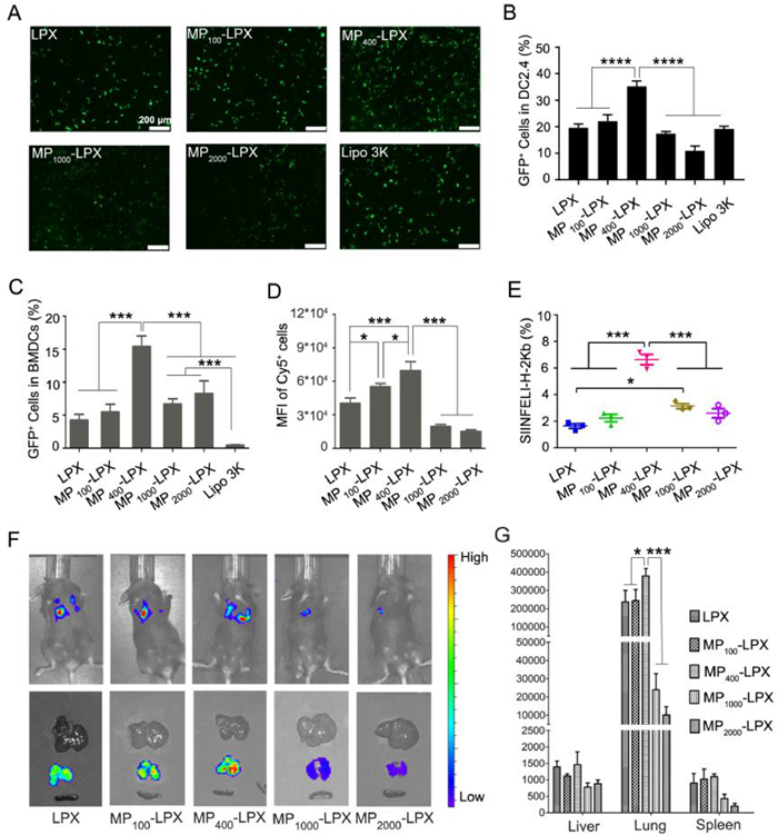
Figure 3. Effects of MPn-LPX with different PEG linker lengths on the mRNA delivery efficiency in vitro and in vivo. (A, B) Transfection capacity of MPn-LPX carrying GFP-mRNA in DC2.4 cells. (A) GFP expression by DC2.4 cells observed by a fluorescence microscope. Scale bar: 200 µm. (B) Transfection efficiency (% GFP+ cells) was quantified by flow cytometry (n = 3). (C) Transfection efficiency of MPn-LPX carrying GFP-mRNA on BMDCs (n = 3). (D) Cellular uptake of MPn-LPX carrying Cy5-mRNA in DC2.4 cells (n = 3). (E) Antigen presentation of MPn-LPX carrying OVA-mRNA in BMDCs (n = 3). (F) Representative whole body and isolated organs images of C57BL/6 mice demonstrated the mRNA expression 6 h after injection of Luc-mRNA loaded MPn-LPX. (G) The quantitative fluorescence intensity of Luc-mRNA expression in liver, lung and spleen (n = 3). Error bars indicate mean ± SEM. Statistical analysis was performed using a one-way ANOVA comparison. P < 0.05, ***P < 0.001, ****P < 0.0001.
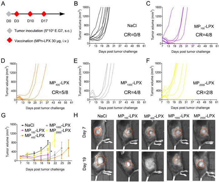
Figure 4. Antitumor effect of MPn-LPX in the E.G7 murine tumor model. (A) Experimental setup of the therapeutic immunization. (B–F) Individual tumor growth curve of each mouse. (G) Average tumor growth curves in each group (n = 8 mice per group). (H) Representative images of tumor size on day 7 and day 19. Error bars indicate mean ± SEM. Statistical analysis was performed using a one-way ANOVA comparison. P < 0.05.
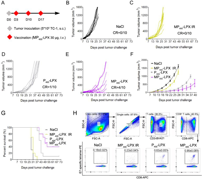
Figure 5. Therapeutic effect of MP400-LPX in the TC-1 tumor model. (A) Experimental setup of the therapeutic immunization. C57BL/6 mice were inoculated subcutaneously with 5 × 10⁵ TC-1 tumor cells on day 0. On days 3, 10, and 17, tumor-bearing mice were treated with indicated formulations containing 30 µg/dose of mRNA. (B–E) Individual tumor growth curves. (F) Average tumor growth curves. (G) Survival curves of each group (n = 10 mice per group). (H) MP400-LPX induced significantly enhanced E7-specific CD8+ T cell responses than P400-LPX (n = 3). The data are shown as mean ± SEM.

 Login In


 DownLoad:

 DownLoad: