Citation: Rong He, Dandan Tang, Ningge Xu, Heng Liu, Kun Dou, Xuejun Zhou, Fabiao Yu. Evaluation of erastin synergized cisplatin anti-nasopharyngeal carcinoma effect with a glutathione-activated near-infrared fluorescent probe[J]. Chinese Chemical Letters, ;2024, 35(2): 108658. doi: 10.1016/j.cclet.2023.108658 shu

Evaluation of erastin synergized cisplatin anti-nasopharyngeal carcinoma effect with a glutathione-activated near-infrared fluorescent probe

Figures(4)

  • Nasopharyngeal carcinoma (NPC), a malignant tumor originating from the nasopharynx, is one of the common malignant tumors of the head and neck. There are significant geographical differences in the incidence of nasopharyngeal carcinoma, with a high incidence in China and Southeast Asian countries. Herein, we designed and synthesized a novel near-infrared fluorescent (NIRF) probe to detect glutathione (GSH) in cellular and tumor environments using semi-naphthofluorescein (SNAFL) as the fluorescent molecular backbone and 2-fluoro-4-nitrobenzenesulfonate as the recognition moiety. Upon reaction with GSH, SNAFL-GSH emitted a fluorescence signal, and its emission wavelength at 650 nm was remarkably enhanced. The results of selectivity experiments indicated that SNAFL-GSH was able to discriminate GSH from Cys, Hcy, and H2S. Moreover, SNAFL-GSH could image both endogenous and exogenous GSH and distinguish normal and cancer cells by fluorescence signal difference. At the cellular level, cisplatin (DDP)-induced ferroptosis and inhibition of proliferation of various NPC cell lines (CNE2, CNE1, 5–8F cells) by erastin combined with DDP were visualized with the help of SNAFL-GSH. In a mouse tumor xenograft model, we successfully employed SNAFL-GSH for the evaluation of the efficacy of erastin combined with DDP in the treatment of NPC. More importantly, the probe could image cancerous tissue sections from NPC patients with an imaging depth of approximately 80 µm. It was foreseen that SNAFL-GSH offered great potential for application in the diagnosis and evaluation of the therapeutic efficacy of NPC, and these results would also provide new ideas for the clinical treatment of NPC.
  • Spintronics is a cutting-edge field of developing new electronic devices by manipulating the electron spin and magnetic moment [1]. Traditional spintronic research mainly focuses on transition metals and inorganic semiconductors, while organic molecules have the advantage of being extremely easy to realize efficient spin control by modifying the specific external conditions for desired electronic structures and magnetic characteristics. Corrole, as a ring-contracted porphyrin, is the aromatic analog of the central macrocycle of vitamin B12. Corrole has a squeezed inner cavity and three inner NHs in its free-base form, making it easier to stabilize high-valent metal ions and thus a promising candidate in spintronics.

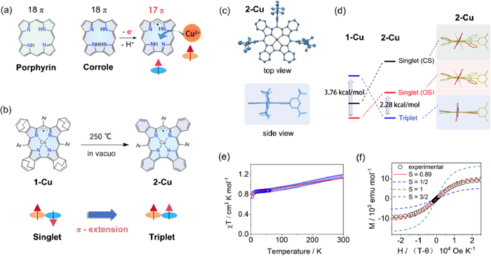
    When coordinated to metal ions such as Cu, Co, and Fe, the electron-rich corrole ligand could be partially oxidized to exhibit radical character, making it difficult to determine the exact oxidation states of central metals and ligands. The most controversial debate was the Cu(Ⅱ)/Cu(Ⅲ) dilemma on copper corrole [2]. The compound was initially thought to be a closed-shell Cu(Ⅲ) complex in 2000, but significant experimental and theoretical evidence over the next twenty years progressively revealed its open-shell singlet state ground state comprised of a Cu(Ⅱ) core and partially oxidized radical ligand (Fig. 1a).

    Figure 1

    Figure 1.  (a) Structure of 18π porphyrin and corrole, and schematic of the 17π corrole radical formation during coordination. (b) Schematic representation of the singlet-to-triplet conversion from 1-Cu to 2-Cu. (c) Single-crystal X-ray diffraction structure of 2-Cu. (d) DFT calculated structures and relative energies of 1-Cu and 2-Cu in triplet, open-shell singlet and closed-shell singlet states. (e) Observed (circle) and simulated (line) χT–T curve of 2-Cu at H = 5000 Oe. (f) Observed (circle) and simulated (line) M-H curves of 2-Cu with different S values.

    Shen Z. and Wu F. from Nanjing University have developed a series of metallocorroles with extended π-conjugation systems, which not only facilitated the formation and stabilization of radical ligands, but also allowed spin configurations of complexes to be easily controlled [3-5]. Recently, Shen, Wu, and co-workers reported that the unambiguous Cu(Ⅱ) corrole with fully oxidized [4n + 1]π radical ligand was obtained through the benzo-fusion at the β-position of corrole ligand [6]. The ground-state conversion of copper corrole radical from singlet to triplet was achieved via a retro-Diels-Alder reaction (Fig. 1b).

    The authors first synthesized a bicyclo[2.2.2]octadiene (BCOD) fused corrole 1-Cu by employing the classic H2O-MeOH approach with starting materials 4, 7-dihydro-4, 7-ethano-2H-isoindole and 3, 5-di-tert-butyl-benzaldehyde. Heating solid 1-Cu at 250 ℃ in vacuo cut the C—C bond in the BCOD bridge, eliminated the ethylene, and quantitatively afforded the benzo-fused 2-Cu. The singlet ground state of 1-Cu was clearly confirmed by the peripheral BCOD protons signals that appeared in the region of 6.60~2.09 ppm, while the signals of benzo protons in 2-Cu were located in a range of −6.2~−25.6 ppm, demonstrating its enhanced paramagnetism.

    The conformations of copper corroles were assumed to be "inherently saddle distorted" owing to the strong d-π interactions of antiferromagnetically coupled Cu(Ⅱ) corrole radicals. When compared to other copper corroles, 2-Cu stood out due to its highly planar macrocycle with a mean plane deviation value of only 0.024 Å (Fig. 1c). The planar structure could perfectly sustain the ferromagnetic coupling (S = 1) between Cu(Ⅱ) and corrole radical.

    The theoretical analysis of 2-Cu was conducted by the authors for three different states, including a close-shell singlet Cu(Ⅲ) (CS), an open-shell singlet antiferromagnetically coupled Cu(Ⅱ) corrole radical (OS) and a triplet ferromagnetically coupled Cu(Ⅱ) corrole radical (T). A lower T state was discovered for 2-Cu than the CS and OS states with a calculated singlet-triplet energy gap of 2.28 kcal/mol, providing theoretical support for the triplet ground state (Fig. 1d). The strongest support for the triplet ground state came from temperature- and field-dependent superconducting quantum interference device (SQUID) magnetometry. The χT value of 2-Cu in 2 K was 0.77 cm3 K/mol, and it reached approximately 1 cm3 K/mol at 300 K. The singlet-triplet energy gap was estimated to be 1.66 kcal/mol by fitting the χT–T plot (Fig. 1e). The field-dependent magnetization plot of 2-Cu at 2 K was fitted to a Brillouin function with S = 0.89, which was close to the value (S = 1) corresponding to the triplet ground state (Fig. 1f). The magnetic hysteresis of 2-Cu was observed at 2 K. Moreover, 2-Cu exhibits remarkable stability in air despite its radical character. The calculated density plots of spin and SOMO both demonstrate that the density is concentrated mostly in the inner corrole ring, which is nicely protected by fused benzenes with low reactivity.

    The research conducted by Shen's group introduces a new approach to the fine-tuning of interactions between metal center and corrole ligand and provides a promising strategy for the creation of stable corrole radical complexes with distinctive high-spin systems. The strategy will further trigger the development of novel functional materials based on corroles and their work will encourage an increasing amount of spintronics research for the use of innovative magnetic and electrical devices.


    1. [1]

      J. Sastre, F.V. Pallardo, J. Viña, Glutathione, in: T. Grune (Ed.), Reactions, Processes: Oxidants and Antioxidant Defense Systems, Springer Berlin Heidelberg, Berlin, Heidelberg, 2005, pp. 91–108.

    2. [2]

      G. Teskey, R. Abrahem, R. Cao, et al., Glutathione as a marker for human disease, in: G.S. Makowski (Ed.), Advances in Clinical Chemistry, Elsevier, London, 2018, pp. 141–159.

    3. [3]

      G.K. Balendiran, R. Dabur, D. Fraser, Cell Biochem. Funct. 22 (2004) 343–352.  doi: 10.1002/cbf.1149

    4. [4]

      M.P. Gamcsik, M.S. Kasibhatla, S.D. Teeter, et al., Biomarkers 17 (2012) 671–691.  doi: 10.3109/1354750X.2012.715672

    5. [5]

      Y. Sun, W.F. Li, N.Y. Chen, et al., Lancet Oncol. 17 (2016) 1509–1520.  doi: 10.1016/S1470-2045(16)30410-7

    6. [6]

      Y.P. Chen, A.T.C. Chan, Q.T. Le, et al., Lancet 394 (2019) 64–80.  doi: 10.1016/S0140-6736(19)30956-0

    7. [7]

      S. Guan, J. Wei, L. Huang, L. Wu, Eur. J. Med. Chem. 207 (2020) 112758.  doi: 10.1016/j.ejmech.2020.112758

    8. [8]

      M.A. Fuertes, C. Alonso, J.M. Pérez, Chem. Rev. 103 (2003) 645–662.  doi: 10.1021/cr020010d

    9. [9]

      J.W. Lv, Z.Y. Qi, G.Q. Zhou, et al., Cancer Sci. 109 (2018) 751–763.  doi: 10.1111/cas.13474

    10. [10]

      M. Song, M. Cui, K. Liu, Eur. J. Med. Chem. 232 (2022) 114205.  doi: 10.1016/j.ejmech.2022.114205

    11. [11]

      T. Jin, W.F. Qin, F. Jiang, et al., Transl. Oncol. 12 (2019) 633–639.  doi: 10.1016/j.tranon.2019.01.002

    12. [12]

      H. Yu, P. Guo, X. Xie, et al., J. Cell. Mol. Med. 21 (2017) 648–657.  doi: 10.1111/jcmm.13008

    13. [13]

      J. Li, F. Cao, H.L. Yin, et al., Cell. Death Dis. 11 (2020) 88.  doi: 10.1038/s41419-020-2298-2

    14. [14]

      B. Lu, X.B. Chen, M.D. Ying, et al., Front. Pharmacol. 8 (2017) 992.

    15. [15]

      X. Xia, X. Fan, M. Zhao, P. Zhu, Curr. Gene Ther. 19 (2019) 117–124.  doi: 10.2174/1566523219666190628152137

    16. [16]

      Y. Zhao, Y. Li, R. Zhang, et al., Onco Targets Ther. 13 (2020) 5429–5441.  doi: 10.2147/ott.s254995

    17. [17]

      D. Giustarini, I. Dalle-Donne, A. Milzani, et al., Nat. Protoc. 8 (2013) 1660–1669.  doi: 10.1038/nprot.2013.095

    18. [18]

      Y. Zhu, J. Wu, K. Wang, J. Xie, et al., Talanta 224 (2021) 121852.  doi: 10.1016/j.talanta.2020.121852

    19. [19]

      D. Sun, Z. Chen, J. Hu, et al., Chin. Chem. Lett. 33 (2022) 4478–4494.  doi: 10.1016/j.cclet.2021.12.043

    20. [20]

      D. Chen, Y. Feng, Crit. Rev. Anal. Chem. 52 (2022) 649–666.  doi: 10.1080/10408347.2020.1819193

    21. [21]

      Z. Xu, T. Qin, X. Zhou, et al., Trends Anal. Chem. 121 (2019) 115672.  doi: 10.1016/j.trac.2019.115672

    22. [22]

      S. Lee, J. Li, X. Zhou, et al., Coord. Chem. Rev. 366 (2018) 29–68.  doi: 10.1016/j.ccr.2018.03.021

    23. [23]

      Y. Zhang, J. Zhang, M. Su, C. Li, Biosens. Bioelectron. 175 (2021) 112866.  doi: 10.1016/j.bios.2020.112866

    24. [24]

      F. Liang, S. Jiao, D. Jin, et al., Spectrochim. Acta A 224 (2020) 117403.  doi: 10.1016/j.saa.2019.117403

    25. [25]

      Z. Xu, X. Huang, X. Han, et al., Chem. 4 (2018) 1609–1628.  doi: 10.1016/j.chempr.2018.04.003

    26. [26]

      N. Li, T. Wang, N. Wang, et al., Angew. Chem. Int. Ed. 62 (2023) e202217326.  doi: 10.1002/anie.202217326

    27. [27]

      W. Shu, J. Yu, H. Wang, et al., Anal. Chim. Acta 1220 (2022) 340081.  doi: 10.1016/j.aca.2022.340081

    28. [28]

      W. Liu, J. Chen, Q. Qiao, et al., Chin. Chem. Lett. 33 (2022) 4943–4947.  doi: 10.1016/j.cclet.2022.03.121

    29. [29]

      F. Chen, J. Zhang, W. Qu, et al., Sens. Actuators B Chem. 266 (2018) 528–533.  doi: 10.1016/j.snb.2018.03.162

    30. [30]

      K. Umezawa, M. Yoshida, M. Kamiya, et al., Nat. Chem. 9 (2017) 279–286.  doi: 10.1038/nchem.2648

    31. [31]

      D. Gong, S.C. Han, A. Iqbal, et al., Anal. Chem. 89 (2017) 13112–13119.  doi: 10.1021/acs.analchem.7b02311

    32. [32]

      S. Hou, Y. Wang, Y. Zhang, et al., Anal. Chim. Acta 1214 (2022) 339957.  doi: 10.1016/j.aca.2022.339957

    33. [33]

      K. Wang, G. Nie, S. Ran, et al., Dyes Pigments 172 (2020) 107837.  doi: 10.1016/j.dyepig.2019.107837

    34. [34]

      P. Hou, J. Sun, H. Wang, et al., Sens. Actuators B Chem. 304 (2020) 127244.  doi: 10.1016/j.snb.2019.127244

    35. [35]

      Z. Zheng, Y. Huyan, H. Li, et al., Sens. Actuators B Chem. 301 (2019) 127065.  doi: 10.1016/j.snb.2019.127065

    36. [36]

      C. Zhang, Y. Qin, C. Deng, et al., Anal. Chim. Acta 1248 (2023) 340933.  doi: 10.1016/j.aca.2023.340933

    37. [37]

      H.M. Jiang, G.X. Yin, Y.B. Gan, et al., Chin. Chem. Lett. 33 (2022) 1609–1612.  doi: 10.1016/j.cclet.2021.09.036

    38. [38]

      X.W. Li, C.Y. Liu, N. Gao, et al., Chin. Chem. Lett. 33 (2022) 2527–2531.  doi: 10.1016/j.cclet.2021.11.080

    39. [39]

      T.X. Jin, M.Y. Cui, D. Wu, et al., Chin. Chem. Lett. 32 (2021) 3899–3902.  doi: 10.1016/j.cclet.2021.06.033

    40. [40]

      Y. Zou, M. Li, Y. Xing, et al., ACS Sens. 5 (2020) 242–249.  doi: 10.1021/acssensors.9b02118

    41. [41]

      N. Ahmed, W. Zareen, Y. Ye, Chin. Chem. Lett. 33 (2022) 2765–2772.  doi: 10.1016/j.cclet.2021.12.092

    42. [42]

      L.R. Jiang, T.H. Chen, E.W. Song, et al., Chem. Eng. J. 427 (2022) 131563.  doi: 10.1016/j.cej.2021.131563

    43. [43]

      S.T. Cai, Q.C. Liu, C. Liu, et al., J. Mater. Chem. B 10 (2022), 1265–1271.  doi: 10.1039/d1tb02639a

    44. [44]

      S. Xu, W.J. Pan, T.B. Ren, et al., Chin. J. Chem. 40 (2021) 1073–1082.  doi: 10.1002/cjoc.202100807

    45. [45]

      C. Duan, M. Won, P. Verwilst, et al., Anal. Chem. 91 (2019) 4172–4178.  doi: 10.1021/acs.analchem.9b00224

    46. [46]

      J.C. Xu, J. Pan, X.M. Jiang, et al., Biosens. Bioelectron. 77 (2016), 725–732.  doi: 10.1016/j.bios.2015.10.049

    47. [47]

      Y.Y. Ma, Z.C. Xu, Q. Sun, et al., Spectrochim. Acta. A 247 (2021) 1386–1425.  doi: 10.1049/icp.2022.0265

    48. [48]

      X.Y. Zhang, W.B. Qu, H. L, et al., Anal. Chim. Acta 1109 (2020) 37–43.  doi: 10.1016/j.aca.2020.02.061

    49. [49]

      J. Dong, G. Lu, Y. Tu, C. Fan, New J. Chem. 46 (2022) 10995–11020.  doi: 10.1039/d1nj06244a

    50. [50]

      M.Y. Lucero, J. Chan, Nat. Chem. 13 (2021) 1248–1256.  doi: 10.1038/s41557-021-00804-0

    51. [51]

      J. Dai, C. Ma, P. Zhang, et al., Dyes Pigments 177 (2020) 108321.  doi: 10.1016/j.dyepig.2020.108321

    52. [52]

      L.Y. Niu, Y.Z. Chen, H.R. Zheng, et al., Chem. Soc. Rev. 44 (2015) 6143–6160.  doi: 10.1039/C5CS00152H

    53. [53]

      J. Hu, W. Gu, N. Ma, et al., Br. J. Pharmacol. 179 (2022) 3991–4009.  doi: 10.1111/bph.15834

    54. [54]

      Q. Cheng, L. Bao, M. Li, et al., J. Obstet. Gynaecol. Res. 47 (2021) 2481–2491.  doi: 10.1111/jog.14779

    55. [55]

      M. Sato, R. Kusumi, S. Hamashima, et al., Sci. Rep. 8 (2018) 968.  doi: 10.1038/s41598-018-19213-4

  • 加载中
    1. [1]

      Chuanfeng FanJian GaoYingkai GaoXintong YangGaoning LiXiaochun WangFei LiJin ZhouHaifeng YuYi HuangJin ChenYingying ShanLi Chen . A non-peptide-based chymotrypsin-targeted long-wavelength emission fluorescent probe with large Stokes shift and its application in bioimaging. Chinese Chemical Letters, 2024, 35(10): 109838-. doi: 10.1016/j.cclet.2024.109838

    2. [2]

      Lin LiBingjun SunJin SunLin ChenZhonggui He . Binary prodrug nanoassemblies combining chemotherapy and ferroptosis activation for efficient triple-negative breast cancer therapy. Chinese Chemical Letters, 2024, 35(10): 109538-. doi: 10.1016/j.cclet.2024.109538

    3. [3]

      Zhendong LiuSainan LiuBin LiuQi MengMeng YuanChunzheng YangYulong BianPing'an MaJun Lin . Fe(Ⅲ)-juglone nanoscale coordination polymers for cascade chemodynamic therapy through synergistic ferroptosis and apoptosis strategy. Chinese Chemical Letters, 2024, 35(11): 109626-. doi: 10.1016/j.cclet.2024.109626

    4. [4]

      Bin FangJiaqi YangLimin WangHaoqin LiJiaying GuoJiaxin ZhangQingyuan GuoBo PengKedi LiuMiaomiao XiHua BaiLi FuLin Li . A mitochondria-targeted H2S-activatable fluorogenic probe for tracking hepatic ischemia-reperfusion injury. Chinese Chemical Letters, 2024, 35(6): 108913-. doi: 10.1016/j.cclet.2023.108913

    5. [5]

      Fan ZhengRunsha XiaoShuai HuangZhikang ChenChen LaiAnyao BiHeying YaoXueping FengZihua ChenWenbin Zeng . Accurate visualization colorectal cancer by monitoring viscosity variations with a novel mitochondria-targeted fluorescent probe. Chinese Chemical Letters, 2025, 36(2): 109876-. doi: 10.1016/j.cclet.2024.109876

    6. [6]

      Meiling ZhaoYao LuYutao ZhangHaoyun XueZhiqian Guo . Ultra-high signal-to-noise ratio near-infrared chemiluminescent probe for in vivo sensing singlet oxygen. Chinese Chemical Letters, 2025, 36(5): 110105-. doi: 10.1016/j.cclet.2024.110105

    7. [7]

      Lixian FuYiyun TanYue DingWeixia QingYong Wang . Water–soluble and polarity–sensitive near–infrared fluorescent probe for long–time specific cancer cell membranes imaging and C. Elegans label. Chinese Chemical Letters, 2024, 35(4): 108886-. doi: 10.1016/j.cclet.2023.108886

    8. [8]

      Jiangshan XuWeifei ZhangZhengwen CaiYong LiLong BaiShaojingya GaoQiang SunYunfeng Lin . Tetrahedron DNA nanostructure/iron-based nanomaterials for combined tumor therapy. Chinese Chemical Letters, 2024, 35(11): 109620-. doi: 10.1016/j.cclet.2024.109620

    9. [9]

      Xing TianDi WuWanheng WeiGuifu DaiZhanxian LiBenhua WangMingming Yu . A lipid droplets-targetable fluorescent probe for polarity detection in cells of iron death, inflammation and fatty liver tissue. Chinese Chemical Letters, 2024, 35(6): 108912-. doi: 10.1016/j.cclet.2023.108912

    10. [10]

      Jiayu ZengMinhui LiuTing YangJia HuangSongjiao LiWanting ZhangDan ChengLongwei HeJia Zhou . Two-dimensional design strategy to construct smart dual-responsive fluorescent probe for the precise tracking of ischemic stroke. Chinese Chemical Letters, 2025, 36(5): 110166-. doi: 10.1016/j.cclet.2024.110166

    11. [11]

      Jin WangXiaoyan PanJunyu ZhangQingqing ZhangYanchen LiWeiwei GuoJie Zhang . Active molecule-based theranostic agents for tumor vasculature normalization and antitumor efficacy. Chinese Chemical Letters, 2024, 35(8): 109187-. doi: 10.1016/j.cclet.2023.109187

    12. [12]

      Yuequan WangCongtian WuChengcheng FengQin ChenZhonggui HeShenwu ZhangCong LuoJin Sun . Spatiotemporally-controlled supramolecular hybrid nanoassembly enabling ferroptosis-augmented photodynamic immunotherapy of cancer. Chinese Chemical Letters, 2025, 36(3): 109902-. doi: 10.1016/j.cclet.2024.109902

    13. [13]

      Yan GaoZi-Lin SongShuang YuXiu-Li ZhaoDa-Wei ChenMing-Xi Qiao . Enhanced ferroptosis by a nanoparticle mimicking hemoglobin coordinate pattern with self-supplying hydrogen peroxide. Chinese Chemical Letters, 2025, 36(5): 110097-. doi: 10.1016/j.cclet.2024.110097

    14. [14]

      Zheng-Biao ZouTai-Zong WuChun-Lan XieYuan WangYan LiGang ZhangRong ChaoLian-Zhong LuoLi-Sheng LiXian-Wen Yangneo-Dicitrinols A–C: Unprecedented PKS-NRPS hybrid citrinin dimers with ferroptosis inhibitory activity from the deep-sea-derived Penicillium citrinum W22. Chinese Chemical Letters, 2024, 35(12): 109723-. doi: 10.1016/j.cclet.2024.109723

    15. [15]

      Yong-Dan ZhaoYidan WangRongrong WangLina ChenHengtong ZuoXi WangJihong QiangGeng WangQingxia LiCanqi PingShuqiu ZhangHao Wang . Reversing artemisinin resistance by leveraging thermo-responsive nanoplatform to downregulating GSH. Chinese Chemical Letters, 2024, 35(6): 108929-. doi: 10.1016/j.cclet.2023.108929

    16. [16]

      Zengchao GuoWeiwei LiuTengfei LiuJinpeng WangHui JiangXiaohui LiuYossi WeizmannXuemei Wang . Engineered exosome hybrid copper nanoscale antibiotics facilitate simultaneous self-assembly imaging and elimination of intracellular multidrug-resistant superbugs. Chinese Chemical Letters, 2024, 35(7): 109060-. doi: 10.1016/j.cclet.2023.109060

    17. [17]

      Yunlong LiXinyu ZhangShuang LiuChunsheng LiQiang WangJin YeYong LuJiating Xu . Engineered iron-based metal-organic frameworks nanoplatforms for cancer theranostics: A mini review. Chinese Chemical Letters, 2025, 36(2): 110501-. doi: 10.1016/j.cclet.2024.110501

    18. [18]

      Haixian RenYuting DuXiaojing YangFangjun HuoLe ZhangCaixia Yin . Development of ESIPT-based specific fluorescent probes for bioactive species based on the protection-deprotection of the hydroxyl. Chinese Chemical Letters, 2025, 36(2): 109867-. doi: 10.1016/j.cclet.2024.109867

    19. [19]

      Hui ZhangRong FengWanyi YuHongbei WeiTianhong WuPeng ZhangWenhai BianXin LiDi GaoGuojun WengZhe YangTony D. JamesXiaolong Sun . Evaluating the global thiols redox state in living cells using a reducing sulfur species responsive fluorescence switching platform. Chinese Chemical Letters, 2025, 36(4): 110528-. doi: 10.1016/j.cclet.2024.110528

    20. [20]

      Yanjun CaiYong JiangYu ChenErzhuo ChengYuan GuYuwei LiQianqian LiuJian ZhangJifang LiuShisong HanBin Yang . Amplifying STING activation and immunogenic cell death by metal-polyphenol coordinated nanomedicines for enhanced cancer immunotherapy. Chinese Chemical Letters, 2025, 36(5): 110437-. doi: 10.1016/j.cclet.2024.110437

Metrics
  • PDF Downloads(3)
  • Abstract views(639)
  • HTML views(30)

通讯作者: 陈斌, bchen63@163.com
  • 1. 

    沈阳化工大学材料科学与工程学院 沈阳 110142

  1. 本站搜索
  2. 百度学术搜索
  3. 万方数据库搜索
  4. CNKI搜索
Address:Zhongguancun North First Street 2,100190 Beijing, PR China Tel: +86-010-82449177-888
Powered By info@rhhz.net

/

DownLoad:  Full-Size Img  PowerPoint
Return