Citation: Yan-Long Ma, Shi Yan, Xiong-Jie Xu, Hua Cao, Ruibing Wang. Synthetic host-guest pairs as novel bioorthogonal tools for pre-targeting[J]. Chinese Chemical Letters, ;2024, 35(2): 108645. doi: 10.1016/j.cclet.2023.108645 shu

Synthetic host-guest pairs as novel bioorthogonal tools for pre-targeting

    This paper is dedicated to the memory of our co-worker Prof. Jiang Wei.
    * Corresponding authors.
    E-mail addresses: caohua@gdpu.edu.cn (H. Cao), rwang@um.edu.mo (R. Wang).
  • Received Date: 17 April 2023
    Revised Date: 25 May 2023
    Accepted Date: 2 June 2023
    Available Online: 4 June 2023

Figures(12)

  • Due to its simplicity, high efficiency, and chemo-selectivity, bioorthogonal chemistry has shown a great application potential in pre-targeting. Currently, four bioorthogonal pairs as targeting tools, including (strept)avidin/biotin, antibody/antigen, oligonucleotide hybridization and IEDDA tools, have been developed and applied in targeted delivery. Nevertheless, all of these tools still suffer from some limitations, such as difficult modification, biochemical fragility and larger molecular weight for biological association tools, as well as chemical instability for IEDDA tools. Synthetic host-guest pairs with relatively small molecular sizes not only possess strong chemical stability, but also have the features of fast conjugation rate, tunable binding affinity, easy modification, and high chemo-selectivity. Consequently, they can be used as a novel non-covalent bioorthogonal tool for pre-targeting. In order to further promote the development of host-guest pairs as novel bioorthogonal tools for pre-targeted delivery, we firstly calculate their conversion rate to make researcher aware of their unique advantages; next, we summarize the recent research progress in this area. The future perspectives and limitations of these unique tools will be discussed. This review will provide a systemic overview of the development of synthetic host-guest pairs as novel bioorthogonal tools for pre-targeting, and may serve as a "go for" resort for researchers who are interested in searching for new synthetic tools to improve pre-targeting.
  • In physiological or pathological conditions, extracellular matrix (ECM) undergoes complex and diverse changes in structural and mechanical properties to support critical interactions with cells. Including stiffness, shear stress, stretching, ligand density, composition, and topography, have been confirmed to regulate cell behaviors. Particularly, tissue stiffening, an unique phenomenon of cancer progression, has attracted plentiful researches on specific cell behaviors in 2D planar systems [1-4]. However, as cells are surrounded by 3D ECM niches in vivo, the dimensionality shift may directly cause differences in ligand distribution, cell adhesion mode and cell stiffness-sensing, which would definitely affect cell behaviors [5,6]. For instance, in context of pathological diseases, cell metastasis is always accelerated by the increasing stiffness of traditional 2D substrates, while observations on 3D ECM mimics may be contradictory [7]. Thus, 3D ECM has been shown to be necessary to further explore the ability of artificial platforms, specifically in cell mechanical sensing.

    To date, the fabrication of diverse 3D models has been achieved by many methods, such as cell spheroid culture, 3D bioprinting, tissue-engineering scaffolds with customizable properties, hydrogels, cell sheets, bioreactors with dynamic loading, and microfluidics [8]. Among them, microfluidics show the advantages of high throughput, stability and automation, making it possible to obtain a great quantity of samples with consistent spatial distribution and structures using low amounts of reagents [9-12]. In terms of materials, hydrogels combine the unique properties of softness, low immunogenicity and tunable mechanical characteristics [13-15]. Thus, monodisperse microgels produced with these elements not only offer exquisite control on homogeneous internal structures, but also shows the advantage in ECM physical features simulation [16]. Additionally, the limited size of microgels can avoid cells inactivation at internal positions caused by deficient oxygen and nutrients [17].

    However, the microgels still present some limitations in the simulation of tumor stiffness due to their nature of high water content and flexibility [18]. To achieve high stiffness, simply increasing the weight concentration is not feasible, as it leads to a simultaneous change in porosity and viscosity, which brings out challenges including microflow shearing and microchip clogging [19]. Moreover, while the mechanical properties can be altered by the amount, type of crosslinking ions or methods, stiffness changes on orders-of-magnitude is hardly achieved [20]. To solve this problem, nanofibers have shown the prospect in simulation of increased collagenous fibrils in hardened tissue [21]. For example, Joshi et al. have successfully combined β-TCP incorporated PLA nanofibers with gelatin for biomimetic bone tissue engineering, the compressive stress was amplified about 8-fold [22]. It showed the nanofiber-hydrogel hybrids contained widely adjustable mechanical properties, which could fit the stiffness of some specific or diseased tissue.

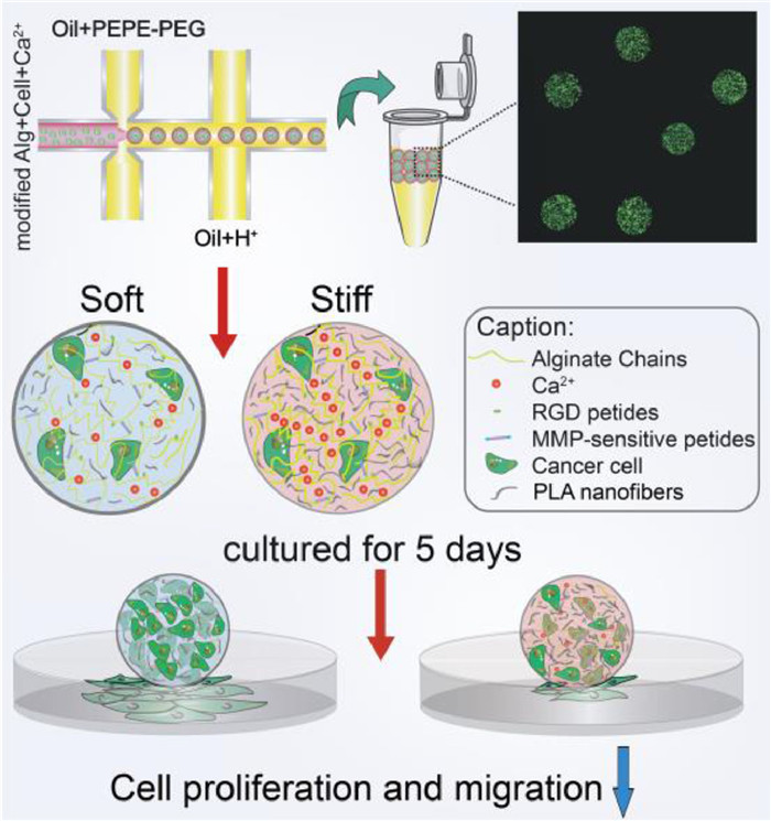
    Herein, we present an innovative strategy to reinforce 3D microgel constructs by formulating composite hydrogel composed of modified alginate and different amount of micron-scale polylactide (PLA) nanofibers for solid carcinoma engineering (Scheme 1). The modified alginate with PVGLIG (proline-valine-glycine-leucine-isoleucine-glycine) and RGD (arginine-glycine-aspartate) peptides provided corresponding sites for cell migration and adhesion. By adding microscale PLA nanofibers uniformly by ultrasound, the composite microgels with increased stiffness showed the similar pore size, proving that we have created a 3D mechanical matrix platform with univariate variables. Following this strategy, solid breast tumors within the pathological breast stiffness range (2–20 kPa) were successfully simulated by using different composite gel ratios and two breast cancer cells with different invasive abilities, epithelial phenotype breast cancer cells MCF-7 and more aggressive mesenchymal SUM-159 [23]. Obviously, high proliferation and invasion ability of both cell types in soft matrix were observed. Furthermore, RT-qPCR analysis revealed the underlying changes at the genetic level. In this study, the proposed 3D microgel with homogeneous microstructure and adjustable stiffness effectively simulates the mechanical microenvironment of solid breast tumors, and demonstrates the advantages in-vitro simulation and tissue engineering.

    Scheme1

    Scheme1.  The synthesis process of microgels and the illustration of cell behavioral differences regulated by the variable stiffness in microgels.

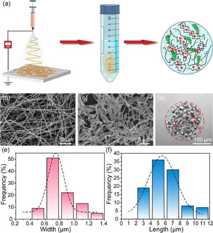
    Firstly, PLA nanofibers with well-controlled diameters were fabricated by electrospinning process (Fig. 1a). Prior to use, morphology of nanofibers should be thoroughly characterized. As shown in Fig. 1b, the SEM micrograph of PLA fibers had random orientation and uniform width after electrospinning, and the average width was about 0.52 ± 0.12 µm, which fitted the Gaussian distribution (Fig. S1 in Supporting information). However, the fibers produced by electrospinning directly are always with irregular length in the tens or hundreds of microns, which might clog the outlets of microchips and hinder the uniform generation of micro-scale microgels. The homogeneous PLA nanofibers that shorter than the diameter of microgels are urgently needed to avoid the large agglomeration. Thus, the electrospun PLA nanofibers were aminolyzed in 5% ethylenediamine to obtain the micro-short scale, in order to disperse it uniformly in hydrogels [24]. And relevant experiments were optimized to discovery the influence of aminolysis time on the degree of fiber breakage. In Fig. S2 (Supporting information), PLA nanofibers became shorter and thicker as the increasing time of ammonolysis. For the 3 h ammonolysis (Fig. 1c), the PLA nanofibers were decomposed into micrometer-scale lengths, which had been suitable for making uniformly sized microgel spheres (Fig. 1d). Therefore, the 3 h ammonolysis PLA fibers with an average width and length of 0.8062 ± 0.1966 µm (Fig. 1e) and 5.9048 ± 2.1419 µm (Fig. 1f) could be employed in the composite hydrogels. And to guarantee the composite hydrogels biocompatible and stability, RGD and PVGLIG (MMP sensitive sequence) were introduced efficiently on alginate in response to cell adhesion and migration (Fig. S3 in Supporting information).

    Figure 1

    Figure 1.  The synthesis and characterization of PLA nanofibers. (a) Schematic diagram for the fabrication of PLA nanofibers and PLA nanofiber reinforced alginate microgels. SEM images of (b) PLA nanofibers and (c) aminolyzed micron-length PLA nanofibers. Scale bars represent 10 µm and 5 µm, respectively. (d) PLA nanofiber reinforced alginate microgels for cell encapsulation, red arrows point out the PLA nanofibers. Scale bar: 100 µm. (e) Width and (f) length distribution of aminolyzed micro-short PLA nanofibers. Fitting performed assuming normal distribution, mean values ± SD: d = 0.8062 ± 0.1966 µm, L = 5.9048 ± 2.1419 µm, both n = 100.

    Next, we fabricated the microfluidic device to produce cell-loaded alginate/PLA hydrogel spheroids by flow-focusing (Fig. S4 in Supporting information) [25]. And the height and length of all microchannels were around 100 µm and 200 µm. Subsequently, the conditions of the continuous phases were optimized by fixing the velocity of dispersed phase. As shown in Videos S1–S4 (Supporting information), when the flow rate of the continuous phase was speeder than 600 µL/h, the dispersed phase at a rate of 40 µL/h could be cut into microgel spheres. To conform to the size requirement (~200 µm), we ultimately chose two continuous phases at the flow rate of 600 µL/h. The obtained microgels were collected in 20% 1H, 1H, 2H, 2H-perfluoro-1-octanol for demulsification.

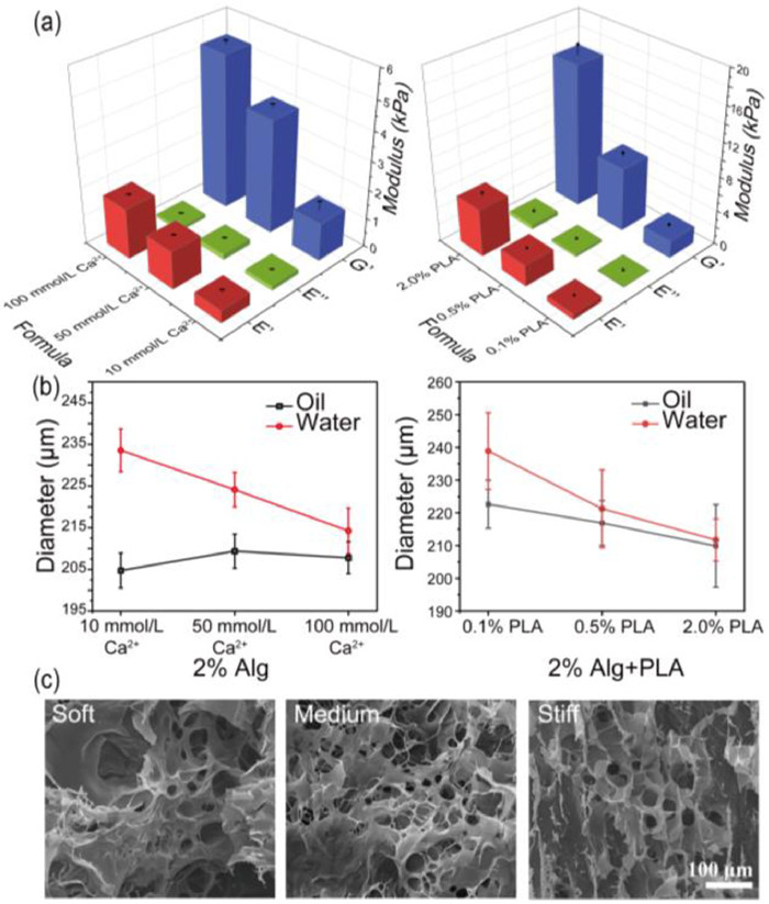
    Subsequently, to simulate varying degrees of stiffening in breast cancer, different amounts of PLA nanofibers added in the compound gels should be explored. And the three groups in Table S1 (Supporting information) were finally selected. As shown in Fig. 2a and Fig. S5 (Supporting information), the viscosity modulus of 2.0% alginate with different concentrations of Ca2+ were far below the elastic modulus, indicating the Young's modulus was mainly affected by the modulus of elasticity. After the corresponding experimental groups that added different amounts of PLA, the viscous modulus of the composite hydrogel still kept the same trend, while the elastic modulus was greatly increased, thereby raising the overall stiffness about 1.54, 1.88, 3.17 times, up to 2.02 ± 0.25 kPa, 7.45 ± 0.49 kPa and 16.76 ± 1.18 kPa, respectively. Meanwhile, the swelling phenomenon during long-term culture also is a crucial feature, and the swelling ratio which reflected microgels cross-linking density was determined by the hydrodynamic diameter calculation of the microgel formulation in oil or water. As shown in Fig. 2b and Fig. S6 (Supporting information), the sizes of pure alginate groups are swelled from 204.73 ± 4.19 µm, 209.39 ± 4.10 µm, 207.80 ± 3.84 µm to 233.57 ± 5.10 µm, 224.14 ± 4.51 µm, 214.25 ± 5.49 µm. Microgels cross-linking with only 10 mmol/L Ca2+ swelled the most remarkable with 14.09% increasing. After adding PLA, the stability of the gel was further improved. The particle sizes are swelled from 222.64 ± 7.34 µm, 216.89 ± 6.82 µm, 209.89 ± 12.65 µm to 238.88 ± 11.73 µm, 221.25 ± 11.87 µm, 211.72 ± 6.41 µm, showing the swelling rates of three gel formulations were reduced to 7.29%, 2.01%, and 0.87%, respectively. Simultaneously, SEM images in Fig. 2c and Fig. S7 (Supporting information) demonstrated the homogeneity of pore size of three formulas, which were about 23.21 ± 7.71 µm, 24.47 ± 4.50 µm and 20.18 ± 8.50 µm, respectively. Statistical results showed that changing the cross-linking state within a certain range Ca2+ concentration would not affect the pore size of gels obviously, thus avoiding the influence in nutrients or the metabolic waste transport. Besides, the distribution of PLA micron nanofibers and the morphology of the cells encapsulated in different gel could be clearly observed from SEM images in Fig. S8 (Supporting information).

    Figure 2

    Figure 2.  Performance characterizations of microgels. (a) Modulus of 2.0% Alginate with different Ca2+ concentration and with PLA nanofibers. N = 3, E'= elastic component, E'' = viscous component, G* = Young's modulus. (b) Swelling performance of different formulations. The microgels generated in oil and soaked in water for 24 h for diameter recording. (c) SEM images of three composite hydrogel formulations, scale bar: 100 µm.

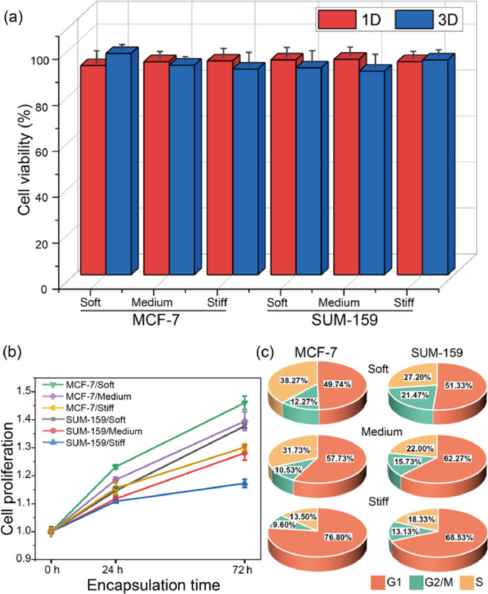
    We proceeded to explore the role of stiffness on cell viability and proliferation in tumor microenvironments simulated by microgels. As shown in Fig. S9 (Supporting information), activity of MCF-7 and SUM-159 cells maintained no significant difference for all three conditions, similar results were obtained for other cell lines as well (Fig. S10 in Supporting information). And through counting and quantitative analysis of encapsulated cells of 1–3 days cultivation in Fig. 3a, the viability was at the range of 88.63% to 96.37%, which proved the high cytocompatible of nanofibers-composited hydrogels. Due to the modification of RGD and PVGLIG motifs, the hydrogel provided suitable sites for cell-matrix intergration and further improved cell growth and proliferation. In Fig. 3b, we estimated cell numbers on day 0, 1, and 3 by CCK8 assay and defined the fold change in proliferation by normalizing to the day 0 data. Two kinds of cells in the soft (2.02 kPa) and medium-stiffness (7.45 kPa) microenvironments showed significantly higher growth than the stiff (16.76 kPa) condition in 72 h. Subsequently, cell cycles were detected by flow cytometry. In Fig. 3c, the cells grown in the soft environments showed more distribution in the G2/M period, indicating the higher proliferation ability, which were consistent with the CCK8 results. In conclusion, both MCF-7 and SUM-159 cells tends to proliferate in a softer 3D microenvironment.

    Figure 3

    Figure 3.  State of MCF-7 and SUM-159 cells within the modified microgels with tunable stiffness. (a) Counting and quantitative viability analysis of encapsulated cell in microgels, n = 50. (b) Proliferation curves of cells encapsulated in heterogeneous environments. N = 3. (c) Analysis of cell cycle in 3D conditions for 48 h by flow cytometry. N = 3.

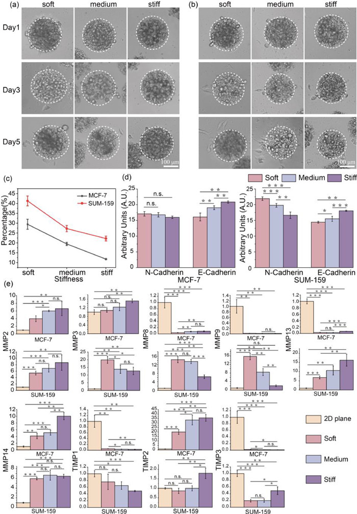
    It is well known that breast cancer cells MCF-7 and SUM-159, exhibiting epithelial phenotype and mesenchymal phenotype, displayed with low and high metastatic potential in 2D plane, respectively (Fig. S11 in Supporting information). To study whether cell migration viability is highly correlated with 3D ECM stiffness, we encapsulated cells in 200 µm microgels and observed cell migrated from the inside of the gel to the outer space in 5 days.

    In Figs. 4a and b, cells exhibited similar tendency of metastatic potential, that were more frequently forming bulges on the surface of spheroids in soft condition. While cells located at the bottom of spheroids could migrate out and attach on the 2D bottom surface. For each culture condition, we measured more than 50 microgels in each independent replicate, and repeated 3 times in Fig. 4c. For MCF-7, the spheroids with surface cell bulges or bottom cell attachment were significantly decreased from 29.44% ± 2.53% to 19.48% ± 0.82% and 11.77% ± 0.23% with the increased stiffness. And SUM-159 presented the migration percentage of 41.30% ± 2.50%, 27.25% ± 1.57% and 22.23% ± 1.21%, respectively, after encapsulated for 72 h under the same condition as MCF-7. It proved that cells with a mesenchymal phenotype still maintained the high migratory capacity in 3D microenvironment. Certainly, with a limited number of encapsulated cells, cell protrusions on the microgel surface could also be observed, but for a longer time (Fig. S12 in Supporting information).

    Figure 4

    Figure 4.  Migration ability and gene expression two kinds of breast cancer cells under different mechanical stiffness stimulations. Images of (a) MCF-7 and (b) SUM-159 migration for 1, 3, 5 days. (c) Efficiency of cell migration after 3 days of culture in varying stiffness 3D constructs. n ≥ 50, N = 3. (d) Immunofluorescence intensity of N-cadherin and E-cadherin in two kinds of cells, N = 3. (e) Gene expression heat map shows several critical protein expression genes associated with cell migration. All gene expression levels are normalized with housekeeping gene GAPDH from each culture condition (-∆∆Ct was applied for normalization), N = 3. * P < 0.05, ** P < 0.01, *** P < 0.001, n.s. is no significance.

    Furthermore, we sought to further testify motility of encapsulated cells by immunofluorescence staining of N-cadherin and E-cadherin. As shown in Fig. S13 (Supporting information), cultured for 72 h, both N-cadherin and E-cadherin were observed in two type of cells. And the expression of E-cadherin was slightly lower in cells within the soft microgels, indicating the weakened intercellular cohesion. Thus, cells easily dispersed and infiltrated to the periphery. Obviously, with the increasing ECM stiffness, the decrease of N-cadherin expression level suggested the weakening of cell migration and invasion ability as well (Fig. 4d). Instead, the expression of epithelial markers E-cadherin in MCF-7 and mesenchymal markers N-cadherin in SUM-159 were more deeply affected by ECM stiffness, respectively, which was determined by cell characteristics. In summary, cell migration became tough in stiff 3D microenvironment. Therefore, we speculated that cells need time to find or pave a way to pass through the ECM with more physical constraints.

    After demonstrating the association between migration and ECM stiffness in encapsulated cells, the changes induced by environmental confinement should be more visualized by gene expression. Matrix metalloproteinases (MMPs) are known to play an important role in developmental and homeostatic remodeling of ECM by mediating the degradation of ECM proteins, while tissue inhibitor of metalloproteinases (TIMPs) can adjust the MMPs expression [26]. From the point of view of migration ability, those genes were selected for further mRNA expression level analysis, and the detail of sequences were shown in Table S2. As shown in Fig. 4e, the expression of MMP2, MMP13, MMP14 and TIMP2 in both cells were significantly upward modulation within stiffer microgels. Meanwhile, the expression of those genes in 2D plane were downward than 3D condition, except the MMP13 in MCF-7 cells. Subsequently, MMP9 and TIMP1 of two kinds of breast cells were significantly down-regulated in stiff 3D microenvironment, and only MMP9 of SUM-159 expressed more in 3D ECM than 2D cultured. These observations were consistent with the known study that MMP2 and MMP9 could be repressed by TIMP1 and TIMP2, respectively. In addition, MMP3 and MMP8 of MCF-7 were significantly up-regulated with ECM stiffness, while the expression of those two genes demonstrated opposite in SUM-159. As for TIMP3, the expression was rapid declined in MCF-7 but raised in SUM-159 as stiffness changed. Those gene expression analysis revealed a complex and sensitive feedback network of cell migration upon 3D mechanical stimulation, which may provide some insights into solid tumor research and therapy.

    In summary, we employed a composite hydrogel of PLA nanofibers and modified alginate with different concentrations of Ca2+, which can typically reinforce the mechanical properties of microgels with similar pore size. And we also investigated the effects of 3D microenvironment stiffness on the cell proliferation, viability, migration, and related gene expression of two types of breast cancer cells. Highly biocompatible and throughput microgels propose a new approach for the construction of solid tumors and observation of tumor migration. We envision that this study will provide an essential foundation for future investigations on ECM mechanisms in breast cancer and spark new approaches for therapy.

    The authors declare that they have no known competing financial interests or personal relationships that could have appeared to influence the work reported in this paper.

    This work was supported by the National Natural Science Foundation of China (Nos. 22034005, 81973569, and 21621003).

    Supplementary material associated with this article can be found, in the online version, at doi:10.1016/j.cclet.2023.108319.


    1. [1]

      J.A. Prescher, C.R. Bertozzi, Nat. Chem. Biol. 1 (2005) 13–21.  doi: 10.1038/nchembio0605-13

    2. [2]

      E. Saxon, C.R. Bertozzi, Science 287 (2000) 2007–2010.  doi: 10.1126/science.287.5460.2007

    3. [3]

      H.C. Hang, C. Yu, D.L. Kato, C.R. Bertozzi, Proc. Natl. Acad. Sci. U. S. A. 100 (2003) 14846–14851.  doi: 10.1073/pnas.2335201100

    4. [4]

      J.A. Prescher, D.H. Dube, C.R. Bertozzi, Nature 430 (2004) 873–877.  doi: 10.1038/nature02791

    5. [5]

      N.J. Agard, J.A. Prescher, C.R. Bertozzi, J. Am. Chem. Soc. 126 (2004) 15046–15047.  doi: 10.1021/ja044996f

    6. [6]

      E.M. Sletten, C.R. Bertozzi, Angew. Chem. Int. Ed. 48 (2009) 6974–6998.  doi: 10.1002/anie.200900942

    7. [7]

      E.M. Sletten, C.R. Bertozzi, Acc. Chem. Res. 44 (2011) 666–676.  doi: 10.1021/ar200148z

    8. [8]

      S.L. Scinto, D.A. Bilodeau, R. Hincapie, et al., Nat. Rev. Methods Prim. 1 (2021) 30.  doi: 10.1038/s43586-021-00028-z

    9. [9]

      C. Bertozzi, ACS Cent. Sci. 9 (2022) 558–559.

    10. [10]

      D.M. Patterson, L. Nazarova, J. Prescher, ACS Chem. Biol. 9 (2014) 592–605.  doi: 10.1021/cb400828a

    11. [11]

      M.L. Smeenk, J. Agramunt, K.M. Bonger, Curr. Opin. Chem. Biol. 60 (2021) 79–88.  doi: 10.1016/j.cbpa.2020.09.002

    12. [12]

      D. Wu, K. Yang, Z. Zhang, et al., Chem. Soc. Rev. 51 (2022) 1336–1376.  doi: 10.1039/D1CS00451D

    13. [13]

      A. Battigelli, B. Almeida, A. Shukla, Bioconjugate Chem. 33 (2022) 263–271.  doi: 10.1021/acs.bioconjchem.1c00564

    14. [14]

      L.I. Willems, W.A. van der Linden, N. Li, et al., Acc. Chem. Res. 44 (2011) 718–729.  doi: 10.1021/ar200125k

    15. [15]

      D.K. Nomura, M.M. Dix, B.F. Cravatt, Nat. Rev. Cancer 10 (2010) 630–638.  doi: 10.1038/nrc2901

    16. [16]

      K.S. Yang, G. Budin, C. Tassa, O. Kister, R. Weissleder, Angew. Chem. Int. Ed. 52 (2013) 10593–10597.  doi: 10.1002/anie.201304096

    17. [17]

      A. Rutkowska, D.W. Thomson, J. Vappiani, et al., ACS Chem. Biol. 11 (2016) 2541–2550.  doi: 10.1021/acschembio.6b00346

    18. [18]

      N.K. Devaraj, R. Weissleder, S.A. Hilderbrand, Bioconjug. Chem. 19 (2008) 2297–2299.  doi: 10.1021/bc8004446

    19. [19]

      N.K. Devaraj, S. Hilderbrand, R. Upadhyay, R. Mazitschek, R. Weissleder, Angew. Chem. Int. Ed. 49 (2010) 2869–2872.  doi: 10.1002/anie.200906120

    20. [20]

      H. Wu, J. Yang, J. Šečkutė, N.K. Devaraj, Angew. Chem. Int. Ed. 53 (2014) 5805–5809.  doi: 10.1002/anie.201400135

    21. [21]

      J. Yang, J. Seckute, C.M. Cole, N.K. Devaraj, Angew. Chem. Int. Ed. 51 (2012) 7476–7479.  doi: 10.1002/anie.201202122

    22. [22]

      Z.J. Gartner, C.R. Bertozzi, Proc. Natl. Acad. Sci. U. S. A. 106 (2009) 4606–4610.  doi: 10.1073/pnas.0900717106

    23. [23]

      G.A. Hudalla, W.L. Murphy, Langmuir 25 (2009) 5737–5746.  doi: 10.1021/la804077t

    24. [24]

      C.H. Kim, J. Axup, A. Dubrovska, et al., J. Am. Chem. Soc. 134 (2012) 9918–9921.  doi: 10.1021/ja303904e

    25. [25]

      Z.Y. Guan, C.Y. Wu, J.T. Wu, et al., ACS Appl. Mater. Interfaces 8 (2016) 13812–13818.  doi: 10.1021/acsami.6b03908

    26. [26]

      H. Koo, S.K. Hahn, S.H. Yun, Bioconjug. Chem. 27 (2016) 2601–2604.  doi: 10.1021/acs.bioconjchem.6b00546

    27. [27]

      H. Koo, M. Choi, E. Kim, et al., Small 11 (2015) 6458–6466.  doi: 10.1002/smll.201502972

    28. [28]

      E. Kim, H. Koo, Chem. Sci. 10 (2019) 7835–7851.  doi: 10.1039/C9SC03368H

    29. [29]

      N.K. Devaraj, ACS Cent. Sci. 4 (2018) 952–959.  doi: 10.1021/acscentsci.8b00251

    30. [30]

      R.E. Bird, S.A. Lemmel, X. Yu, Q.A. Zhou, Bioconjugate Chem. 32 (2021) 2457–2479.  doi: 10.1021/acs.bioconjchem.1c00461

    31. [31]

      R. Rossin, M.S. Robillard, Curr. Opin. Chem. Biol. 21 (2014) 161–169.  doi: 10.1016/j.cbpa.2014.07.023

    32. [32]

      C.L. Schreiber, B.D. Smith, Nat. Rev. Chem. 3 (2019) 393–400.  doi: 10.1038/s41570-019-0095-1

    33. [33]

      E.J.L. Stéen, P.E. Edem, K. Nørregaard, et al., Biomaterials 179 (2018) 209–245.  doi: 10.1016/j.biomaterials.2018.06.021

    34. [34]

      G. Liu, E.A. Wold, J. Zhou, Curr. Top. Med. Chem. 19 (2019) 892–897.  doi: 10.2174/1568026619666190510091921

    35. [35]

      W. Yi, P. Xiao, X. Liu, et al., Sig. Transduct. Target. Ther. 7 (2022) 386.  doi: 10.1038/s41392-022-01250-1

    36. [36]

      M. Handula, K.T. Chen, Y. Seimbille, Molecules 26 (2021) 4640.  doi: 10.3390/molecules26154640

    37. [37]

      T. Liang, Z. Chen, H. Li, Z. Gu, Trends Chem. 4 (2022) 157–168.  doi: 10.1016/j.trechm.2021.11.008

    38. [38]

      B.L. Oliveira, Z. Guo, G.J.L. Bernardes, Chem. Soc. Rev. 46 (2017) 4895–4950.  doi: 10.1039/C7CS00184C

    39. [39]

      R. Zhang, J. Gao, G. Zhao, et al., Nanoscale 15 (2023) 461–469.  doi: 10.1039/D2NR06056F

    40. [40]

      R. Rossin, S.M. van den Bosch, W. ten Hoeve, et al., Bioconjugate Chem. 24 (2013) 1210–1217.  doi: 10.1021/bc400153y

    41. [41]

      R. Selvaraj, J.M. Fox, Curr. Opin. Chem. Biol. 17 (2013) 753–760.  doi: 10.1016/j.cbpa.2013.07.031

    42. [42]

      K.N. Houk, A.G. Leach, S.P. Kim, X. Zhang, Angew. Chem. Int. Ed. 42 (2003) 4872–4897.  doi: 10.1002/anie.200200565

    43. [43]

      A. Rondon, F. Degoul, Bioconjug. Chem. 31 (2020) 159–173.  doi: 10.1021/acs.bioconjchem.9b00761

    44. [44]

      R.J. McMahon, Avidin-Biotin Interactions: Methods and Applications, Humana Press, Totowa, NJ, 2008.

    45. [45]

      www.clinicaltrials.gov/ct2/show/study/NCT00988715.

    46. [46]

      G. Yu, K. Jie, F. Huang, Chem. Rev. 115 (2015) 7240–7303.  doi: 10.1021/cr5005315

    47. [47]

      X. Ma, Y. Zhao, Chem. Rev. 115 (2015) 7794–7839.  doi: 10.1021/cr500392w

    48. [48]

      M.A. Beatty, F. Hof, Chem. Soc. Rev. 50 (2021) 4812–4832.  doi: 10.1039/D0CS00495B

    49. [49]

      J. Zhou, G. Yu, F. Huang, Chem. Soc. Rev. 46 (2017) 7021–7053.  doi: 10.1039/C6CS00898D

    50. [50]

      W.C. Geng, J.L. Sessler, D.S. Guo, Chem. Soc. Rev. 49 (2020) 2303–2315.  doi: 10.1039/C9CS00622B

    51. [51]

      L. Taiariol, C. Chaix, C. Farre, E. Moreau, Chem. Rev. 122 (2022) 340–384.  doi: 10.1021/acs.chemrev.1c00484

    52. [52]

      M. Altai, R. Membreno, B. Cook, V. Tolmachev, B.M. Zeglis, J. Nucl. Med. 58 (2017) 1553–1559.  doi: 10.2967/jnumed.117.189944

    53. [53]

      F.C.J. van de Watering, M. Rijpkema, M. Robillard, W.J.G. Oyen, O.C. Boerman, Front. Med. 1 (2014) 44.

    54. [54]

      C.J. Addonizio, B.D. Gates, M.J. Webber, Bioconjugate Chem. 32 (2021) 1935–1946.  doi: 10.1021/acs.bioconjchem.1c00326

    55. [55]

      S.M. Cheal, S.K. Chung, B.A. Vaughn, N.K.V. Cheung, S.M. Larson, J. Nucl. Med. 63 (2022) 1302–1315.  doi: 10.2967/jnumed.121.262186

    56. [56]

      J. Murray, J. Sim, K. Oh, et al., Angew. Chem. Int. Ed. 56 (2017) 2395–2398.  doi: 10.1002/anie.201611894

    57. [57]

      S.K. Ghosh, A. Dhamija, Y.H. Ko, et al., J. Am. Chem. Soc. 141 (2019) 17503–17506.  doi: 10.1021/jacs.9b09639

    58. [58]

      G. Sung, S.Y. Lee, M.G. Kang, et al., Chem. Commun. 56 (2020) 1549–1552.  doi: 10.1039/C9CC09699J

    59. [59]

      W. Cao, X. Qin, T. Liu, ChemBioChem 22 (2021) 2914–2917.  doi: 10.1002/cbic.202100357

    60. [60]

      K.L. Kim, G. Sung, J. Sim, et al., Nat. Commun. 9 (2018) 1712.  doi: 10.1038/s41467-018-04161-4

    61. [61]

      J. An, S. Kim, A. Shrinidhi, et al., Nat. Biomed. Eng. 4 (2020) 1044–1052.  doi: 10.1038/s41551-020-0589-7

    62. [62]

      D.V.D.W. Kankanamalage, J.H.T. Tran, N. Beltrami, et al., J. Am. Chem. Soc. 144 (2022) 16502–16511.  doi: 10.1021/jacs.2c05726

    63. [63]

      W. Cao, X. Qin, Y. Wang, et al., Angew. Chem. Int. Ed. 60 (2021) 11196–11200.  doi: 10.1002/anie.202100916

    64. [64]

      A. Kataki-Anastasakou, S. Hernandez, E.M. Sletten, ACS Chem. Biol. 16 (2021) 2124–2129.  doi: 10.1021/acschembio.1c00494

    65. [65]

      R. Sasmal, N.D. Saha, M. Pahwa, et al., Anal. Chem. 90 (2018) 11305–11314.  doi: 10.1021/acs.analchem.8b01851

    66. [66]

      A. Som, M. Pahwa, S. Bawari, et al., Chem. Sci. 12 (2021) 5484–5494.  doi: 10.1039/D0SC06860H

    67. [67]

      M. Li, A. Lee, K.L. Kim, et al., Angew. Chem. Int. Ed. 57 (2018) 2120–2125.  doi: 10.1002/anie.201711629

    68. [68]

      A. Lee, M. Li, Y.H. Ko, et al., Chem. Commun. 57 (2021) 12179–12182.  doi: 10.1039/D1CC04779E

    69. [69]

      B.D. Gates, J.B. Vyletel, L. Zou, M.J. Webber, Bioconjug. Chem. 33 (2022) 2262–2268.  doi: 10.1021/acs.bioconjchem.2c00242

    70. [70]

      F. Huang, J. Liu, Y. Liu, Chem. Sci. 13 (2022) 8885–8894.  doi: 10.1039/D2SC02797F

    71. [71]

      H. Tang, D. Fuentealba, Y.H. Ko, et al., J. Am. Chem. Soc. 133 (2011) 20623–20633.  doi: 10.1021/ja209266x

    72. [72]

      A. Douhal, Chem. Rev. 104 (2004) 1955–1976.  doi: 10.1021/cr020669j

    73. [73]

      A.A. Epenetos, D. Snook, H. Durbin, P.M. Johnson, J. Taylor-Papadimitriou, Cancer Res. 46 (1986) 3183–3191.

    74. [74]

      G.G. Bornstein, AAPS J. 17 (2015) 525–534.  doi: 10.1208/s12248-015-9738-4

    75. [75]

      I. Mahmood, Antibodies 10 (2021) 20.  doi: 10.3390/antib10020020

    76. [76]

      Y. Matsuda, B.A. Mendelsohn, Chem. Pharm. Bull. 69 (2021) 976–983.  doi: 10.1248/cpb.c21-00258

    77. [77]

      S.Y. Wu, F.G. Wu, X. Chen, Adv. Mater. 34 (2022) 2109210.  doi: 10.1002/adma.202109210

    78. [78]

      X. Shu, K. Xu, D. Hou, C. Li, Isr. J. Chem. 58 (2018) 1230–1240.  doi: 10.1002/ijch.201800115

    79. [79]

      T. Ogoshi, T.-a. Yamagishi, Y. Nakamoto, Chem. Rev. 116 (2016) 7937–8002.  doi: 10.1021/acs.chemrev.5b00765

    80. [80]

      W. Liu, E.M. Peck, K.D. Hendzel, B.D. Smith, Org. Lett. 17 (2015) 5268–5271.  doi: 10.1021/acs.orglett.5b02633

    81. [81]

      C.F.A. Gómez-Durán, W. Liu, M.L. Betancourt-Mendiola, B.D. Smith, J. Org. Chem. 82 (2017) 8334–8341.  doi: 10.1021/acs.joc.7b01486

    82. [82]

      M.V. Rekharsky, Y. Inoue, Chem. Rev. 98 (1998) 1875–1918.  doi: 10.1021/cr970015o

    83. [83]

      J. Lagona, P. Mukhopadhyay, S. Chakrabarti, L. Isaacs, Angew. Chem. Int. Ed. 44 (2005) 4844–4870.  doi: 10.1002/anie.200460675

    84. [84]

      A. Urbach, V. Ramalingam, Isr. J. Chem. 51 (2011) 664–678.  doi: 10.1002/ijch.201100035

    85. [85]

      K.I. Assaf, W.M. Nau, Chem. Soc. Rev. 44 (2015) 394–418.  doi: 10.1039/C4CS00273C

    86. [86]

      S.J. Barrow, S. Kasera, M.J. Rowland, J. del Barrio, O.A. Scherman, Chem. Rev. 115 (2015) 12320–12406.  doi: 10.1021/acs.chemrev.5b00341

    87. [87]

      H. Yao, H. Ke, X. Zhang, et al., J. Am. Chem. Soc. 140 (2018) 13466–13477.  doi: 10.1021/jacs.8b09157

    88. [88]

      L.L. Wang, Z. Chen, W.E. Liu, et al., J. Am. Chem. Soc. 139 (2017) 8436–8439.  doi: 10.1021/jacs.7b05021

    89. [89]

      L.P. Yang, X. Wang, H. Yao, W. Jiang, Acc. Chem. Res. 53 (2020) 198–208.  doi: 10.1021/acs.accounts.9b00415

    90. [90]

      Y.L. Ma, M. Quan, X.L. Lin, et al., CCS Chem. 3 (2021) 1078–1092.  doi: 10.31635/ccschem.020.202000288

    91. [91]

      Z. Liu, S.K.M. Nalluri, J.F. Stoddart, Chem. Soc. Rev. 46 (2017) 2459–2478.  doi: 10.1039/C7CS00185A

    92. [92]

      H. Yao, S.Y. Li, H. Zhang, et al., Chem. Commun. 59 (2023) 5411–5414.  doi: 10.1039/D2CC06622J

    93. [93]

      L. Cao, M. Šekutor, P.Y. Zavalij, et al., Angew. Chem. Int. Ed. 53 (2014) 988–993.  doi: 10.1002/anie.201309635

    94. [94]

      G. Hettiarachchi, D. Nguyen, J. Wu, et al., PLoS One 5 (2010) e10514.  doi: 10.1371/journal.pone.0010514

    95. [95]

      X. Zhang, X. Xu, S. Li, et al., Sci. Rep. 8 (2018) 8819.  doi: 10.1038/s41598-018-27206-6

    96. [96]

      M.G. Strebl, J. Yang, L. Isaacs, J.M. Hooker, Mol. Imaging 17 (2018), doi: 10.1177/1536012118799838.  doi: 10.1177/1536012118799838

    97. [97]

      S. Moghaddam, C. Yang, M. Rekharsky, et al., J. Am. Chem. Soc. 133 (2011) 3570–3581.  doi: 10.1021/ja109904u

    98. [98]

      L. Zou, A.S. Braegelman, M.J. Webber, ACS Cent. Sci. 5 (2019) 1035–1043.  doi: 10.1021/acscentsci.9b00195

    99. [99]

      M. Li, S. Kim, A. Lee, et al., ACS Appl. Mater. Interfaces 11 (2019) 43920–43927.  doi: 10.1021/acsami.9b16283

    100. [100]

      V.I.J. Jallinoja, B.D. Carney, M. Zhu, et al., Bioconjugate Chem. 32 (2021) 1554–1558.  doi: 10.1021/acs.bioconjchem.1c00280

    101. [101]

      V.I.J. Jallinoja, B.D. Carney, K. Bhatt, et al., Mol. Pharmaceut. 19 (2022) 2268–2278.  doi: 10.1021/acs.molpharmaceut.2c00102

    102. [102]

      A. Forero, P.L. Weiden, J.M. Vose, et al., Blood 104 (2004) 227–236.

    103. [103]

      S. Shen, A. Forero, A.F. LoBuglio, et al., J. Nucl. Med. 46 (2005) 642–651.

    104. [104]

      W. Wei, Z.T. Rosenkrans, J. Liu, et al., Chem. Rev. 120 (2020) 3787–3851.  doi: 10.1021/acs.chemrev.9b00738

    105. [105]

      V.I.J. Jallinoja, J.L. Houghton, J. Nucl. Med. 62 (2021) 1200–1206.  doi: 10.2967/jnumed.120.260687

    106. [106]

      H.C. Huang, S. Barua, G. Sharma, S.K. Dey, K. Rege, J. Control. Release 155 (2011) 344–357.  doi: 10.1016/j.jconrel.2011.06.004

    107. [107]

      N. Fernandes, C.F. Rodrigues, A.F. Moreira, I.J. Correia, Biomater. Sci. 8 (2020) 2990–3020.  doi: 10.1039/D0BM00222D

    108. [108]

      H. Zhu, B. Li, C.Y. Chan, et al., Adv. Drug Deliver. Rev. 192 (2023) 114644.  doi: 10.1016/j.addr.2022.114644

    109. [109]

      G. Yang, S.Z.F. Phua, A.K. Bindra, Y. Zhao, Adv. Mater. 31 (2019) 1805730.  doi: 10.1002/adma.201805730

    110. [110]

      Q. Cheng, L. Yue, J. Li, et al., Small 17 (2021) 2101332.  doi: 10.1002/smll.202101332

    111. [111]

      Q. Cheng, M. Xu, C. Sun, et al., Mater. Horiz. 9 (2022) 934–941.  doi: 10.1039/D1MH01813B

    112. [112]

      W. Li, K. Dong, H. Wang, et al., Biomaterials 217 (2019) 119310.  doi: 10.1016/j.biomaterials.2019.119310

    113. [113]

      G. Crini, Chem. Rev. 114 (2014) 10940–10975.  doi: 10.1021/cr500081p

    114. [114]

      Y. Chen, Y. Liu, Chem. Soc. Rev. 39 (2010) 495–505.  doi: 10.1039/B816354P

    115. [115]

      K.A. Connors, Chem. Rev. 97 (1997) 1325–1357.  doi: 10.1021/cr960371r

    116. [116]

      M.E. Brewster, T. Loftsson, Adv. Drug Deliver. Rev. 59 (2007) 645–666.  doi: 10.1016/j.addr.2007.05.012

    117. [117]

      S.V. Kurkov, T. Loftsson, Int. J. Pharmaceut. 453 (2013) 167–180.  doi: 10.1016/j.ijpharm.2012.06.055

    118. [118]

      T. Loftsson, M.E. Brewster, J. Pharm. Pharmacol. 62 (2010) 1607–1621.  doi: 10.1111/j.2042-7158.2010.01030.x

    119. [119]

      C. Kahle, U. Holzgrabe, Chirality 16 (2004) 509–515.  doi: 10.1002/chir.20068

    120. [120]

      Z. Yang, R. Breslow, Tetrahedron Lett. 38 (1997) 6171–6172.  doi: 10.1016/S0040-4039(97)01427-5

    121. [121]

      S.S. Agasti, M. Liong, C. Tassa, et al., Angew. Chem. Int. Ed. 51 (2012) 450–454.  doi: 10.1002/anie.201105670

    122. [122]

      E. Garin, Y. Rolland, S. Laffont, J. Edeline, Eur. J. Nucl. Med. Mol. I. 43 (2016) 559–575.  doi: 10.1007/s00259-015-3157-8

    123. [123]

      J.F. Prince, R. van Diepen, R. van Rooij, M.G. Lam, Nucl. Med. Commun. 37 (2016) 218–219.  doi: 10.1097/MNM.0000000000000424

    124. [124]

      S.J. Spa, M.M. Welling, M.N. van Oosterom, et al., Theranostics 8 (2018) 2377–2386.  doi: 10.7150/thno.23567

    125. [125]

      M.M. Welling, S.J. Spa, D.M. van Willigen, et al., J. Control. Release 293 (2019) 126–134.  doi: 10.1016/j.jconrel.2018.11.020

    126. [126]

      M.M. Welling, N. Duszenko, D.M. van Willigen, et al., Bioconjug. Chem. 32 (2021) 607–614.  doi: 10.1021/acs.bioconjchem.1c00061

    127. [127]

      M. Xu, J. Tao, Z. Wei, et al., Nano Today 43 (2022) 101450.  doi: 10.1016/j.nantod.2022.101450

    128. [128]

      B.J. Shorthill, C.T. Avetta, T.E. Glass, J. Am. Chem. Soc. 126 (2004) 12732–12733.  doi: 10.1021/ja047639d

    129. [129]

      G.B. Huang, S.H. Wang, H. Ke, L.P. Yang, W. Jiang, J. Am. Chem. Soc. 138 (2016) 14550–14553.  doi: 10.1021/jacs.6b09472

    130. [130]

      H. Ke, L.P. Yang, M. Xie, et al., Nat. Chem. 11 (2019) 470–477.  doi: 10.1038/s41557-019-0235-8

    131. [131]

      H. Chai, Z. Chen, S.H. Wang, et al., CCS Chem. 2 (2020) 440–452.  doi: 10.31635/ccschem.020.202000160

    132. [132]

      W.E. Liu, Z. Chen, L.P. Yang, H.Y. Au-Yeung, W. Jiang, Chem. Commun. 55 (2019) 9797–9800.  doi: 10.1039/C9CC04603H

    133. [133]

      L.M. Bai, H. Yao, L.P. Yang, W. Zhang, W. Jiang, Chin. Chem. Lett. 30 (2019) 881–884.  doi: 10.1016/j.cclet.2018.11.033

    134. [134]

      H. Yao, X. Wang, M. Xie, et al., Org. Biomol. Chem. 18 (2020) 1900–1909.  doi: 10.1039/D0OB00290A

    135. [135]

      Y.L. Ma, C. Sun, Z. Li, et al., CCS Chem. 4 (2022) 1977–1989.  doi: 10.31635/ccschem.021.202101178

    136. [136]

      S. Wilhelm, A.J. Tavares, Q. Dai, et al., Nat. Rev. Mater. 1 (2016) 16014.  doi: 10.1038/natrevmats.2016.14

    137. [137]

      Q. Dai, S. Wilhelm, D. Ding, et al., ACS Nano 12 (2018) 8423–8435.  doi: 10.1021/acsnano.8b03900

    138. [138]

      J. Liu, M. Li, Z. Luo, et al., Nano Today 15 (2017) 56–90.  doi: 10.1016/j.nantod.2017.06.010

    139. [139]

      K.T. Xenaki, S. Oliveira, P.M.P. van Bergen en Henegouwen, Front. Immunol. 8 (2017) 1287.  doi: 10.3389/fimmu.2017.01287

    140. [140]

      J.M. Mejia Oneto, I. Khan, L. Seebald, M. Royzen, ACS Cent. Sci. 2 (2016) 476–482.  doi: 10.1021/acscentsci.6b00150

    141. [141]

      Q. Jin, Z. Liu, Q. Chen, J. Control. Release 329 (2021) 882–893.  doi: 10.1016/j.jconrel.2020.10.019

    142. [142]

      Y.L. Liu, D. Chen, P. Shang, D.C. Yin, J. Control. Release 302 (2019) 90–104.  doi: 10.1016/j.jconrel.2019.03.031

    143. [143]

      M. Saadat, M.K.D. Manshadi, M. Mohammadi, et al., J. Control. Release 328 (2020) 776–791.  doi: 10.1016/j.jconrel.2020.09.017

    144. [144]

      L.A.L. Fliervoet, E. Mastrobattista, Adv. Drug Deliv. Rev. 106 (2016) 63–72.  doi: 10.1016/j.addr.2016.04.021

    145. [145]

      A.S. Timin, M.M. Litvak, D.A. Gorin, et al., Adv. Healthc. Mater. 7 (2018) 1700818.  doi: 10.1002/adhm.201700818

    146. [146]

      T. Zhang, R. Lin, H. Wu, X. Jiang, J. Gao, Adv. Drug Deliv. Rev. 185 (2022) 114300.  doi: 10.1016/j.addr.2022.114300

    147. [147]

      S. Abbina, E.M.J. Siren, H. Moon, J.N. Kizhakkedathu, ACS Biomater. Sci. Eng. 4 (2018) 3658–3677.  doi: 10.1021/acsbiomaterials.7b00514

  • 加载中
    1. [1]

      Hongyi LiHuiyun WenHe ZhangJin LiXiang CaoJiaqing ZhangYutao ZhengSaipeng HuangWeiming XueXiaojun Cai . Polymeric micelle-hydrogel composites design for biomedical applications. Chinese Chemical Letters, 2025, 36(5): 110072-. doi: 10.1016/j.cclet.2024.110072

    2. [2]

      Fang-Yuan ChenWen-Chao GengKang CaiDong-Sheng Guo . Molecular recognition of cyclophanes in water. Chinese Chemical Letters, 2024, 35(5): 109161-. doi: 10.1016/j.cclet.2023.109161

    3. [3]

      Shuaiwen LiZihui ChenFeng YangWanqing Yue . The age of vanadium-based nanozymes: Synthesis, catalytic mechanisms, regulation and biomedical applications. Chinese Chemical Letters, 2024, 35(4): 108793-. doi: 10.1016/j.cclet.2023.108793

    4. [4]

      Cheng-Da ZhaoHuan YaoShi-Yao LiFangfang DuLi-Li WangLiu-Pan Yang . Amide naphthotubes: Biomimetic macrocycles for selective molecular recognition. Chinese Chemical Letters, 2024, 35(4): 108879-. doi: 10.1016/j.cclet.2023.108879

    5. [5]

      Yuanjiao LiuXiaoyang ZhaoSongyao ZhangYi WangYutuo ZhengXinrui MiaoWenli Deng . Site-selection and recognition of aromatic carboxylic acid in response to coronene and pyridine derivative. Chinese Chemical Letters, 2024, 35(8): 109404-. doi: 10.1016/j.cclet.2023.109404

    6. [6]

      Qihan LinJiabin XingYue-Yang LiuGang WuShi-Jia LiuHui WangWei ZhouZhan-Ting LiDan-Wei ZhangtaBOX: A water-soluble tetraanionic rectangular molecular container for conjugated molecules and taste masking for berberine and palmatine. Chinese Chemical Letters, 2024, 35(5): 109119-. doi: 10.1016/j.cclet.2023.109119

    7. [7]

      Dan LuoJinya TianJianqiao ZhouXiaodong Chi . Anthracene-bridged "Texas-sized" box for the simultaneous detection and uptake of tryptophan. Chinese Chemical Letters, 2024, 35(9): 109444-. doi: 10.1016/j.cclet.2023.109444

    8. [8]

      Xia LiYandie LiuZhenglin DuQiangsheng ZhangQing ChenJialin XieKelong Zhu . Bowl-in-bowl encapsulation of corannulene by herteroatom-bridged nanobelts. Chinese Chemical Letters, 2025, 36(5): 110249-. doi: 10.1016/j.cclet.2024.110249

    9. [9]

      Ran ZhuPan ZhangYitong XuJiutong MaQiong Jia . Design of host-guest interaction based molecularly imprinted polymers: Targeting recognition of the epitope of neuron-specific enolase via a SERS assay. Chinese Chemical Letters, 2025, 36(6): 110259-. doi: 10.1016/j.cclet.2024.110259

    10. [10]

      Jie YangXin-Yue LouDihua DaiJingwei ShiYing-Wei Yang . Desymmetrized pillar[8]arenes: High-yield synthesis, functionalization, and host-guest chemistry. Chinese Chemical Letters, 2025, 36(1): 109818-. doi: 10.1016/j.cclet.2024.109818

    11. [11]

      Caihong MaoYanfeng HeXiaohan WangYan CaiXiaobo Hu . Synthesis and molecular recognition characteristics of a tetrapodal benzene cage. Chinese Chemical Letters, 2024, 35(8): 109362-. doi: 10.1016/j.cclet.2023.109362

    12. [12]

      Zhimin SunXin-Hui GuoYue ZhaoQing-Yu MengLi-Juan XingHe-Lue Sun . Dynamically switchable porphyrin-based molecular tweezer for on−off fullerene recognition. Chinese Chemical Letters, 2024, 35(6): 109162-. doi: 10.1016/j.cclet.2023.109162

    13. [13]

      Chuan-Zhi NiRuo-Ming LiFang-Qi ZhangQu-Ao-Wei LiYuan-Yuan ZhuJie ZengShuang-Xi Gu . A chiral fluorescent probe for molecular recognition of basic amino acids in solutions and cells. Chinese Chemical Letters, 2024, 35(10): 109862-. doi: 10.1016/j.cclet.2024.109862

    14. [14]

      Bingwei WangYihong DingXiao Tian . Benchmarking model chemistry composite calculations for vertical ionization potential of molecular systems. Chinese Chemical Letters, 2025, 36(2): 109721-. doi: 10.1016/j.cclet.2024.109721

    15. [15]

      Jianmei Guo Yupeng Zhao Lei Ma Yongtao Wang . Ultra-long room temperature phosphorescence, intrinsic mechanisms and application based on host-guest doping systems. Chinese Journal of Structural Chemistry, 2024, 43(9): 100335-100335. doi: 10.1016/j.cjsc.2024.100335

    16. [16]

      Cheng HeRenlan HuangLingling WeiQiuhui HeJinbo LiuJiao ChenGe GaoCheng YangWanhua Wu . Uncovering the mask of sensitizers to switch on the TTA-UC emission by supramolecular host-guest complexation. Chinese Chemical Letters, 2025, 36(4): 110103-. doi: 10.1016/j.cclet.2024.110103

    17. [17]

      Shengyong LiuHui LiWei ZhangYan ZhangYan DongWei Tian . Multiple host-guest and metal coordination interactions induce supramolecular assembly and structural transition. Chinese Chemical Letters, 2025, 36(6): 110465-. doi: 10.1016/j.cclet.2024.110465

    18. [18]

      Xixian SunShengke LiRuibing WangLeyong Wang . Functional macrocyclic arenes with active binding sites inside cavity for biomimetic molecular recognition. Chinese Chemical Letters, 2025, 36(4): 110806-. doi: 10.1016/j.cclet.2024.110806

    19. [19]

      Zhen DaiLinzhi TanYeyu SuKerui ZhaoYushun TianYu LiuTao Liu . Site-specific incorporation of reduction-controlled guest amino acids into proteins for cucurbituril recognition. Chinese Chemical Letters, 2024, 35(5): 109121-. doi: 10.1016/j.cclet.2023.109121

    20. [20]

      Binhan ZhaoZheng LiLan ZhengZhichao YeYuyang YuanShanshan ZhangBo LiangTianyu Li . Recent progress in the biomedical application of PEDOT:PSS hydrogels. Chinese Chemical Letters, 2024, 35(10): 109810-. doi: 10.1016/j.cclet.2024.109810

Metrics
  • PDF Downloads(5)
  • Abstract views(708)
  • HTML views(37)

通讯作者: 陈斌, bchen63@163.com
  • 1. 

    沈阳化工大学材料科学与工程学院 沈阳 110142

  1. 本站搜索
  2. 百度学术搜索
  3. 万方数据库搜索
  4. CNKI搜索
Address:Zhongguancun North First Street 2,100190 Beijing, PR China Tel: +86-010-82449177-888
Powered By info@rhhz.net

/

DownLoad:  Full-Size Img  PowerPoint
Return