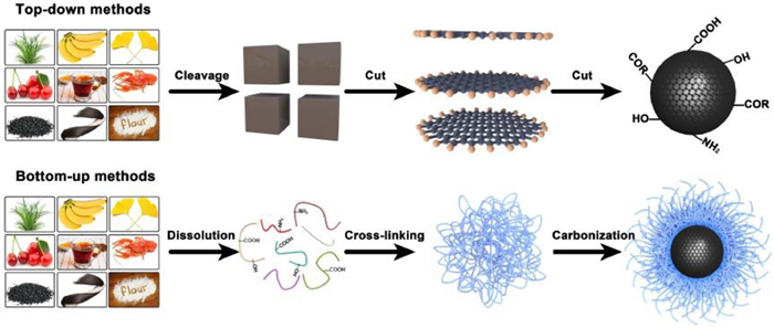
Figure 1.
Methods used to synthesize biomass-derived CDs.
State-of-the-art of biomass-derived carbon dots: Preparation, properties, and applications
Mengyuan Fang , Boyang Wang , Xiaoli Qu , Senrui Li , Jinsheng Huang , Jiangnan Li , Siyu Lu , Nan Zhou

Biomass, which is a natural, abundant, and renewable high-carbon resource, includes animals, plants, and microorganisms [1-5]. Biomass is ubiquitous, and a large amount of waste biomass should be recycled to prevent pollution. In most countries, waste biomass is typically disposed of via incineration. Only a few countries use high-tech waste processing methods to diminish carbon emissions; however, high-tech processing methods are costly. Recently, with the goals of carbon neutrality and peaking, reducing the pollution caused by waste biomass and converting biomass into inexpensive high-value-added products with high efficiency has attracted considerable attention [6-9].
As a new class of fluorescent nanocarbon materials, carbon dots (CDs) have become critical materials in nanomedicine [10,11], energy conversion and storage [12,13], and optoelectronic devices [14,15] because of their high stability, simple preparation methods, and excellent optical and electrical properties [16-18]. Unlike traditional carbon-based nanomaterials, CDs are diverse and are primarily regulated by their precursors and preparation methods [19-21]. Recently, biomass has become increasingly valuable as a precursor for CDs because its rich heteroatoms can easily achieve self-doping and the properties of the fabricated CDs are excellent [22-24]. Compared with artificial carbon sources, biomass is less toxic, which is crucial for bioapplications. Additionally, the low cost of biomass renders it useful for the large-scale preparation of CDs [25-27]. Therefore, conversion of biomass into CDs can reduce environmental pollution and expand the development and applications of CDs.
In the past three years, numerous reviews on the synthesis and chemical and physical properties of biomass-derived CDs have been published. However, most reviews focused only on the application fields of CDs (energy or biological applications) rather than providing comprehensive overviews. Therefore, summarizing the recent advances in biomass-derived CDs is critical for determining the future direction of this popular research field.
This review critically comments on the recent advances in biomass-derived CDs. We analyze and compare various preparation methods, particularly concerning the carbonation transition reaction. With this in mind, we highlight the latest achievements in controlling reactivity, tailoring optical properties, and introducing new functionalities. Furthermore, we describe several emerging applications, such as nanomedicine, energy, and optoelectronic devices. Lastly, we provide insight into the future direction and foreseeable challenges of biomass-derived CDs.
Currently, many methods can be used to convert biomass precursors into fluorescent CDs. According to the formation mechanism and reaction conditions, the methods can be divided into top-down (hydrothermal, solvothermal, and microwave-assisted) and bottom-up methods (pyrolysis) (Fig. 1). In addition, synergistic preparation methods, such as the microwave-assisted-hydrothermal and pyrolysis-oxidation methods, are commonly used to increase the yield and improve the optical properties of CDs. In this section, we summarize the most recently developed preparation methods and analyze their advantages and disadvantages. Moreover, several large-scale methods used to accelerate the industrialization of CDs are described [28-30].
The hydrothermal method is the most commonly used method for synthesizing biomass-derived CDs. This is primarily because biomass contains abundant water; therefore, the organic molecules and polymers comprising biomass present excellent water solubility [31-33]. Under high-temperature and high-pressure conditions, the organic molecules in biomass self-polymerize or polymerize with each other to form various crosslinked structures [34-36]. CDs are subsequently formed via carbonization. For example, we used avocado peel and sarcocarp as raw materials, adjusted the reaction temperature and time, and prepared CDs with quantum yields (QYs) of 9.56% and 8.97%, respectively [37]. The emission wavelengths of the avocado-peel- and sarcocarp-derived CDs are located in the blue–green spectral region, which was attributed to the primary components of the avocado peel and sarcocarp being different. Light-emitting CDs with emission wavelengths in the blue–green region has also been prepared using a hydrothermal method and beef as the carbon source [38-40]. Moreover, CDs have been fabricated using microorganisms as carbon sources [41-43]. We analyzed the reason for the blue–green emission and determined that the amino acids, proteins, and polysaccharides in biomass participate in hydrothermal reactions [44-46]. The hydrothermal products of the precursors typically present blue emission; therefore, the CDs fabricated using the hydrothermal methods will always emit light in the blue–green spectral region.
The solvothermal method is similar to the hydrothermal method; however, the reaction solvent is organic (e.g., ethanol, ethyl acetate, and acetone) to dissolve the water-insoluble molecules in biomass and promote CDs synthesis [47-49]. For example, curcumin, an active ingredient in ginger, exhibits poor solubility in water [50]. Therefore, a solvothermal method can be used to accelerate the conversion of curcumin into CDs. The as-prepared CDs exhibited blue–green emission and a QY of 8.6%. Currently, the solvothermal method is the only method used to convert biomass (green plants) into red-emitting CDs. This is because green plants are rich in chlorophyll. During CDs synthesis, the chlorophyll embedded into the carbon core or connected to the surface of CDs can induce a chlorophyll-like emission behavior. Chlorophyll is highly soluble in alcohol, acetone, and dimethyl sulfoxide; therefore, these solvents can be used to fabricate CDs using solvothermal reactions. CDs fabricated using Taxus leaves and acetone as the carbon source and solvent, respectively, exhibited a half-peak width of 20 nm. Furthermore, the QYs of the CDs with excitation wavelengths of 413 and 660 nm were 59% and 31%, respectively [51]. The CDs fabricated using Wedelia trilobata and ethanol as the carbon source and solvent, respectively, emitted red light at 645 nm, and their QY was 18.16% [52]. The water solubility of the CDs prepared using solvothermal methods is typically poor; moreover, their emission wavelengths change in aqueous media. To increase the water solubility of CDs, a small amount of water can be added to the reaction mixture during the solvothermal process. This does not change the red emission of the CDs fabricated using green plants. For example, the CDs fabricated using Spermatophyta, Pteridophyta, Bryophytes, and Algae exhibited excellent red emission in a mixture of ethanol and water (70:30, v/v) [53]. It should be emphasized that biomass-derived CDs prepared by the hydrothermal method cannot appear red emission after being dispersed in the organic solvent. After the CDs are successfully prepared, they will be purified by a filter membrane (0.22 µm). The CDs with red light-emitting molecules in the shell are water-insoluble and will be removed in this step. The contents of other preparation methods of biomass-derived CDs were summarized in Supporting information, and outlines their large-scale preparation
Biomass-derived CDs are widely used nanomaterials. Therefore, an in-depth understanding of their properties would allow researchers to develop CDs with tailored properties for specific applications. The optical properties of biomass-derived CDs are critical because almost all biomass-derived CDs exhibit photoluminescence, stability promotes application expansion, and negligible toxicity, which is crucial for bioapplications. Therefore, an in-depth understanding of the properties of biomass-derived CDs is necessary for the development of efficient synthesis methods and expansion of the applicability of CDs. The properties including optical properties, stability, and toxicity are summarized in Supporting information.
The excellent physical and chemical properties of biomass-derived CDs are related to the structure of CDs, such as element and shell types. Judicious regulation strategies can be used to adjust the properties and promote the efficient application of biomass-derived CDs.
Heteroatom doping is commonly used to adjust the optical properties of biomass-derived CDs. This is necessary because, typically, unmodified CDs present shortcomings, such as low QY and single surface shell structure, which considerably limit their application value [54-56]. Addition of other precursors during synthesis is the most effective strategy for fabricating heteroatom-doped CDs (Fig. 2a). The most common dopants are non-metals, such as N, S, B, and P, and metals, such as Mg and Fe [57-59]. Biomass-derived CDs doped with non-metals, and in particular N, present stronger light absorption and better QYs than unmodified CDs. N easily forms bonds with C. The energy required for electrons to transition from the π orbitals of C=N to the n orbitals is low; therefore, new radiative recombination pathways can easily form [60]. Dopamine hydrochloride and melamine were used as N sources to prepare N-doped biomass-derived CDs. Melamine promoted the formation of pyridinic- and amine-N, thereby decreasing the band gap of the CDs and promoting a red shift of the emission wavelength. In addition, the N content and QY (35.39%) of the N-doped biomass-derived CDs fabricated using melamine were higher than the N content and QY (2.95%) of the N-doped biomass-derived CDs fabricated using dopamine hydrochloride [61]. Wang et al. used S-rich durian as the carbon source to fabricate S-doped biomass-derived CDs. The as-prepared CDs comprised thiophene moieties, which improved the optical and chemical stability of the CDs and increased their QY [62]. CDs prepared using empty fruit bunches and sodium thiosulfate were also highly stable, and their fluorescence properties remained unchanged after more than eight months of storage [63]. Shen et al. used 3-aminophenylboronic acid and alkali lignin as carbon sources to prepare CDs with three emission wavelengths [64]. Theoretical calculations demonstrated that the bandgap of the CDs increased gradually with increasing number of C–B bonds. Moreover, the emission wavelength of the CDs was modulated via B-doping. The charge density of CDs can be changed via metal doping; moreover, the CDs can be endowed with new functions. Pure bougainvillea plant leaves and FeCl2 were used as the C and Fe sources, respectively, to synthesize Fe-doped CDs. Structure characterization revealed that single Fe atoms were loaded onto CDs, and they exhibited nanozyme-like effect. Fe-doped CDs regulated tumor microenvironments through reactive oxygen species (ROS) regulation and lysosome-mediated autophagy, thereby effectively inhibiting tumor growth in a mouse model of drug-resistant glioblastoma [65]. CDs prepared using Lycium ruthenicum and MgCl2·6H2O as the carbon and Mg sources, respectively, were highly biocompatible with the pre-osteoblast cell line MC3T3-E1 (subclone 4). The experimental results demonstrated that an appropriate concentration of Mg-doped CDs was suitable for long-term cell growth, and that the Mg-doped CDs promoted cell growth, proliferation, and osteogenic differentiation capabilities [66].
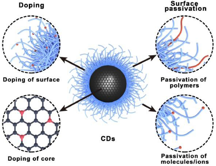
In addition to heteroatom doping, CDs can be modified through surface passivation by changing the chemical state of the surface of CDs without changing the intrinsic properties of the carbon cores. Owing to the abundant functional groups on the surface of CDs, the optical properties and solubility of CDs can be changed via modification with metal ions, organic molecules, and polymers (Fig. 2b) [67-69]. Modification of CDs with metal ions and organic molecules induces quenching of their optical properties, which lays the foundation for developing CDs as sensors. CD fluorescence was quenched and CDs were used to develop dual-function detectors for detecting sunset yellow dye and Mn2+ ions [70]. Moreover, the surface of CDs can be modified with organic molecules to confer CDs tumor-targeting or anticancer functions. For example, lycorine-adjusted CDs derived from mulberry leaves presented remarkable anticancer activity and were used for targeted drug delivery to tumor sites [71]. Biomass-derived CDs prepared using water as the solvent typically emit blue light. Organic solvents should be used to extend the emission wavelengths of CDs. However, the bioapplications of CDs in the organic phase are limited; therefore, hydrophilic polymers should be used to modify CDs to improve their water dispersibility. Honeysuckle, turmeric, and perilla were used as carbon sources to fabricate CDs with blue, green, and red fluorescence, respectively. Poly(ethylene glycol) (PEG) was added to a dispersion of CDs in dichloromethane to achieve surface modification, and the modified CDs were used as stains for bacterial imaging. PEG, which was used to increase the hydrophilicity of CDs, is an environmentally friendly and degradable lubricant [72]. PEG-modified ginkgo-biloba-derived CDs exhibited remarkable lubricity, good load-carrying capacity, and long service life under boundary lubrication [73]. The antiwear and antifriction properties of PEG added with 0.20 wt% CDs were 70.5% and 34.7% higher, respectively, than those of pure PEG.
Biomass-derived CDs present unique applications in biomedicine because of their high biocompatibility and low toxicity. Therefore, they are widely used in bioimaging, sensing, tumor therapy, and as drug carriers. Moreover, owing to their good electrical conductivity, large surface area, and high mobility, biomass-derived CDs present remarkable prospects in catalysis, energy conversion, and storage [74-76]. This section describes the numerous applications of biomass-derived CDs. The most widely used field of biomass-derived CDs is optical applications including, optical imaging, sensors, and phototherapy. The application in the drug delivery and energy fields are summarized in the supporting information
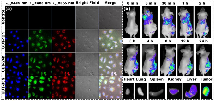
Optical imaging plays a critical role in basic research and clinical settings because it allows researchers and clinicians to comprehensively monitor biological processes to analyze cell morphology and physiological processes. The most attractive feature of CDs is their outstanding fluorescence. Biomass-derived CDs exhibit stable and tunable emission wavelengths, rendering them promising candidates for optical imaging agents. CDs enter cells through endocytosis during the co-incubation of cells and biomass-derived CDs. To eliminate the interference of excess CDs in the medium, remove the excess CDs by centrifugation and washing, then disperse the cells in a new medium for imaging [77,78]. Recently, researchers have developed numerous methods to tailor the fluorescence properties of biomass-derived CDs to meet the increasing demands of optical imaging [79-82]. CDs derived from corn stalk shells emitted blue light with a QY of 16% [83]. A549 lung cancer cells were co-incubated with CDs, and the cells exhibited strong blue fluorescence under excitation at 405 nm. PC12 cells incubated in a medium containing 500 µg/mL cyanobacteria-derived CDs for 24 h emitted green fluorescence after excitation at 405 nm [84]. However, almost no fluorescence was observed in the center of the cells, indicating that the CDs were quickly taken up by the cells and mainly located in the cytoplasm. Spinach-derived red-emission CDs co-incubated with A459 lung cancer cells emitted intense red fluorescence under excitation at 543 nm. The emission was localized in the cytoplasm, and a transparent nuclear region originated in the cell [85]. In addition to single-color imaging, multi-channel imaging can be achieved owing to the excitation-dependent properties of biomass-derived CDs. Upon red-shifting the excitation wavelength of CDs derived from the dwarf banana peel from 395 nm to 505 nm, the emission wavelength extended from the blue–violet to the yellow–green spectral region. After incubating CDs with clone 9 hepatocyte cells, the cells emitted blue, green, and red light upon excitation at 405, 488, and 555 nm, respectively. After 12 h of incubation, weak and strong fluorescent signals were emitted by the cytoplasm and nucleus, respectively. Furthermore, after a longer incubation time of 36 h, strong fluorescent signals were emitted by the nucleus and cytoplasm (Fig. 3a) [86]. Yong et al. used biorefining by-products as the carbon source to fabricate blue-emission CDs for in vivo imaging, and the QY of the CDs was 13%. The as-fabricated CDs were injected into tumor-bearing mice, and fluorescent imaging revealed that 5 min after CDs injection, fluorescent signals were emitted from the mouse head. The CDs were almost exclusively located at the tumor sites 12 h after injection, demonstrating that they can be used for long-term in vivo tumor imaging (Fig. 3b) [87]. Biomass-derived CDs have also been used for analyzing in vivo metabolic processes. Ding et al. fabricated red-emission CDs using pulp-free lemon juice and utilized them as an imaging agent [88]. They intravenously injected a CD aqueous dispersion into nude mice through the tail vein, and observed bright fluorescence in the bladder 15 min after injection. Their results indicate that the CDs were primarily excreted through urine.
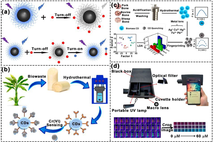
Biomass-derived CDs can coordinate and detect specific analytes because of their rich surface structures. In the presence of a target analyte, the initial CDs system is destroyed via adsorption or chemical reactions. This changes the fluorescence emission of CDs, promoting the detection of the target analyte. To date, biomass-derived CDs have been used as sensors for various materials, such as metal ions [89-93] and molecules [94-98], based on fluorescence enhancement or quenching (Fig. 4a). To detect metal ions, due to the coordination of CDs to metals, the coordination between CDs and metal ions triggers the transfer of non-radiative electrons from excited CDs to the unfilled d orbitals of metal ions, resulting in fluorescence quenching. The detection mechanism has been confirmed using several metal ions (i.e., Fe3+, Cu2+, Hg2+, and Pb2+) [99-103]. Poa pratensis-derived CDs exhibited good hydrophilic fluorescence and strong cyan–blue emission and were used for highly selective detection of Mn2+ ions in aqueous media. The fluorescence of the CDs was quenched in the presence of Mn2+ ions, and the detection limit was 1.2 µmol/L [104]. The CD-based sensor constructed using banana stems detected Cr5+ in the linear detection range of 10–30 µmol/L with a detection limit of 2.4 µmol/L. Density functional theory calculations and lifetime decay analysis demonstrated that the fluorescence quenching mechanism of CDs was photoinduced electron transfer (Fig. 4b) [105]. Added metal ions typically quench the fluorescence of CDs. Upon adding other analytes to replace the metal ions on the surface of CDs, fluorescence can be restored or enhanced. The utilization rate of CDs was considerably improved by the ability of CDs to detect various substances [106]. The fluorescence of grape-seed-derived CDs was quenched in the presence of Cu2+ ions, and it gradually recovered upon the addition of ascorbic acid to the system. Under optimal conditions, the linear detection ranges for Cu2+ ions and ascorbic acid were 150–500 µmol/L and 0.1–400 µmol/L, respectively and the limits of detection were 0.048 and 0.036 mmol/L, respectively [107]. The detection of organic molecules by biomass-derived CDs is based on the adsorption of organic molecules on the surface shell, using hydrogen bonding or π–π stacking. When the emission spectrum of CDs overlaps with the absorption spectrum of organic molecules, fluorescence resonance energy transfer occurs between the CDs in the excited state and the organic molecules in the ground state, which quenches the fluorescence of CDs. In addition, fluorescence quenching occurs when interactions between CDs and organic molecules form non-fluorescent ground-state complexes, or when there is charge transfer [108,109]. Kang et al. used tobacco leaves as the carbon source to fabricate CDs that were then used to detect organic molecules [110]. They reported that the as-synthesized CDs were selectively quenched by tetracycline. This was attributed to tetracycline reacting with the hydroxyl and carboxyl groups on the surface of the CDs leading to static quenching. CDs derived from waste tobacco stems were used to detect other tetracycline antibiotics in real water samples using the internal filtering effect. Biomass-derived-CD-based array detectors with good detection efficiency were developed. Molecularly imprinted polymers were formed on the surface of catalpa-walnut-shell-derived CDs using a sol–gel method, and the as-fabricated CDs precisely captured 5-nitroimidazole antibiotics. Using the changes in fluorescence to construct fingerprints, CDs were used to distinguish secnidazole, ornidazole, metronidazole, and tinidazole [111]. This fluorescent sensor array sensitively identified 5-nitroimidazole antibiotics over a wide concentration range. Niu et al. demonstrated that CDs fabricated using various raw materials (e.g., sheep, bovine, and pork bones) interacted with heavy metal ions to varying degrees and could be used as probes to distinguish Pb2+, Ag+, Fe3+, Hg2+, and Cu2+ ions (Fig. 4c) [112]. According to the fingerprints, Ag+ ions exhibited the strongest quenching effect on the sheep-bone-derived CDs, whereas Fe3+ ions presented the strongest quenching effect on all three types of CDs. Using hierarchical cluster and linear discriminant analyses, it was determined that array detection was effective for single ions and binary and ternary mixtures. Moreover, the accuracy of the array for binary and ternary mixtures was 100%. In addition to array detection, smartphone-based portable detection platforms can be developed for efficient real-time detection of analytes. A fluorescent sensing system and detection platform based on cinnamon-leaf-derived CDs exhibited excellent performance for analysis of tap water and wine samples. Light and color signals were collected using a smartphone, and the standard response curves of Al3+ ions in solutions at various concentrations were obtained through color recognition using specialized applications and equipment (Fig. 4d) [53].
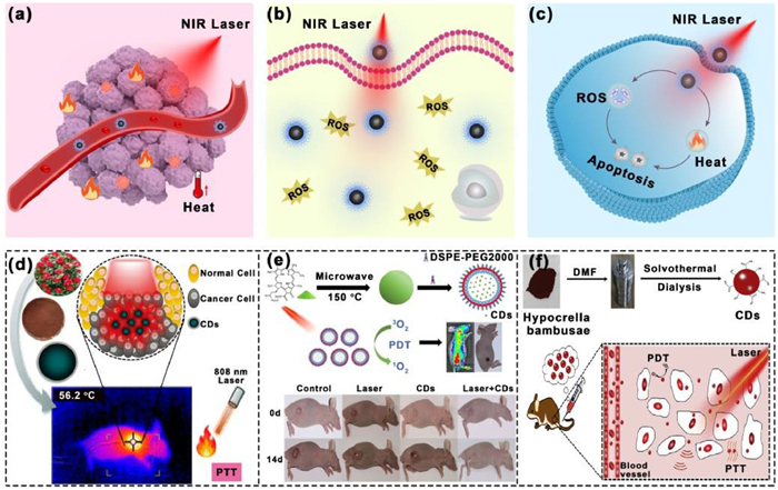
Phototherapy is a highly selective and minimally invasive treatment that involves focusing light beams on target areas to minimize side effects [113-115]. Typically, phototherapy requires a light source and a phototherapeutic agent that can be converted into thermal energy [photothermal therapy (PTT)] (Fig. 5a), chemical energy [photodynamic therapy (PDT)] (Fig. 5b), or both (Fig. 5c) [116]. Recently, researchers have demonstrated that biomass-derived CDs can be used as effective photosensitizers to generate ROS upon light excitation and as photothermal agents to convert light energy into heat. This promoted their application in PTT and PDT. Photothermal therapy can be divided into diathermia, hyperthermia and thermal ablation according to the temperature, among which the temperature of diathermia is the lowest (< 41 ℃), and the temperature of thermal ablation is the highest (> 46 ℃) [117-119]. Watermelon-derived CDs exhibited near-infrared emission with a QY of 0.4% [120]. The survival rate of cells co-incubated with CDs (20 mg/mL) was high. Upon irradiating the cells with an 808 nm laser light under the same conditions, cell survival rate was considerably lower (> 10%). This demonstrated that the CDs efficiently ablated cancer cells via a photothermal process. Furthermore, upon injecting tumor-bearing mice with CDs, the tumor sites heated rapidly under 808 nm light irradiation, and after 10 min of daily irradiation for 6 d, the tumors almost disappeared. CDs derived from Camellia japonica flowers exhibited efficient PTT performance and a high photothermal conversion efficiency of 55.4% at a moderate laser power (808 nm, 1.1 W/cm2). After 5 min of continuous irradiation, the temperature of the CD-treated tumor sites increased considerably to ~58 ℃, which was sufficient to kill cancer cells without damaging surrounding healthy tissue (Fig. 5d) [121]. In vivo experiments demonstrated that CDs and laser irradiation presented an excellent PTT effect in a group of mice, with complete tumor ablation and no significant recurrence within 10 d. A microwave-assisted method and pheophytin as the carbon source were used to synthesize hydrophobic CDs that emitted light at 680 nm and produced abundant singlet oxygen (1O2) species with a QY of 0.62. Subsequently, PEGylated derivative of 1,2-distearoyl-sn-glycero-3-PE (DSPE)-mPEG2000 was used to modify the CDs. The modified CDs presented higher hydrophilicity and retained their high 1O2 generation efficiency (Fig. 5e) [122]. The PDT efficacy of CDs was examined using 4T1 tumor-bearing mice, and tumor growth was completely inhibited after laser irradiation. This indicated that the CDs successfully accumulated at the tumor sites and produced 1O2 species under 671 nm laser irradiation. Moreover, the mice survived 40 d after treatment. In addition to using the abundant functional groups on the surface of pure CDs, photosensitizers can be grafted onto the surface of CDs. This increases the photosensitizer concentration per unit of volume and promotes the phototherapeutic effect. CDs fabricated using osmanthus leaves as the raw material were modified with polyoxyethylene diamine and photosensitized with folic acid. The as-produced CDs targeted tumor cells and mitochondria and generated 1O2 species in mitochondria under near-infrared laser irradiation [123]. Flow cytometry was used to analyze the photodynamic therapeutic effect of CDs on T24 cancer cells and 7702 healthy cells. The apoptosis rate of 7702 cells incubated with CDs (11.56%) was lower than that of T24 cells (41.8%). These findings suggest that CDs can be used for targeting cancer cells via PDT. Cancerous cells produce high levels of biothiols, which reduces the efficacy of PDT. To mitigate these challenges, Zhao et al. developed Cu2+-modified spinach-derived CDs for effective PDT. They demonstrated that binding chlorophyll and Cu2+ ions onto the surface of CDs decreased the difference between the energy levels of chlorophyll molecules. This led to an increase in number of generated 1O2 species, thereby enhancing PDT efficacy. Conversely, the Cu2+ ions on the surface of CDs can combine with biothiols to decrease the concentration of free biothiols in tumor cells and further enhance PDT efficacy [124]. In addition, Wang et al. used Hypocrella bambusae as the precursor to fabricate CDs and used the CDs in dual-functional PDT–PTT [125]. The tumors in the control and PDT groups were not eliminated after 14 d of treatment. In contrast, the tumors in the PDT–PTT group were entirely suppressed after 14 d of treatment (Fig. 5f).
From the above description, we can see that biomass-derived CDs play a huge role in the fields of biology, sensing, energy conversion and storage, which mainly depends on the special characteristics of the precursors and structures. Biomass itself has low toxicity, so the prepared CDs also have low toxicity, which is very beneficial to its imaging, treatment and other bioapplications. Meanwhile, the abundant organic precursors in biomass make the derived CDs have more abundant surface states. The rich polymer structure makes it easier to be passivated for loading drugs and improving drug loading. In tumor therapy, biomass-derived CDs still play an indispensable role. Biomass is rich in heteroatoms. The introduction of heteroatoms, especially N, S, and P, can expand the absorption range and improve the photothermal conversion efficiency.
Compared with organic compound-derived CDs, the main disadvantages of biomass-derived CDs focus on the emission wavelength and low quantum yield, which will affect their heir range of application. The low quantum yield makes it difficult to localize in cells and in vivo. There are few studies on biomass-derived CDs two-photon/upconversion luminescence, which makes it only possible to use blue-violet light sources for localization in vivo, and there will inevitably be interference from background fluorescence. In phototherapy, it is easy to develop CDs with high photothermal conversion efficiency or high reactive oxygen species from organic compound precursors, because the precursors are rich in choice and can achieve directional design. Although biomass-derived CDs are also rich in precursors, the molecules contained in them are single, so it is difficult to break through.
The diversity of precursors used to prepare CDs has led to the development of various types of CDs. The use of biomass as a carbon source has promoted the inexpensive and large-scale preparation of CDs. Owing to the abundance and sustainability of biomass, the commercialization prospects of biomass-derived CDs have recently improved. However, because of the diversity of precursors and their highly variable structures, small changes in reaction conditions can yield different results. Therefore, to increase the application value of biomass-derived CDs, their optical properties should be thoroughly analyzed and stabilized.
In this review, we describe the preparation and applications of biomass-derived CDs by correlating their regulation and physicochemical properties. CDs prepared using different methods and solvent environments exhibit various characteristics, and their excellent optical stability, anti-drifting properties, and high ionic strength promote their applications. We describe doping and surface passivation methods for regulating the properties of biomass-derived CDs. Lastly, the latest progress in biomass-derived CDs for biology, sensing, and energy applications is summarized. Herein, we suggest several promising directions for future research.
1. Increasing yield: Biomass is the most affordable and extensively used precursor for CDs fabrication. Several researchers have reported kilogram-scale preparation of biomass-derived CDs. However, the large-scale fabrication of CDs still requires industrialization. Therefore, suitable carbonization methods should be selected and adequate reactors should be designed for the large-scale preparation of biomass-derived CDs.
2. Regulation of optical properties: The CDs derived from organic molecules exhibit high QY emission in the violet-to-near-infrared spectral range. The CDs derived from biomass should be further studied and methods for expanding the emission wavelength and QY should be developed. Introduction of N precursors through doping can improve QY; however, this effect can be further improved. Development of violet- and near-infrared-emitting CDs should expand the application value of CDs.
3. Mechanism of formation: CDs synthesis involves dehydration, crosslinking, and carbonization of precursors with polycondensation sites. However, biomass is rich in amino acids, polysaccharides, vitamins, and other components, which hinder the formation of biomass-derived CDs. The temperature and reaction time should be judiciously controlled because the precipitation of each molecule that participates in the reaction is regulated by temperature.
4. Diversification of applications: Owing to the limitations of their self-generated optical properties, biomass-derived CDs present limited applications in optical imaging. CDs are critical for imaging specific organelles or subcellular structures; moreover, displays and lighting are critical parts of application transformation. Increasing attention has been dedicated to, and the rapid development of biomass-derived CDs will expand their applications.
In summary, the remarkable number of recent reports on the fabrication, properties, and applications of CDs has led to the development of biomass-derived CDs. Researchers should continue to develop biomass-derived CDs with excellent selectivity, efficiency, and applicability. Furthermore, scholars should gain a deeper understanding of biomass conversion processes, identify more favorable reactions and combinations, and establish a feasible plan.
The authors declare that they have no known competing financial interests or personal relationships that could have appeared to influence the work reported in this paper.
This work was supported by the National Natural Science Foundation of China (Nos. 52122308, 21905253, 51973200).
Supplementary material associated with this article can be found, in the online version, at doi:
T.C. Wareing, P. Gentile, A.N. Phan, ACS Nano 15 (2021) 15471–15501. doi: 10.1021/acsnano.1c03886
Y. Wang, M. Zhang, X. Shen, et al., Small 17 (2021) 2008079. doi: 10.1002/smll.202008079
G. Zhang, X. Liu, L. Wang, et al., J. Mater. Chem. A 10 (2022) 9277–9307. doi: 10.1039/D2TA01442D
Y. Chai, M. Bai, A. Chen, et al., Chem. Eng. J. 453 (2023) 139814. doi: 10.1016/j.cej.2022.139814
F. Shan, L. Fu, X. Chen, et al., Chin. Chem. Lett. 33 (2022) 2942–2948. doi: 10.1016/j.cclet.2021.12.094
C. Li, J. Li, L. Qin, et al., ACS Catal. 11 (2021) 11336–11359. doi: 10.1021/acscatal.1c02551
J. Shi, R. Zhang, X. Liu, et al., Carbohyd. Polym. 301 (2023) 120323. doi: 10.1016/j.carbpol.2022.120323
S.S. Sekhon, J. Lee, J. -S. Park, J. Energy Chem. 65 (2022) 149–172. doi: 10.1016/j.jechem.2021.05.052
W. Yang, X. Li, L. Fei, et al., Green Chem. 24 (2022) 675–700. doi: 10.1039/D1GC02964A
B. Wang, H. Song, X. Qu, et al., Coord. Chem. Rev. 442 (2021) 214010. doi: 10.1016/j.ccr.2021.214010
L. Ðorđević, F. Arcudi, M. Cacioppo, et al., Nat. Nanotechnol. 17 (2022) 112–130. doi: 10.1038/s41565-021-01051-7
Y. Zhai, B. Zhang, R. Shi, et al., Adv. Energy Mater. 12 (2022) 2103426. doi: 10.1002/aenm.202103426
T. Feng, S. Tao, D. Yue, et al., Small 16 (2020) 2001295. doi: 10.1002/smll.202001295
Y. Ru, G.I.N. Waterhouse, S. Lu, Aggregate 3 (2022) e296. doi: 10.1002/agt2.296
B. Zhao, Z. a. Tan, Adv. Sci. 8 (2021) 2001977. doi: 10.1002/advs.202001977
B. Wang, S. Lu, Matter 5 (2022) 110–149. doi: 10.1016/j.matt.2021.10.016
B. Wang, G.I.N. Waterhouse, S. Lu, Trend. Chem. 5 (2023) 76–87. doi: 10.1016/j.trechm.2022.10.005
B. Wang, H. Cai, G.I.N. Waterhouse, et al., Small Sci. 2 (2022) 2200012. doi: 10.1002/smsc.202200012
X. Yang, X. Li, B. Wang, et al., Chin. Chem. Lett. 33 (2022) 613–625. doi: 10.1016/j.cclet.2021.08.077
B. Wang, J. Yu, L. Sui, et al., Adv. Sci. 8 (2021) 2001453. doi: 10.1002/advs.202001453
B. Wang, Z. Wei, L. Sui, et al., Light Sci. Appl. 11 (2022) 172. doi: 10.1038/s41377-022-00865-x
Y. Xu, B. Wang, M. Zhang, et al., Adv. Mater. 34 (2022) 2200905. doi: 10.1002/adma.202200905
W. Li, Z. Wei, B. Wang, et al., Mater. Chem. Front. 4 (2020) 277–284. doi: 10.1039/C9QM00618D
W. Meng, X. Bai, B. Wang, et al., Energy Environ. Mater. 2 (2019) 172–192. doi: 10.1002/eem2.12038
S. Xiang, M. Tan, Environ. Sci. Nano 9 (2022) 3206–3225. doi: 10.1039/D2EN00435F
P. Kaur, G. Verma, Mater. Today Sustain. 18 (2022) 100137. doi: 10.1016/j.mtsust.2022.100137
M. Moradi, R. Molaei, S.A. Kousheh, et al., Crit. Rev. Food Sci. Nutr. 63 (2023) 1943–1959. doi: 10.1080/10408398.2021.2015283
X. Niu, W. Zheng, T. Song, et al., Chin. Chem. Lett. 34 (2023) 107560. doi: 10.1016/j.cclet.2022.05.074
X. Niu, T. Song, H. Xiong, Chin. Chem. Lett. 32 (2021) 1953–1956. doi: 10.1016/j.cclet.2021.01.006
W. Li, Y. Liu, B. Wang, et al., Chin. Chem. Lett. 30 (2019) 2323–2327. doi: 10.1016/j.cclet.2019.06.040
Q. Zeng, T. Feng, S. Tao, et al., Light Sci. Appl. 10 (2021) 142. doi: 10.1038/s41377-021-00579-6
R. Fu, H. Song, X. Liu, et al., Chin. J. Chem. 41 (2023) 1007. doi: 10.1002/cjoc.202200736
B. Zhao, H.Y. Ma, M.Y. Zheng, et al., Carbon Energy 4 (2022) 88–114. doi: 10.1002/cey2.175
L. Ai, Y. Yang, B. Wang, et al., Sci. Bull. 66 (2021) 839–856. doi: 10.1016/j.scib.2020.12.015
J. Zhang, A. Xia, X. Zhu, et al., Fuel Process. Technol. 232 (2022) 107276. doi: 10.1016/j.fuproc.2022.107276
Y. Hu, M. Li, Z. Gao, et al., Mater. Today Chem. 20 (2021) 100423. doi: 10.1016/j.mtchem.2021.100423
W. Meng, B. Wang, L. Ai, et al., J. Colloid Interface Sci. 598 (2021) 274–282. doi: 10.1016/j.jcis.2021.04.022
J. Geng, X. Song, X. Zhang, et al., J. Agric. Food Chem. 67 (2019) 6995–7004. doi: 10.1021/acs.jafc.9b01372
K. Liu, Y. Song, M. Tan, J. Agric. Food Chem. 68 (2020) 9789–9795. doi: 10.1021/acs.jafc.0c03499
H. Wang, W. Su, M. Tan, Innovation 1 (2020) 100009.
S. Zhang, D. Zhang, Y. Ding, et al., Analyst 144 (2019) 5497–5503. doi: 10.1039/C9AN01103J
F. Lin, C. Li, Z. Chen, Front. Microbiol. 9 (2018) 259. doi: 10.3389/fmicb.2018.00259
K. Qin, D. Zhang, Y. Ding, et al., Analyst 145 (2020) 177–183. doi: 10.1039/C9AN01753D
Q. Huang, Q. Bao, C. Wu, et al., J. Pharm. Anal. 12 (2022) 104–112. doi: 10.1016/j.jpha.2021.04.004
S. Li, F. Li, Y. Dong, et al., Small 18 (2022) 2106269. doi: 10.1002/smll.202106269
M. Zhang, W. Zhang, X. Fan, et al., Nano Lett. 22 (2022) 7203–7211. doi: 10.1021/acs.nanolett.2c02674
X. Tong, Y. Zhu, C. Tong, et al., Anal. Chim. Acta 1178 (2021) 338829. doi: 10.1016/j.aca.2021.338829
C. Tang, R. Long, X. Tong, et al., Microchem. J. 164 (2021) 106000. doi: 10.1016/j.microc.2021.106000
Z. Liu, W. Jin, F. Wang, et al., Sens. Actuat. B: Chem. 296 (2019) 126698. doi: 10.1016/j.snb.2019.126698
T. Pal, S. Mohiyuddin, G. Packirisamy, ACS Omega 3 (2018) 831–843. doi: 10.1021/acsomega.7b01323
J. Liu, Y. Geng, D. Li, et al., Adv. Mater. 32 (2020) 1906641. doi: 10.1002/adma.201906641
C. Liang, X. Xie, D. Zhang, et al., J. Mater. Chem. B 9 (2021) 5670–5681. doi: 10.1039/D0TB02979C
H. Rao, W. Liu, K. He, et al., ACS Sustain. Chem. Eng. 8 (2020) 8857–8867. doi: 10.1021/acssuschemeng.0c03354
S. Miao, K. Liang, J. Zhu, et al., Nano Today 33 (2020) 100879. doi: 10.1016/j.nantod.2020.100879
F. Li, D. Yang, H. Xu, Chem. Eur. J. 25 (2019) 1165–1176. doi: 10.1002/chem.201802793
Z. Wei, B. Wang, M. Xie, et al., Chin. Chem. Lett. 33 (2022) 751–756. doi: 10.1016/j.cclet.2021.08.014
X. Yang, S. Hou, T. Chu, et al., Ind. Crop. Prod. 167 (2021) 113507. doi: 10.1016/j.indcrop.2021.113507
H. Ren, F. Qi, A. Labidi, et al., ACS ES&T Engg. 3 (2023) 260–270.
W. Wang, J. Chen, D. Wang, et al., Anal. Methods 13 (2021) 789–795. doi: 10.1039/D0AY02186E
Y. Jia, Z. Cheng, G. Wang, et al., Food Chem. 402 (2023) 134245. doi: 10.1016/j.foodchem.2022.134245
Y. Liu, C. Yong, B. Tong, et al., Opt. Mater. 134 (2022) 113144. doi: 10.1016/j.optmat.2022.113144
G. Wang, Q. Guo, D. Chen, et al., ACS Appl. Mater. Interfaces 10 (2018) 5750–5759. doi: 10.1021/acsami.7b16002
U. Abd Rani, L.Y. Ng, C.Y. Ng, et al., Mater. Today Proc. 46 (2021) 1934–1939. doi: 10.1016/j.matpr.2021.02.225
X. Gu, L. Zhu, D. Shen, et al., Polymers 14 (2022) 2779. doi: 10.3390/polym14142779
P. Muhammad, S. Hanif, J. Li, et al., Nano Today 45 (2022) 101530. doi: 10.1016/j.nantod.2022.101530
Y. Hou, R. Zhang, H. Cheng, et al., Colloid. Surface. A 656 (2023) 130264. doi: 10.1016/j.colsurfa.2022.130264
Y. Li, Z. Tang, Z. Pan, et al., ACS Nano 16 (2022) 4357–4370. doi: 10.1021/acsnano.1c10556
L. Wang, W. Li, B. Wu, et al., Chem. Eng. J. 300 (2016) 75–82. doi: 10.1016/j.cej.2016.04.123
Y. Zheng, J. Hao, K. Arkin, et al., Food Chem. 403 (2023) 134415. doi: 10.1016/j.foodchem.2022.134415
B.K. Korah, B. Mathew, Mater. Today Sustain. 21 (2023) 100273. doi: 10.1016/j.mtsust.2022.100273
Y. Shao, C. Zhu, Z. Fu, et al., J. Nanopart. Res. 22 (2020) 229. doi: 10.1007/s11051-020-04917-4
W. Zhao, Y. Wang, K. Liu, et al., Chin. Chem. Lett. 33 (2022) 798–802. doi: 10.1016/j.cclet.2021.08.084
Z. Mou, Q. Yang, B. Zhao, et al., ACS Sustain. Chem. Eng. 9 (2021) 14997–15007. doi: 10.1021/acssuschemeng.1c05678
P. Li, L. Sun, S. Xue, et al., SmartMat 3 (2022) 226–248. doi: 10.1002/smm2.1131
G. Hu, Y. Wang, S. Zhang, et al., Carbon 203 (2023) 1–10. doi: 10.1016/j.carbon.2022.11.048
H. Li, Q. Qing, L. Zheng, et al., Chin. Chem. Lett. 33 (2022) 3573–3576. doi: 10.1016/j.cclet.2022.01.050
H. Ren, Y. Yuan, A. Labidi, et al., Chin. Chem. Lett. 34 (2023) 107998. doi: 10.1016/j.cclet.2022.107998
Y. Xiao, X. Yin, P. Sun, et al., Chin. Chem. Lett. 33 (2022) 5051–5055. doi: 10.1016/j.cclet.2022.03.109
P. Jayasekhar Babu, S. Saranya, Y.D. Singh, et al., Opt. Mater. 117 (2021) 111120. doi: 10.1016/j.optmat.2021.111120
J. Zhang, G. Zheng, Y. Tian, et al., Inorg. Chem. Commun. 144 (2022) 109837. doi: 10.1016/j.inoche.2022.109837
J. Qin, X. Gao, Q. Chen, et al., RSC Adv. 11 (2021) 31791–31794. doi: 10.1039/D1RA05199G
L. Wang, B. Wang, E. Liu, et al., Chin. Chem. Lett. 33 (2022) 4111–4115. doi: 10.1016/j.cclet.2022.01.042
Z. Li, Q. Wang, Z. Zhou, et al., Microchem. J. 166 (2021) 106250. doi: 10.1016/j.microc.2021.106250
X. Wang, P. Yang, Q. Feng, et al., Polymers 11 (2019) 616. doi: 10.3390/polym11040616
L. Li, R. Zhang, C. Lu, et al., J. Mater. Chem. B 5 (2017) 7328–7334. doi: 10.1039/C7TB00634A
R. Atchudan, T.N.J.I. Edison, S. Perumal, et al., Fuel 275 (2020) 117821. doi: 10.1016/j.fuel.2020.117821
C. Huang, H. Dong, Y. Su, et al., Nanomaterials 9 (2019) 387. doi: 10.3390/nano9030387
H. Ding, X. Zhou, B. Qin, et al., J. Lumin. 211 (2019) 298–304. doi: 10.1016/j.jlumin.2019.03.064
M. Chen, J. Zhai, Y. An, et al., Front. Chem. 10 (2022) 940398. doi: 10.3389/fchem.2022.940398
X. Huang, J. Liu, B. Zhao, et al., Front. Energy Res. 10 (2022) 253.
L. Xia, X. Li, Y. Zhang, et al., Molecules 27 (2022) 1258. doi: 10.3390/molecules27041258
S. Tang, D. Chen, G. Guo, et al., Sci. Total Environ. 825 (2022) 153913. doi: 10.1016/j.scitotenv.2022.153913
H. Lu, C. Li, H. Wang, et al., ACS Omega 4 (2019) 21500–21508. doi: 10.1021/acsomega.9b03198
H. Liu, J. Ding, K. Zhang, et al., Talanta 209 (2020) 120508. doi: 10.1016/j.talanta.2019.120508
R. Gao, Z. Wu, L. Wang, et al., Nanomaterials 10 (2020) 1561. doi: 10.3390/nano10081561
N. Pourmahdi, A.H.M. Sarrafi, A. Larki, J. Fluoresc. 29 (2019) 887–897. doi: 10.1007/s10895-019-02400-5
X. Sun, Y. Liu, N. Niu, et al., Anal. Bioanal. Chem. 411 (2019) 5519–5530. doi: 10.1007/s00216-019-01930-y
H. Liu, J. Ding, L. Chen, et al., J. Photochem. Photobiol. A 400 (2020) 112724. doi: 10.1016/j.jphotochem.2020.112724
Y. Qiu, D. Li, Y. Li, et al., Cellulose 29 (2022) 367–378. doi: 10.1007/s10570-021-04314-7
S. Jing, Y. Zhao, R. -C. Sun, et al., ACS Sustain. Chem. Eng. 7 (2019) 7833–7843. doi: 10.1021/acssuschemeng.9b00027
Y. Zhao, S. Jing, X. Peng, et al., Cellulose 27 (2020) 415–428. doi: 10.1007/s10570-019-02807-0
Y. Xie, D. Cheng, X. Liu, et al., Sensors 19 (2019) 3169. doi: 10.3390/s19143169
R.S. Costa, M.O. de Castro, G.H. da Silva, et al., Carbon Trends 5 (2021) 100133. doi: 10.1016/j.cartre.2021.100133
P. Krishnaiah, R. Atchudan, S. Perumal, et al., Chemosphere 286 (2022) 131764. doi: 10.1016/j.chemosphere.2021.131764
J. Goswami, S.S. Rohman, A.K. Guha, et al., Mater. Chem. Phys. 286 (2022) 126133. doi: 10.1016/j.matchemphys.2022.126133
C. Ji, Y. Zhou, R.M. Leblanc, et al., ACS Sens. 5 (2020) 2724–2741. doi: 10.1021/acssensors.0c01556
J. Li, O. Xu, X. Zhu, RSC Adv. 11 (2021) 34107–34116. doi: 10.1039/D1RA05656E
T. Rasheed, TrAC Trend, Anal. Chem. 160 (2023) 116957.
A. Döring, E. Ushakova, A.L. Rogach, Light Sci. Appl. 11 (2022) 75. doi: 10.1038/s41377-022-00764-1
Y. -M. Liang, H. Yang, B. Zhou, et al., Anal. Chim. Acta 1191 (2022) 339269. doi: 10.1016/j.aca.2021.339269
Q. Sun, L. Fu, C. Yin, et al., Sensor. Actuat. B: Chem. 373 (2022) 132716. doi: 10.1016/j.snb.2022.132716
L. Fu, T. Liu, F. Yang, et al., J. Photochem. Photobiol. A 424 (2022) 113638. doi: 10.1016/j.jphotochem.2021.113638
X. Wang, L. Zhu, Z. Gu, et al., Nanophotonics 11 (2022) 4955–4976. doi: 10.1515/nanoph-2022-0574
B. Li, S. Zhao, L. Huang, et al., Chem. Eng. J. 408 (2021) 127245. doi: 10.1016/j.cej.2020.127245
C.L. Shen, H.R. Liu, Q. Lou, et al., Theranostics 12 (2022) 2860–2893. doi: 10.7150/thno.70721
M. Li, M. Zhao, J. Li, Wiley Interdiscip. Rev. Nanomed. Nanobiotechnol. 15 (2023) e1865. doi: 10.1002/wnan.1865
Z. Xie, T. Fan, J. An, et al., Chem. Soc. Rev. 49 (2020) 8065–8087. doi: 10.1039/D0CS00215A
Q. Zheng, X. Liu, Y. Zheng, et al., Chem. Soc. Rev. 50 (2021) 5086–5125. doi: 10.1039/D1CS00056J
X. Yi, Q.Y. Duan, F.G. Wu, Research (2021) 9816594.
Y. Li, G. Bai, S. Zeng, et al., ACS Appl. Mater. Interfaces 11 (2019) 4737–4744. doi: 10.1021/acsami.8b14877
D. Kim, G. Jo, Y. Chae, et al., Nanoscale 13 (2021) 14426–14434. doi: 10.1039/D1NR03999G
Y. Wen, Q. Jia, F. Nan, et al., Chem. Asian J. 14 (2019) 2162–2168. doi: 10.1002/asia.201900416
J. Zhao, M. Zou, M. Huang, et al., Chem. Sci. 11 (2020) 3636–3643. doi: 10.1039/D0SC00757A
R. Liu, L. Zhang, J. Zhao, et al., Adv. Ther. 2 (2019) 1900011. doi: 10.1002/adtp.201900011
Q. Jia, X. Zheng, J. Ge, et al., J. Colloid Interface Sci. 526 (2018) 302–311. doi: 10.1016/j.jcis.2018.05.005

Figure 2 Modification methods for biomass-derived CDs: (a) heteroatom doping of the surface and carbon core and (b) surface passivation using polymers, ions, and molecules.


Figure 4 Use of biomass-derived CDs for sensing applications. (a) Sensing mechanism of CDs. (b) Cr6+ ion detection (Reproduced with permission [105]. Copyright 2022, Elsevier). (c) Array detector based on biomass-derived CDs (Reproduced with permission [53]. Copyright 2022, Elsevier). (d) Smartphone-based portable detection platform (Reproduced with permission [53]. Copyright 2020, American Chemical Society).

Figure 5 Use of biomass-derived CDs for phototherapy. Schematics of the (a) PTT, (b) PDT, and (c) PTT/PDT mechanisms (Reproduced with permission [116]. Copyright 2022, Wiley-VCH). (d) Camellia-japonica-derived CDs for highly efficient PTT (Reproduced with permission [121]. Copyright 2021, Royal Society of Chemistry). (e) Pheophytin-derived CDs for PDT (Reproduced with permission [122]. Copyright 2019, Wiley-VCH). (f) Hypocrella-bambusae-derived CDs for PDT–PTT (Reproduced with permission [125]. Copyright 2018, Elsevier).

 扫一扫看文章

扫一扫关注我们
 DownLoad:

 下载:
下载:
 下载:
下载: