
 Login In
Oral delivery of polyester nanoparticles for brain-targeting: Challenges and opportunities
-
* Corresponding author.
E-mail address: jxgou_syphu@163.com (J. Gou).
Figures(6)
Citation:
Ping Wang, Yue Wang, Ping Li, Chen Chen, Siman Ma, Linxuan Zhao, Haibing He, Tian Yin, Yu Zhang, Xing Tang, Jingxin Gou. Oral delivery of polyester nanoparticles for brain-targeting: Challenges and opportunities[J]. Chinese Chemical Letters,
;2023, 34(4): 107691.
doi:
10.1016/j.cclet.2022.07.034

Figures(6)


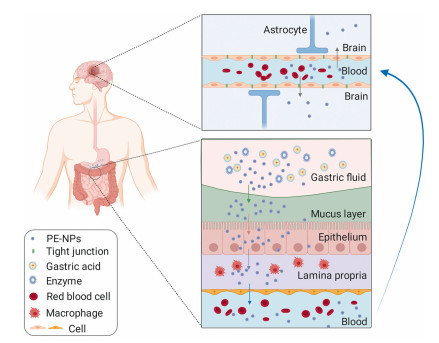
Therapeutic agents for brain diseases are usually administered by injection, including intracerebral, intracerebroventricular or intravenous injection. However, patients receiving injections via these routes would suffer from lots of adverse effects including pain, allergic reactions and inflammation, especially bacterial infections and some surgical complications [1-3]. Therefore, a more suitable administration route of brain drugs has to be explored. Among all the administration routes, oral delivery is considered the most desirable because of better patient compliance, ease of drug administration and low occupation on medical resources [2]. However, it has been reported that oral delivery of active pharmaceutical ingredients (APIs) to the brain is confronted with problems including gastrointestinal (GI) side effects, poor bioavailability of the drug, short half-lives in the blood circulation and incapability to cross the blood brain barrier (BBB) [4, 5]. Nanocarriers, including nanocrystals (NCs), solid lipid nanoparticles (SLNs), and polyester nanoparticles (PE-NPs), are effective delivery tools that can facilitate drug accumulation in the brain. Compared with free APIs, oral NCs increased drug brain accumulation by 2~6 times [6-8], while oral SLNs increased up to 2~16-fold [9-11] and oral PE-NPs increased up to 1.5~17-fold [12-14]. Oral NCs showed lowest increase in brain uptake probably due to accelerated dissolution during absorption and transportation [6-8] while oral SLNs and PE-NPs with slower drug leakage [15, 16] showed higher increase. Notably, polyester is the type of synthetic polymer that has been used clinically for its excellent safety profile with more than 10 polyester-based products available commercially on the market [17]. For example, Genexol-PM®, PEG-PLA micelle loading paclitaxel (PTX), was the first FDA-approved polymeric micelle formulation [18]. Subsequently, Nanoxel-PM®, which was a docetaxel loaded PEG-PLA micelle, was also approved for the treatment of breast cancer and small cell lung cancer [19]. Besides, PE-NPs (Table 1) were reported to cross intestinal and brain membranes after oral administration [20], and were maintained intact in the intestine and brain [21], allowing the successful transport of brain drugs through those biological barriers and promoting therapeutic effect [22-34]. Thus the absorption of PE-NPs 'as a whole' strategy is preferred. To promote the development of next-generation oral PE-NPs for brain-targeting, greater knowledge regarding the process of overcoming biological barriers for enhanced oral absorption and brain penetration is required. Thus, the status quo will be summarized with emphasis on challenges, key factors and strategies taken to adapt oral PE-NPs for brain drugs.
In order to achieve therapeutic concentrations of drugs in the brain, a highly efficient absorption of PE-NPs is preferred. For therapeutics to reach the brain after oral administration, intact PE-NPs must transit through the GI tract, enter into the bloodstream, and then reach the brain across the BBB [35, 36]. In doing so, brain-targeting absorption of the PE-NPs is limited by various physiological barriers and remains a major problem in drug development (Fig. 1).
NPs with small size are featured with huge surface area, leading to better contact with the biological barrier to promote penetration through the GI tract and brain [37]. However, NPs are also thermodynamically unstable, which promotes agglomeration or crystal growth [38]. Thus, stability issues related to NPs deserve great concern during the process of oral delivery for brain-targeting.
The GI tract acts as the first defensive barrier against exogenous toxins and pathogens at the same time [39]. NPs should better remain intact in the GI tract to guarantee their eventual entrance into the brain. Therefore, the GI tract has posed a great challenge for brain drug delivery.
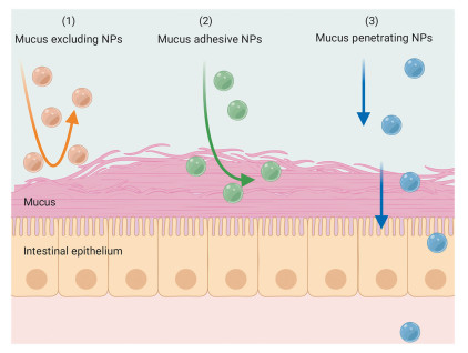
The mucus layer is a dynamic semipermeable barrier composed of a tenacious hydrogel material [40]. It prevents certain agents from reaching the epithelial surface [41]. In addition, through electrostatic or hydrophobic interactions, the mucus layer may trap NPs [42]. Fig. 2 summarized the fate of NPs in mucus, including exclusion, adhesion and penetration of NPs. Penetration of this mucus barrier is necessary so that NPs reach epithelial cells.
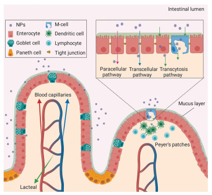
After successfully penetrating through the mucus layer, NPs are faced with the intestinal epithelium [43], which is the major transport barrier. It has been reported that NPs can be taken up in particulate form in the intestinal epithelium [44]. However the specific mechanism remains to be illustrated. Currently, there are three known uptake mechanisms (Fig. 3): (1) transcytosis; (2) transcellular uptake; and (3) paracellular transport [37]. Different mechanisms of absorption may coexist, depending on the drug delivery system [43]. The intestinal uptake of NPs can be optimized by adapting their physicochemical properties or using technique based on receptor-and transporter-mediated endocytosis [45].
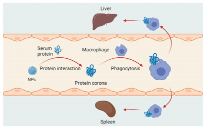
After passage of the intestinal epithelial barrier, NPs can enter the bloodstream or lymph circulation. NPs, that entered the lymphatic circulation, can avoid presystemic hepatic first-pass metabolism and finally enter into the bloodstream [46]. Once NPs are exposed to blood, they may disassociate in blood, resulting leakage of payload [47] since the biological environment in blood is also complex [48]. Besides, the intact NPs would face two main obstacles (Fig. 4): (1) adsorption of proteins; (2) interactions with phagocytes [49]. Apparently, this process may decrease amounts of NPs available for accumulation in the brain.
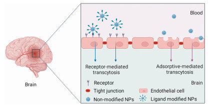
Part of the orally administered NPs eventually reaches the BBB. The BBB is responsible for brain protection and homeostasis. It refuses the access of exotic substances to the brain and only small molecules (usually lipid soluble molecules whose molecular weight < 400 Da) can penetrate the BBB [50]. According to current research, two strategies are mainly employed for NPs to cross the BBB (Fig. 5): (1) receptor-mediated transcytosis based on specific ligands [51]; (2) adsorption-mediated transcytosis based on the electrostatic attraction [52]. However, the detailed understanding on the mechanism of trans-BBB NPs delivery was still limited. Besides, the efficiency of oral NPs delivery to the brain was very low, almost 0.1%–0.3% of the initial oral dose [35, 36] while the best NPs formulation delivered up to 5% of the initial dose penetrating the BBB effectively through intravenous injection [51]. Regarding this issue, designing a strategy for more effective BBB penetration is important.
Parameters like size, shape, and surface charge are potential factors influencing the performance of PE-NPs and have gained interests or application in oral brain drugs (Table 2). Recommended size, shape and surface charge of oral delivery of PE-NPs for brain-targeting are 50–200 nm in diameter, spherical in shape and −1 mV ~−15 mV in zeta potential, respectively (Fig. S1 in Supporting information). And the reasons why these values are recommended can be found in the supporting information. Yet little was known about the crucial parameters of PE-NPs and their connections determining oral PE-NPs efficiency of brain targeting. For instance, it was speculated that only NPs up to 64 nm with neutral or negative charges could access the brain tissue and reach neuronal somata by passive diffusion [53], indicating that NPs size was a key parameter for bypassing of the BBB [54]. However, some studies found that neither charge nor size of NPs, but surface modification of NPs was the decisive factor determining NPs penetration through the BBB in vivo [55]. For example, Voigt et al. [56] carried out real-time imaging of retinal blood vessels and retinal tissue by in vivo confocal neuroimaging after administration of a variety of NPs which differed in size (67–464 nm) and zeta-potential (−51–20 mV). Their findings demonstrated that NPs fabricated with cationic or non-ionic surfactants achieved successful BBB penetration. In their work, the effect of surfactants was found dependent on charge: (1) non-ionic surfactants induced BBB penetration to enhance drug delivery; (2) anionic surfactants prevented BBB penetration to reduce NPs brain entrance. With this in mind, it was important to understand the physiology and pathophysiology of the GI tract and BBB modifications for the purpose of utilizing these traits to improve current oral PE-NPs technologies capable of successfully targeting lesioned areas of the brain.
In spite of challenges mentioned above, various PE-NPs have been developed for oral delivery of brain drugs (Fig. 6). Those carrier systems are required to remain intact after bypassing majority of the biological barriers and achieve desired delivery. To improve the integrity of PE-NPs in vivo, various methods improving stability and overcoming barriers of oral PE-NPs have been reported. Then, brain-targeting efficiency and therapeutic effects have been enhanced.
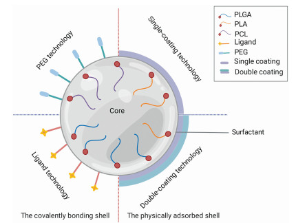
PLGA is one of the most widely used material for oral brain-targeting delivery owing to its favorable safety profile, sustained-release characteristics and easy manipulation for NPs size, thus offering more control over drug delivery [28, 57]. PLA, obtained by the polymerization of lactic acid, is biocompatible polyester [58, 59]. PCL, a semi-crystalline formed by ring-opening polymerization of ε-caprolactone, is also biodegradable polyester [57, 60]. PLA or PCL conjugated with PEG have been demonstrated to be promising carriers for oral delivery of brain drugs.
Generally, PE-NPs demonstrated a certain degree of stability in vitro and in vivo, and protected brain drugs from the erosive pH environment, enzyme degradation [61-64]. For example, when exposed to the digestive environment of the GI tract, epigallocatechin-3-gallate (EGCG) stability was improved by encapsulation in PLGA NPs [65], and the formulation displayed characteristics of sustained release. Then intact NPs crossed the BBB by absorptive transcytosis [14, 66, 67] and reduced neuro-inflammation and Aβ plaque/peptide burden in APP/PS1 mice [14]. Similarly, previous studies reported by Mitta's group showed that estradiol-loaded PLGA NPs exhibited 8.5-fold increase in brain-targeting efficiency than free estradiol after oral administration [24]. They found that the amount of the drug released in vitro and the Cmax of the drug in the plasma increased with a decrease in molecular weight and lactide content of PLGA [68]. Therefore, it was feasible to alter properties of PE-NPs by altering polyester molecular characteristics [69]. For instance, star-shaped PLGA NPs were reported with higher drug loading efficiency than linear PLGA NPs owing to their ordered branched structure with multiple arms organized around a core [70]. While choosing six-armed star-shaped PLGA NPs (6-s-PLGA) to encapsulate puerarin (PU) (Fig. S2a in Supporting information), cellular internalization and permeation were significantly improved. Then, a 6.1-fold and 2.3-fold enhancement of the Cmax in plasma and brain, respectively, was achieved compared with free PU administered orally, thus improving neuroprotective effects [27]. And to investigate whether the NPs were transported as a whole, NPs labeled with FRET dyes were used, and intact NPs were observed in the intestines and brain following oral administration via FRET imaging (Fig. S2b in Supporting information) [27], indicating the promotive effect brought by branched PLGA.
To improve the stability of PE-NPs, surfactants, such as Tween-80 (T-80), polyvinyl alcohol (PVA) and sodium dodecyl sulfate, have been added into PE-NPs [71, 72]. Those adsorbed surfactants further stabilized PE-NPs via electrostatic and steric repulsion [73]. For instance, poloxamer 188 stabilized PEG-PCL NPs loading Ginkgolide B (GB) [34] by the formation of protective hydrophilic coating layers which increased inter-particle steric repulsion [74] and preserved the structural integrity of the PE-NPs [75]. These PEG-PCL NPs were internalized into Madin-Darby canine kidney cells mainly by clathrin-dependent endocytosis and lipid raft/caveolae-mediated endocytosis, and then accumulated in the endoplasmic reticulum and lysosomes. Further study confirmed that those PEG-PCL NPs readily transported across the chorion, GI barriers and BBB in zebrafish [34]. Accordingly, PEG-PCL NPs increased Cmax of GB in the brain (0.17 ± 0.01 µg/g) by nearly 2 folds vs. free GB (0.09 ± 0.01 µg/g) via oral administration and achieved superior therapeutic efficacy by using murine Parkinson's disease model [34].
Currently, the application of different surfactants on the same nano-system was reported to have various benefits [76]. Eventually, the concentration and therapeutic effect of PE-NPs in the brain changed accordingly. For example, adding surfactants to PLGA NPs formulation of superparamagnetic iron oxide nanoparticles (SPION) was reported to stabilize PLGA NPs, and then influenced their bio-distribution [77]. SPION loaded PLGA NPs stabilized by didodecyl-dimethyl-ammonium-bromide (DMAB) (SPION-DMAB NPs) were found with better colloidal stability than those stabilized by α-tocopheryl-polyetheleneglycol-succinate (TPGS) (SPION-TPGS NPs) [23] due to the positive charge brought by DMAB [78] while TPGS was an efficient emulsifier to help cellular uptake and extend blood circulation time of drugs [79, 80]. In the in vivo study, they found that oral administration of SPION-DMAB NPs on day 7 resulted in ~3.3-fold higher concentration of Fe accumulating in the brain (9.73 ± 1.54 mg/kg tissue weight) compared with SPION-TPGS NPs (3.05 ± 0.28 mg/kg tissue weight) [23]. This improvement in brain-targeting may be attributed to the better stability of SPION-DMAB NPs and higher diffusive capability [81]. Interestingly, SPION-DMAB NPs were found mainly internalized in the brain while SPION-TPGS NPs were in the liver on day 7 (Fig. S3 in Supporting information) [23]. However, the theoretical connection between the stability of PE-NPs and the efficacy of the surfactant was unknown. The traditional selection was based trial and error approach, which required vast amounts of effort and wasted a lot of time [72]. Fortunately, colloidal probe microscopy has been commonly used to investigate interactions between colloidal particles and is expected to become a helpful tool for surfactant screening [82].
There have been mainly two different approaches overcoming the mucus barrier: the use of mucus-penetrating materials and mucoadhesive materials [37]. Mucoadhesive materials endowed NPs with prolonged residence time in the mucus layer [46], which may result to degradation of the NPs [83]. Besides, NPs covered by muco-adhesive materials were often trapped in mucus layer [84], and their oral absorption was further reduced by fast turnover (between 50 and 270 min) and continuously secreted mucus [1]. Therefore, muco-adhesive strategy may not be suitable for enhancing the efficiency of oral brain drugs delivery. Mucus-penetrating materials were biomaterials designed to help PE-NPs diffuse through the mucus layers [85] by reducing the retention time of PE-NPs in mucus. Intact PE-NPs interacted directly with the epithelial cells, thus enhancing the intestinal absorption and brain uptake of PE-NPs [37]. For example, compared with unmodified PLGA NPs, Pluronic F127 modified PLGA NPs showed a 2-fold increase in the efficiency of mucus penetration [86] while plasma concentrations of NPs were significantly enhanced by 1.5-fold, which resulted in an increase of NPs concentration in the brain on day 7 after oral administration [87]. PEGylation is one feasible way in improving mucus-penetrating ability, which reduces electrostatic and hydrophobic interaction between mucins and PE-NPs [83]. For instance, the model photosensitizer (mTHPP) loaded PLGA-PEG NPs increased 17-fold accumulation of mTHPP in mucus-secreting HT-29-MTX cells compared to PLGA NPs. Fluorescence microscopy further confirmed the effect of PLGA-PEG NPs by higher intensity of red fluorescence (Fig. S4 in Supporting information) [88]. Generally, NPs modified with low molecular weight PEG were often found with concentration-dependent reduction on mucus adhesion [89-91]. It should be noted that mucus-penetrating materials usually were hydrophilic polymers while NPs with hydrophobic surface was easier to be taken by M cells, enterocytes [37] and BBB [51]. Therefore the hydrophilic/hydrophobic balance needs to be further considered for the subsequent uptake.
Evidence is accumulating to support that integral NPs are able to be delivered across the intestinal epithelia [92]. For example, Tang et al. used fluorescence imaging tools to obtain evidence of trans-epithelial transport of intact PLGA NPs. Their results suggested that intact NPs crossed the intestinal barrier into circulation and then accumulated at C. neoformans infected lungs [93]. Similarly, Yin et al. employed fluorescent probes to illustrate the fate of lectin-conjugated PLGA NPs and found the absorption and transportation of integral NPs into the systematic circulation. Subsequently, a significant amount of lectin-conjugated PLGA NPs was detected in the liver and spleen (0.16%–0.37% and 0.15%–0.27% of dose) while just a small amount was observed in the heart and stomach (< 0.04% of dose) [94]. With intact NPs penetrate across the intestinal epithelia and reach the systemic circulation, the targeting to the brain is highly expectable [95]. To enhance the intestinal absorption of conventional PE-NPs, whole PE-NPs were functionalized with different targeting ligands [96]. However, receptor-and transporter-mediated brain-targeting must be considered at the same time. Otherwise, PE-NPs would be diverted to other tissues rather than the brain after efficient oral absorption. For example, various bile acids, proved to enhance the oral deliverability of PE-NPs, usually targeted to liver via oral route due to active transportation of bile acids by their transporters from the GI tract to liver [97].
Modification of NPs with ligands has been proved to promote cellular targeting and intestinal uptake via receptor-mediated endocytosis [96]. Various types of peptide ligands, including lectins [98], transferrin [99], neonatal Fc [100] and RGD (arginine–glycine–aspartate) [101] were utilized for the said purpose. For instance, transferrin (Tf) binds to the transferrin receptor (TfR) expressed in both the GI tract [102] and the BBB [103], making it a possible ligand for oral PE-NPs delivery to the brain. However, TfR is almost saturated with endogenous Tf in the bloodstream [52], indicating a compromised delivery efficiency [103].
It should be noted that some peptide ligands may have stability problem in the GI tract [83]. Therefore, non-peptide ligands, including gambogic acid (GA), were employed to improve uptake of NPs across the GI tract. To further enhance the efficiency of brain-targeting, the selected ligands are required to bind with receptors of the BBB. GA is a naturally occurring xanthone which can bind to TfR [104]. This process was carrier-mediated and non-competitive active transport via active targeting [105, 106]. For instance, cyclosporine (CsA) was formulated into a series of GA-conjugated PLGA NPs (GA-PLGA NPs which differed in surface GA density). The intestinal uptake of GA-PLGA NPs increased with increasing GA density on the NPs surface (Figs. S5a in Supporting information). And at least 2-fold increase in plasma concentration at 6 h and brain concentration at 72 h of CsA was observed compared to PLGA NPs after oral administration (Figs. S5b and c in Supporting information) [25], indicating the transport efficiency of GA.
In view of the heterogeneity and variability of membrane receptors, membrane transporters are preferred for efficient drug delivery as new targeting sites [107] as inspired by the transport of nutrients [102]. More importantly, these transporters also expressed on BBB [103]. Some examples included natural substrates and derivatives, such as amino acids [108], vitamins [109] and carnitine [110]. For instance, Luo et al. successfully prepared ascorbate (As)-conjugated NPs (As-PLGA NPs) (Fig. S6a in Supporting information) to improve oral delivery of PTX. The result showed that As-PLGA NPs loading PTX improved membrane permeability (Peff) and absorption rate (Ka) more effectively than PLGA NPs, and highest improvement of Peff and Ka was 2.26-fold and 2.6-fold respectively (Fig. S6b in Supporting information). The possible mechanism was attributed to the interaction of As with sodium-dependent vitamin C transporter 1 (SVCT1) on the epithelial cells (Fig. S6c in Supporting information). The accumulation of As-PLGA NPs in the villi and their penetration to the basolateral side remarkably increased intestinal absorption [111]. The Na+-coupled organic cation/carnitine transporter 2 (OCTN2) was also used for targeting PTX delivery [110]. OCTN2 was expressed in the small intestine for L-carnitine (LC) transportation [112]. And Kou et al. developed LC conjugated PLGA NPs for oral delivery of PTX. Intestinal absorption experiments demonstrated that the apparent permeability coefficient (Papp) and Ka for the drug increased almost 2-fold when LC was conjugated to PLGA NPs. Further studies suggested that the OCTN2-mediated endocytosis/transcytosis enhanced intestinal absorption and cellular uptake of PTX rather than the traditional transmembrane transport (Fig. S7a in Supporting information) [110]. Interestingly, it was found that the predominant transportation route for oral absorption of NPs was the lymphatic system. In the subsequent pharmacokinetics study, the Cmax of PTX for LC-PLGA NPs in plasma was 5.3-fold higher than for PLGA NPs [110]. Besides, the OCTN2 transporter was also highly expressed on brain capillary endothelial cells, indicating its potential for brain-targeting [110]. For example, Kou et al. then found that LC-PLGA NPs enhanced the brain accumulation of PTX up to 11-fold compared to PLGA NPs after intravenous injection. The enhanced brain accumulation was attributed to LC-mediated cellular recognition and internalization [113]. They confirmed that OCTN2 significantly promoted the transcytosis of NPs across the BBB, and then improved the uptake of NPs in brain and anti-glioma efficacy (Fig. S7b in Supporting information) [113]. The results mentioned above suggested that LC conjugated PE-NPs hold bright prospects for oral delivery of brain drugs across the intestinal wall and BBB. However, oral delivery systems based on LC conjugated PLGA NPs for brain-targeting have not been investigated, which is a promising nano-platform in the future.
PEGylation, a strategy originally designed to extend the half-life of drugs in the bloodstream [114], has also been widely applied in the oral delivery of PE-NPs for brain-targeting to overcome obstacles by the bloodstream [12, 29]. For instance, PLGA-PEG NPs significantly increased T1/2 of schisantherin A (SA) in plasma up to 20.87 h, which was 3.31-fold longer than free SA (T1/2 of 6.31 h), indicating less elimination of SA loaded PLGA-PEG NPs in plasma. Intact PLGA-PEG NPs was internalized into cells, and fluorescence resonance energy transfer (FRET) imaging further suggested that PLGA-PEG NPs were maintained intact in the intestine and brain after oral administration for 2 h (Figs. S8a and b in Supporting information) [21]. And the PLGA-PEG NPs significantly enhanced the Cmax of SA in plasma and brain by 6.3-fold and 10.8-fold, respectively, versus free SA via oral administration (Fig. S8c in Supporting information), and thus improved the anti-parkinsonian activity of SA. The result was ascribed to increased systematic circulation time. Similar results were reported by Elena et al. who found that oral administration of dexibuprofen (DEX)-loaded PLGA-PEG NPs particularly improved memory impairment and induced a significant reduction in amylogenesis in the treatment of Alzheimer's disease compared to free DEX [29]. Generally, longer circulation time laid the foundation to better brain-targeting. Specifically, PEG chains on PE-NPs surface formed a protective layer [115], not only protecting NPs in the GI tract, but also minimizing interactions with protein or macrophages to overcome concerns of clearance by the mononuclear phagocyte system (MPS), thus prolonging circulation time [116]. And the extended circulation time enhanced the possibility of the PE-NPs to interact with the BBB [117]. For example, PE-NPs prepared by the blend of PEG and PLGA increased 16.8-fold curcumin (CUR) concentration in the brain 12 h after oral administration compared to free CUR [13], while PLGA NPs increased 3-fold [30]. Besides, it was found that the molecular weight and the density of PEG were important with respect to MPS uptake [118]. For instance, less interaction with the MPS was observed with an increase in PEG molecular [69].
The technology of surface coating has stabilized and modified PE-NPs, and then enhanced BBB penetration. For instance, T-80 has been widely used in oral delivery for brain-targeting for many years [24]. For example, 0.1 mg/rat dose of estradiol loaded PLGA NPs coated with T-80 showed approximately 2.3-fold and 2-fold higher concentrations in plasma and brain 24 h after oral administration, respectively, when using uncoated PLGA NPs as control [24]. T-80 in blood mimicked low density lipoproteins (LDL) and facilitated adsorption of apolipoprotein, which acted as targeting ligands to promote brain targeting by receptor-mediated endocytosis [119]. Interestingly, T-80 was reported to provide protective action of PE-NPs in simulated gastric fluids (SGF) and simulated intestinal fluids (SIF) due to forming a protective layer [75]. Besides, it was proposed that T-80 contributed in brain-targeting by inhibition of efflux transporters such as P-glycoproteins [120]. Another example was chitosan (CS) coated PE-NPs. CS modified NPs were found with advantages like preventing degradation by enzymes, controlled release and thus improving bioavailability in the brain [3]. For example, the cumulative release of irinotecan (IRN) from CS coated PLGA NPs in pH 7.4 PBS was 76.68% in 48 h [28] while PLGA NPs was 100% [121]. In pharmacokinetic study, CS coated PLGA NPs increased 3.53-fold and 8.03-fold the IRN concentration in plasma and brain respectively compared with free IRN after oral administration [28]. However, CS was also a muco-adhesive material. To decrease the adhesion of CS with mucus, strategies like cationic-shielding coating, surface charge regulation and chemically modified CS derivatives, were developed [43]. For example, the partially positive charge of CS was shielded by conjugating with Pluronic F127 [122]. Even some CS derivatives, such as alkylglyceryl CS, were proved to improve blood compatibility and brain targeting efficiency by crossing BBB [123].
To fully exert advantages of active targeting and enhance brain uptake, strategies like double coating of T-80 and apolipoprotein E (ApoE) were developed. ApoE, a protein with 299 amino acid residues [124], acted as a ligand for LDL receptors [125]. It was found that ApoE endowed PE-NPs with active brain targeting by binding to LDL receptors in BBB [126]. For example, by stirring/incubation method of T-80 (inner coating) and ApoE (exterior coating), Krishna et al. [33] successfully prepared double-coated PEG-PCL NPs loading DNP (D3-NP) (Fig. S9a in Supporting information). In SGF and SIF, D3-NP enhanced 2 folds of a percentage of surfactant coating remaining (89.48% ± 6.4% in SGF and 86.54% ± 3.9% in SIF) compared with that of T-80 coating PEG-PCL NPs (D2-NP) (46.8% ± 5.3% in SGF and 39.72% ± 5.4% in SIF), indicating ApoE stabilized PEG-PCL NPs in the biological fluids. More importantly, in the subsequent pharmacokinetic test, the Cmax and AUC0-t of DNP in the brain for D3-NP were 2- and 2.5-fold, 3-fold and 3.5-fold higher than that of D2-NP and PEG-PCL NPs (D1-NP), respectively (Figs. S9b and c in Supporting information) [33]. Accordingly, D3-NP showed the highest AChE (the major biological marker for Alzheimer's disease) inhibitory activity in the pharmacodynamic studies. And main mechanism to cross BBB was attributed to receptor-mediated transport (Fig. S9d in Supporting information). These results indicated that PE-NPs with double-coating were promising. However, the interaction between double coating and PE-NPs was not considered in the studies, which may lead to a deviation from the constant size ranges of PE-NPs formulations [127]. Apart from double coating of T-80 and ApoE, other types of double coating can also be added to the surface of NPs. For example, double coating of T-80 and PEG on NPs for oral delivery enhanced their blood circulation and brain uptake [127].
Because surface coating was adsorbed to PE-NPs surface via reversible physical adhesion [128], decoating would occur upon drastic environment change [46], resulting in particle aggregation or reduced brain targeting. Therefore, PE-NPs system with covalent conjugated materials would be a potential approach to avoid decoating issues. It was proved that covalent bonding of PE-NPs was superior to physical attachment [129] for the reason that covalent interaction was stronger than the van der Waals interaction.
Conjugation of ligands onto NPs surface, such as ApoE, Tf and lactoferrin, has been widely used to improve brain uptake of NPs [130]. For example, Raudszus et al. coupled ApoE to the surface of PLA NPs (ApoE-PLA NPs) to increase BBB targeting. They compared the cellular uptake of Lumogen®Red loaded ApoE-PLA NPs and PLA NPs with human brain microvascular endothelial cells (HBMEC) by fluorescence microscopy. Their results showed a 2-fold enhancement in cellular uptake of ApoE-PLA NPs (Fig. S10a in Supporting information) [131], indicating the necessity of ligand coupling. Girotra and Singh confirmed that ApoE conjugated NPs have higher brain targeting ability and anti-migraine activity than T-80 coated NPs, which was ascribed to facilitated BBB trespassing achieved by ApoE grafting [129]. Another example was Tf. For instance, Tf-grafted PLGA-PEG NPs (Tf-PEG-PLGA NPs) were designed and prepared to deliver clofazimine to treat central nervous system tuberculosis. And the Tf-PEG-PLGA NPs were found to interact with brain endothelial hCMEC/D3 cell by TfR-mediated mechanism (Fig. S10b in Supporting information), suggesting the BBB targeting potential of Tf-PEG-PLGA NPs [132]. However, the action of NPs may be hampered by competitive binding of endogenous Tf [103], which presents some limitation on the Tf-based strategy. As an alternative, GA [25] and antibodies against TfR [133] were developed to avoid ligand competition. For instance, OX26 monoclonal antibody conjugated PLGA NPs was successfully applied for brain delivery and exhibited better analgesic effects compared with PLGA NPs [134]. But Tf and antibodies against TfR probably encountered GI stability issues [96] as compared to GA, indicating the necessity of ligand stabilization in the GI tract. Furthermore, brain-targeting ability of GA conjugated PE-NPs via oral route has been confirmed by Saini et al. [25], which was mentioned before.
Another major mechanism for brain delivery was transporter-mediated transcytosis [107]. For example, a recent study constructed galantamine (GLM)-loaded PLGA-PEG NPs with ascorbic acid (Asc) conjugated at the outer terminus of PEG to target the sodium dependent-vitamin C transporters (SVCT2). Specific uptake of Asc-PEG-PLGA NPs via SVCT2 transporters was confirmed by fluorescence microscopy in SVCT2 expressing NIH/3T3 cells. The results demonstrated significantly higher uptake of Asc-PEG-PLGA NPs compared to PLGA-PEG NPs (Figs. S11a and b in Supporting information). Subsequently, biodistribution studies demonstrated that Asc-PEG-PLGA NPs increased 3.9-fold the brain concentration of GLM as compared to PEG-PLGA NPs, and thus showed higher acetyl choline activity, retention of memory and AChE inhibitory activity as compared with the non-modified group in vivo pharmacodynamic studies [135]. However, transporters were also expressed on normal cells and avoiding the off-target distribution of PE-NPs was still a problem [107]. It was noteworthy that, in certain situations such as brain tumor, overexpression of transporters like amino acid transporters was noticed [136]. Thus, the possible solution was finding certain transporters overexpressed in particular brain diseases, which can reduce drug side effects. Besides, dual-ligand based on receptor-and/or transporter-targeting may be another alternative to overcome the disadvantages of receptor or transporter saturation [103]. For example, the strategy of crosslinking lactoferrin (Lf) and folic acid (FA) to the surface of PLGA NPs (Lf/FA/PLGA NPs) was already developed (Fig. S11c in Supporting information) to treat glioma [137].
Each barrier has its own microenvironment, which imposes different requirements on the physicochemical properties of PE-NPs, thus further complicating the delivery process. Accordingly, there were multiple contradictions concerning NPs size, charge and surface modification. For example, NPs larger than 130 nm had higher possibility of being absorbed through M cells [138], and NPs between 50 nm and 500 nm were identified as ideal for absorption by enterocytes [139], whereas NPs under 100 nm were easier to be uptake by BBB [140-142]. NPs with hydrophobic surface had significantly more uptake by M cells but also enhanced interactions with mucus [37]. Another example was that positively charged NPs may be trapped in the mucus [143] while they could be more readily taken up by cells [144, 145]. Neutral NPs tend to exhibit prolonged blood circulation [146], whereas positively charged NPs can be more effectively internalized by cells in the GI tract and BBB [147, 148]. In order to balance these contradictions, 'smart' drug delivery systems, which were capable of changing their properties to adapt to the environment of each barrier, were a potential strategy. Fortunately, studies on intravenous administration of anti-cancer drug delivery systems have provided abundant examples for resolving the contradiction in the oral brain-targeting process [149-151]. For instance, Huang et al. developed a 'smart' nanoparticle system decorating with dtACPP (an activatable CPP) to treat glioma (Fig. S12a in Supporting information) [150]. The 'smart' nanoparticle system "turned off" their internalization ability during circulation. However, the enhanced cellular uptake was readily activated in a tumor microenvironment. By using a real time fluorescence imaging technique, they confirmed that dtACPP modified NPs showed higher accumulation in the brain compared with non-smart nanoparticle system (non-modified NPs and CPP modified NPs) (Fig. S12b in Supporting information). Accordingly, dtACPP modified NPs induced more tumor cell apoptosis and possessed better antitumor efficacy [150]. This finding demonstrated that 'smart' drug delivery systems were promising in addressing the contradictory designs needed for oral delivery of PE-NPs to the brain.
Although brain diseases differ in pathogeny, they share common obstacles in terms of oral therapy. Those obstacles are posed by the GI tract, the bloodstream and the BBB. Among them, BBB plays the most challenging part in oral drug delivery to the brain. Fortunately, intact PE-NPs have the ability to deliver drugs to the brain in a controlled and sustained way. These PE-NPs systems presented versatility in their size, shape, charge, and surface functionalization, which allowed them to be stable in the GI tract, circulate in the bloodstream, penetrate the BBB, and then influence the efficiency of oral delivery for brain-targeting. By tuning those parameters, many attempts have been made to obtain more stable PE-NPs in vivo, like shape-switching PE-NPs and 'smart' drug delivery systems.
During the transit of PE-NPs from the GI tract to the brain, PE-NPs are gradually broken down. Only PE-NPs that survive the harsh GI environment, manage to be absorbed by intestinal epithelia, and then are carried through the blood circulation can reach the BBB and being absorbed thereafter. To improve the BBB penetration of intact PE-NPs via oral rote, the initial challenge for oral PE-NPs delivery to the brain was to maintain the integrity of PE-NPs during the oral absorption process, thereby enhancing PE-NPs concentration in the blood. Then, stealth technology of PE-NPs, like PEGylation, allowed them to circulate long enough in blood, and thus enhanced the amounts of PE-NPs to cross the BBB. Recent advances focused on surface modification with coating material and ligand to modulate their brain-targeted ability after oral administration. Among those methods, surface coating technology was most studied because of its simple process based on the stirring/incubation method, while polyester-ligand strategy was a relatively new area that was based on complex synthesis process and was harder to achieve. They can both enhanced the stability and target ability of PE-NPs. However, surface coating strategy was based on physical adsorption, and reduced brain targeting happens when pre-mature decoating occurs. Therefore, covalent ligand conjugation technology may be a better method. Besides, the selection of benign material to form the core was also important. Because brain drugs encapsulated in the polyester matrix were released from the polyester, and the types of polyester played a critical role in the stability of PE-NPs.
In the treatment of neurodegenerative diseases, such as Alzheimer's disease and Parkinson's disease, oral medications are preferred. Oral delivery PE-NPs strategies potentially have implications for the management of those diseases. However, single strategy was difficult to achieve desirable therapeutic effect. The combination of two or more strategies was promising yet necessary. Besides, the influence of some key parameters of PE-NPs on the treatment effect of brain disease, like size, shape and surface charge, should be considered. However, those carrier systems encapsulated drugs via physical blending, which may lead to payload leakage in the process and reduce concentrations of drugs in the brain. Recently, the conjugation of brain drugs with synthetic polymers to assemble into NPs has the potential to overcome this limitation. Additionally, brain-targeting clinical trials have included studies on brain-targeting immune cells and monoclonal antibodies, such as intracerebroventricular injection of EGFRvIII-CAR T cells in patients with leptomeningeal disease secondary to glioblastoma and intravenous infusions of PRX002/RG7935 (PRX002) in patients with Parkinson's disease. Administering patients with EGFRvIII-CAR T cells may aid in the recognition and destruction of brain tumor cells in patients. PRX002/RG7935 (PRX002) inhibits the transfer of presumed pathogenic forms of α-synuclein from neuron to neuron, thereby potentially protecting neurons and slowing disease progression. Under the above circumstances, adjunctive therapy with oral medications is a potential approach. In the future, it may be possible to combine PE-NPs with immunotherapy to enhance the therapeutic effect of a variety of brain diseases.
The authors declare that they have no known competing financial interests or personal relationships that could have appeared to influence the work reported in this paper.
This work was supported by the National Key R & D Program of China (No. 2020YFE0201700), the National Mega-project for Innovative Drugs (No. 2019ZX09721001), the National Natural Science Foundation of China (No. 81673378), the Liaoning Revitalization Talents Program (No. XLYC1908031), the Project of Liaoning Provincial Department of Education (No. 2019LQN07), and the PhD Research Startup Foundation of Liaoning Province (No. 2020-BS-128).
Supplementary material associated with this article can be found, in the online version, at doi:
X. Tan, X. Liu, Y. Zhang, et al., Expert Opin. Drug Deliv. 15 (2018) 805–820.
doi: 10.1080/17425247.2018.1503250
N. Aggarwal, Z. Qamar, S. Rehman, S. Baboota, J. Ali, Curr. Pharm. Des. 26 (2020) 2280–2290.
doi: 10.2174/1381612826666200406072451
S. Yu, X. Xu, J. Feng, M. Liu, K. Hu, Int. J. Pharm. 560 (2019) 282–293.
doi: 10.1016/j.ijpharm.2019.02.012
F. Erdo, L.A. Bors, D. Farkas, A. Bajza, S. Gizurarson, Brain Res. Bull. 143 (2018) 155–170.
doi: 10.1016/j.brainresbull.2018.10.009
M.M. Wen, N.S. El-Salamouni, W.M. El-Refaie, et al., J. Control. Release 245 (2017) 95–107.
doi: 10.1016/j.jconrel.2016.11.025
S. Xiong, W. Liu, D. Li, et al., Mol. Pharm. 16 (2019) 1444–1455.
doi: 10.1021/acs.molpharmaceut.8b01012
C. Chen, L. Wang, F. Cao, et al., Int. J. Pharm. 497 (2016) 239–247.
doi: 10.1016/j.ijpharm.2015.12.014
T. Chen, C. Li, Y. Li, et al., Mol. Pharm. 13 (2016) 3864–3875.
doi: 10.1021/acs.molpharmaceut.6b00644
C.F. Luo, M. Yuan, M.S. Chen, et al., Int. J. Pharm. 410 (2011) 138–144.
doi: 10.1016/j.ijpharm.2011.02.064
V. Kakkar, S.K. Muppu, K. Chopra, I.P. Kaur, Eur. J. Pharm. Biopharm. 85 (2013) 339–345.
doi: 10.1016/j.ejpb.2013.02.005
P. Mohammadi, R. Mahjub, M. Mohammadi, et al., Drug. Dev. Ind. Pharm. 47 (2021) 146–152.
doi: 10.1080/03639045.2020.1862172
E. Sanchez-Lopez, M. Ettcheto, M.A. Egea, et al., J. Nanobiotechnol. 16 (2018) 32.
doi: 10.1186/s12951-018-0356-z
A. Mukherjee, S. Sarkar, S. Jana, S. Swarnakar, N. Das, Brain Res. 1704 (2019) 164–173.
doi: 10.1016/j.brainres.2018.10.016
A. Cano, M. Ettcheto, J.H. Chang, et al., J. Control. Release 301 (2019) 62–75.
doi: 10.1016/j.jconrel.2019.03.010
F.Q. Hu, Y. Hong, H. Yuan, Int. J. Pharm. 273 (2004) 29–35.
doi: 10.1016/j.ijpharm.2003.12.016
I. Posadas, S. Monteagudo, V. Cena, Nanomedicine 11 (2016) 833–849.
doi: 10.2217/nnm.16.15
T.M. Allen, P.R. Cullis, Science 303 (2004) 1818–1822.
doi: 10.1126/science.1095833
S. Kalepu, V. Nekkanti, Acta Pharm. Sin. B 5 (2015) 442–453.
doi: 10.1016/j.apsb.2015.07.003
T. Wang, D. Zhang, D. Sun, J. Gu, J. Pharm. Anal. 10 (2020) 221–232.
doi: 10.1016/j.jpha.2020.05.002
C. Fernandes, C. Martins, A. Fonseca, et al., ACS Appl. Mater. Interfaces 10 (2018) 39557–39569.
doi: 10.1021/acsami.8b17224
T. Chen, C.W. Li, Y. Li, et al., ACS Appl. Mater. Interfaces 9 (2017) 9516–9527.
doi: 10.1021/acsami.7b01171
A. Paul, S. Das, J. Das, A. Samadder, A.R. Khuda-Bukhsh, Toxicol. Lett. 222 (2013) 10–22.
doi: 10.1016/j.toxlet.2013.07.006
S. Ghosh, I. Ghosh, M. Chakrabarti, A. Mukherjee, Food Chem. Toxicol. 136 (2020) 110989.
doi: 10.1016/j.fct.2019.110989
G. Mittal, H. Carswell, R. Brett, S. Currie, M.N.V.R. Kumar, J. Control. Release 150 (2011) 220–228.
doi: 10.1016/j.jconrel.2010.11.013
P. Saini, R. Ganugula, M. Arora, M.N.V.R. Kumar, Sci. Rep. 6 (2016) 29501.
doi: 10.1038/srep29501
H.Y. Zhang, C.K. Firempong, Y.W. Wang, et al., Acta Pharmacol. Sin. 37 (2016) 834–844.
doi: 10.1038/aps.2016.37
T.K. Chen, W. Liu, S. Xiong, et al., ACS Appl. Mater. Interfaces 11 (2019) 45276–45289.
doi: 10.1021/acsami.9b16047
N. Ahmad, M.A. Alam, R. Ahmad, S. Umar, F. Jalees Ahmad, J. Microencapsul. 35 (2018) 327–343.
doi: 10.1080/02652048.2018.1485755
E. Sanchez-Lopez, M. Ettcheto, M.A. Egea, et al., Nanomedicine 13 (2017) 1171–1182.
doi: 10.1016/j.nano.2016.12.003
C. Dende, J. Meena, P. Nagarajan, et al., Sci. Rep. 7 (2017) 10062.
doi: 10.1038/s41598-017-10672-9
H. Shen, X.Y. Hu, M. Szymusiak, Z.J. Wang, Y. Liu, Mol. Pharmaceut. 10 (2013) 4546–4551.
doi: 10.1021/mp400358z
R. Pandey, G.K. Khuller, J. Antimicrob. Chemoth. 57 (2006) 1146–1152.
doi: 10.1093/jac/dkl128
K.V. Krishna, G. Wadhwa, A. Alexander, et al., ACS Chem. Neurosci. 10 (2019) 4124–4135.
doi: 10.1021/acschemneuro.9b00343
Y. Zhao, S. Xiong, P. Liu, et al., Int. J. Nanomed. 15 (2020) 10453–10467.
doi: 10.2147/IJN.S272831
A. Lalatsa, N.L. Garrett, T. Ferrarelli, et al., Mol. Pharmaceut. 9 (2012) 1764–1774.
doi: 10.1021/mp300068j
A. Lalatsa, V. Lee, J.P. Malkinson, et al., Mol. Pharmaceut. 9 (2012) 1665–1680.
doi: 10.1021/mp300009u
E.M. Pridgen, F. Alexis, O.C. Farokhzad, Expert Opin. Drug Del. iv 12 (2015) 1459–1473.
doi: 10.1517/17425247.2015.1018175
L.B. Wu, J. Zhang, W. Watanabe, Adv. Drug Deliv. Rev. 63 (2011) 456–469.
doi: 10.1016/j.addr.2011.02.001
C. Li, T.T. Bei, Z.H. Niu, et al., Curr. Microbiol. 76 (2019) 896–903.
doi: 10.1007/s00284-019-01706-8
S.H. Bakhru, S. Furtado, A.P. Morello, E. Mathiowitz, Adv. Drug Deliv. Rev. 65 (2013) 811–821.
doi: 10.1016/j.addr.2013.04.006
J. Griesser, G. Hetenyi, H. Kadas, et al., Int. J. Pharmaceut. 538 (2018) 159–166.
doi: 10.1016/j.ijpharm.2018.01.018
M. Boegh, H.M. Nielsen, Basic Clin. Pharmacol. 116 (2015) 179–186.
doi: 10.1111/bcpt.12342
M.C. Chen, K. Sonaje, K.J. Chen, H.W. Sung, Biomaterials 32 (2011) 9826–9838.
doi: 10.1016/j.biomaterials.2011.08.087
D.I.A. Pereira, N.I. Mohammed, O. Ofordile, et al., Gates Open Res. 2 (2018) 48.
doi: 10.12688/gatesopenres.12866.2
K.S. Kim, K. Suzuki, H. Cho, Y.S. Youn, Y.H. Bae, ACS Nano 12 (2018) 8893–8900.
doi: 10.1021/acsnano.8b04315
Y. Wang, X. Tan, X. Fan, et al., Expert Opin. Drug Deliv. (2021) 1–18.
H. Fasehee, G. Zarrinrad, S.M. Tavangar, S.H. Ghaffari, S. Faghihi, Mater. Sci. Eng. C: Mater. Biol. Appl. 63 (2016) 587–595.
doi: 10.1016/j.msec.2016.03.023
V.H. Nguyen, B.J. Lee, Int. J. Nanomed. 12 (2017) 3137–3151.
doi: 10.2147/IJN.S129300
J.Y. Oh, H.S. Kim, L. Palanikumar, et al., Nat. Commun. 9 (2018) 4548.
doi: 10.1038/s41467-018-06979-4
X.W. Dong, Theranostics 8 (2018) 1481–1493.
doi: 10.7150/thno.21254
C. Saraiva, C. Praca, R. Ferreira, et al., J. Control. Release 235 (2016) 34–47.
doi: 10.1016/j.jconrel.2016.05.044
G. Tosi, L. Costantino, B. Ruozi, F. Forni, M.A. Vandelli, Expert Opin. Drug Del. 5 (2008) 155–174.
doi: 10.1517/17425247.5.2.155
R.G. Thorne, C. Nicholson, Proc. Natl. Acad. Sci. U. S. A. 103 (2006) 5567–5572.
doi: 10.1073/pnas.0509425103
S.W.L. Lee, M. Campisi, T. Osaki, et al., Adv. Healthc. Mater. 9 (2020) 1901486.
doi: 10.1002/adhm.201901486
G. Tosi, J.T. Duskey, J. Kreuter, Expert Opin. Drug Del. 17 (2020) 23–32.
doi: 10.1080/17425247.2020.1698544
N. Voigt, P. Henrich-Noack, S. Kockentiedt, et al., Eur. J. Pharm. Biopharm. 87 (2014) 19–29.
doi: 10.1016/j.ejpb.2014.02.013
Z. Xu, D. Wang, Y. Cheng, M. Yang, L.P. Wu, Mater. Sci. Eng. C: Mater. Biol. Appl. 92 (2018) 1006–1015.
doi: 10.1016/j.msec.2018.05.031
M. McKenzie, D. Betts, A. Suh, et al., Molecules 20 (2015) 20397–20408.
doi: 10.3390/molecules201119705
E. Niza, A. Ocana, J.A. Castro-Osma, I. Bravo, C. Alonso-Moreno, Cancers 13 (2021) 3387.
doi: 10.3390/cancers13143387
F. Molavi, M. Barzegar-Jalali, H. Hamishehkar, J. Control. Release 320 (2020) 265–282.
doi: 10.1016/j.jconrel.2020.01.028
P.R. Rauta, N.M. Das, D. Nayak, S. Ashe, B. Nayak, IET Nanobiotechnol. 10 (2016) 254–261.
doi: 10.1049/iet-nbt.2015.0021
M. Duran-Lobato, L. Martin-Banderas, L.M.D. Goncalves, M. Fernandez-Arevalo, A.J. Almeida, J. Nanopart. Res. 17 (2015) 61.
doi: 10.1007/s11051-015-2875-y
I.M. Kalogeras, Eur. J. Pharm. Sci. 42 (2011) 470–483.
doi: 10.1016/j.ejps.2011.02.003
B. Eshaghi, N. Alsharif, X. An, et al., Adv. Sci. 7 (2020) 2000649.
doi: 10.1002/advs.202000649
G.J. Zhang, J.F. Zhang, Drug Des. Dev. Ther. 12 (2018) 2509–2518.
doi: 10.2147/DDDT.S172919
Y. Zhou, Z. Peng, E.S. Seven, R.M. Leblanc, J. Control. Release 270 (2018) 290–303.
doi: 10.1016/j.jconrel.2017.12.015
M. Silva-Abreu, A.C. Calpena, P. Andres-Benito, et al., Int. J. Nanomed. 13 (2018) 5577–5590.
doi: 10.2147/IJN.S171490
G. Mittal, D.K. Sahana, V. Bhardwaj, M.N.V.R. Kumar, J. Control. Release 119 (2007) 77–85.
doi: 10.1016/j.jconrel.2007.01.016
M.L. Hans, A.M. Lowman, Curr. Opin. Solid. State Mater. 6 (2002) 319–327.
doi: 10.1016/S1359-0286(02)00117-1
J. Hadar, S. Skidmore, J. Garner, et al., J. Control. Release 304 (2019) 75–89.
doi: 10.1016/j.jconrel.2019.04.039
A. Mante, M. Heider, C. Zlomke, K. Mader, Eur. J. Pharm. Biopharm. 108 (2016) 32–40.
doi: 10.1016/j.ejpb.2016.08.009
Y.C. Wang, Y. Zheng, L. Zhang, Q.W. Wang, D.R. Zhang, J. Control. Release 172 (2013) 1126–1141.
doi: 10.1016/j.jconrel.2013.08.006
H. ShamsiJazeyi, C.A. Miller, M.S. Wong, J.M. Tour, R. Verduzco, J. Appl. Polym. Sci. 131 (2014) 40576.
X.H. Tian, X.N. Lin, F. Wei, et al., Int. J. Nanomed. 6 (2011) 445–452.
M. Bagad, Z.A. Khan, Int. J. Nanomed. 10 (2015) 3921–3935.
C. Bouissou, J.J. Rouse, R. Price, C.F. van der Walle, Pharm. Res. 23 (2006) 1295–1305.
doi: 10.1007/s11095-006-0180-2
M. Filippousi, M. Angelakeris, M. Katsikini, et al., J. Phys. Chem. C 118 (2014) 16209–16217.
doi: 10.1021/jp5037266
R. Gossmann, K. Langer, D. Mulac, PloS One 10 (2015) e0127532.
doi: 10.1371/journal.pone.0127532
M.A. Farooq, X.Y. Huang, A. Jabeen, et al., Colloids Surf. B 199 (2021) 111523.
doi: 10.1016/j.colsurfb.2020.111523
M.R. Vijayakumar, K.Y. Vajanthri, C.K. Balavigneswaran, et al., Colloids Surf. B 145 (2016) 479–491.
doi: 10.1016/j.colsurfb.2016.05.037
C. Curtis, D. Toghani, B. Wong, E. Nance, Colloids Surf. B 170 (2018) 673–682.
doi: 10.1016/j.colsurfb.2018.06.050
L.B. Wu, S.R.P. da Rocha, Langmuir 23 (2007) 12104–12110.
doi: 10.1021/la702108x
H.S. He, Y. Lu, J.P. Qi, et al., Acta Pharm. Sin. B 9 (2019) 36–48.
doi: 10.1016/j.apsb.2018.06.005
J. Witten, T. Samad, K. Ribbeck, Curr. Opin. Biotech. 52 (2018) 124–133.
doi: 10.1016/j.copbio.2018.03.010
M. Liu, J. Zhang, W. Shan, Y. Huang, Asian J. Pharm. Sci. 10 (2015) 275–282.
doi: 10.1016/j.ajps.2014.12.007
X. Zhou, Y. Liu, Y.M. Huang, et al., J. Drug Deliv. Sci. Technol 52 (2019) 157–164.
doi: 10.1016/j.jddst.2019.04.030
B. Semete, L. Booysen, L. Kalombo, et al., Int. J. Pharm. 424 (2012) 115–120.
doi: 10.1016/j.ijpharm.2011.12.043
L. Mahlert, J. Anderski, D. Mulac, K. Langer, Eur. J. Pharm. Sci. 133 (2019) 28–39.
doi: 10.1016/j.ejps.2019.03.010
M. Yang, S.K. Lai, Y.Y. Wang, et al., Angew. Chem. Int. Ed. 50 (2011) 2597–2600.
doi: 10.1002/anie.201006849
Y.Y. Wang, S.K. Lai, J.S. Suk, et al., Angew. Chem. Int. Ed. 47 (2008) 9726–9729.
doi: 10.1002/anie.200803526
Q. Xu, L.M. Ensign, N.J. Boylan, et al., ACS Nano 9 (2015) 9217–9227.
doi: 10.1021/acsnano.5b03876
Z. Zhang, J. Qi, Y. Lu, W. Wu, H. Yuan, Med. Res. Rev. 41 (2021) 2590–2598.
doi: 10.1002/med.21797
Y. Tang, S. Wu, J. Lin, et al., Nano Lett. 18 (2018) 6207–6213.
doi: 10.1021/acs.nanolett.8b02229
Y. Yin, D. Chen, M. Qiao, X. Wei, H. Hu, J. Control. Release 123 (2007) 27–38.
doi: 10.1016/j.jconrel.2007.06.024
J. Ding, Y. Sun, J. Li, H. Wang, S. Mao, J. Drug Target. 25 (2017) 532–540.
doi: 10.1080/1061186X.2017.1289541
X. Zhang, W. Wu, Drug Discov. Today 19 (2014) 898–904.
doi: 10.1016/j.drudis.2014.03.001
K. Thanki, R.P. Gangwal, A.T. Sangamwar, S. Jain, J. Control. Release 170 (2013) 15–40.
doi: 10.1016/j.jconrel.2013.04.020
C. Wang, P.C. Ho, L.Y. Lim, Int. J. Pharm. 400 (2010) 201–210.
doi: 10.1016/j.ijpharm.2010.08.023
L. Zhang, Y. Shi, Y. Song, et al., J. Drug Target. 26 (2018) 931–940.
doi: 10.1080/1061186X.2018.1455839
E.M. Pridgen, F. Alexis, T.T. Kuo, et al., Sci. Transl. Med. 5 (2013) 213ra167.
M. Garinot, V. Fievez, V. Pourcelle, et al., J. Control. Release 120 (2007) 195–204.
doi: 10.1016/j.jconrel.2007.04.021
A.B. Shreya, S.Y. Raut, R.S. Managuli, N. Udupa, S. Mutalik, AAPS PharmSciTech 20 (2018) 15.
M. Agrawal, D.K. Tripathi Ajazuddin, et al., J. Control. Release 260 (2017) 61–77.
doi: 10.1016/j.jconrel.2017.05.019
Y. Liu, Y. Chen, L. Lin, H. Li, Int. J. Nanomed. 15 (2020) 10385–10399.
doi: 10.2147/IJN.S277645
R. Ganugula, M. Arora, P. Saini, M. Guada, M. Kumar, J. Am. Chem. Soc. 139 (2017) 7203–7216.
doi: 10.1021/jacs.6b13231
M. Arora, R. Ganugula, N. Kuma, et al., ACS Appl. Bio Mater. 2 (2019) 3540–3550.
doi: 10.1021/acsabm.9b00419
H. Su, Y. Wang, S. Liu, et al., Acta Pharm. Sin. B 9 (2019) 49–58.
doi: 10.1016/j.apsb.2018.10.005
A. Jain, S.K. Jain, Acta Diabetol. 52 (2015) 663–676.
doi: 10.1007/s00592-015-0714-3
S.S. Feng, L. Mei, P. Anitha, C.W. Gan, W. Zhou, Biomaterials 30 (2009) 3297–3306.
doi: 10.1016/j.biomaterials.2009.02.045
L. Kou, Q. Yao, M. Sun, et al., Adv. Healthc. Mater. 6 (2017) 1700165.
doi: 10.1002/adhm.201700165
Q. Luo, M. Jiang, L. Kou, et al., Artif. Cells Nanomed. Biotechnol. 46 (2018) 198–208.
doi: 10.1080/21691401.2017.1417864
G. Wang, L. Zhao, Q. Jiang, et al., Asian J. Pharm. Sci. 15 (2020) 158–173.
doi: 10.1016/j.ajps.2020.02.002
L. Kou, Y. Hou, Q. Yao, et al., Artif. Cells Nanomed. Biotechnol. 46 (2018) 1605–1616.
A.S. Hoffman, Acta Biomater. 40 (2016) 1–5.
doi: 10.1016/j.actbio.2016.05.029
V. Bourganis, T. Karamanidou, E. Samaridou, et al., Eur. J. Pharm. Biopharm. 97 (2015) 239–249.
doi: 10.1016/j.ejpb.2015.01.021
H. Baharifar, M. Khoobi, S.A. Bidgoli, A. Amani, Int. J. Biol. Macromol. 143 (2020) 181–189.
doi: 10.1016/j.ijbiomac.2019.11.157
W.S. Zhang, A. Mehta, Z.Q. Tong, L. Esser, N.H. Voelcker, Adv. Sci. 8 (2021) 2003937.
doi: 10.1002/advs.202003937
J.M. Rabanel, P.A. Piec, S. Landri, S.A. Patten, C. Ramassamy, J. Control. Release 328 (2020) 679–695.
doi: 10.1016/j.jconrel.2020.09.042
K.H. Lin, S.T. Hong, H.T. Wang, et al., Int. J. Mol. Sci. 17 (2016) 1998.
doi: 10.3390/ijms17121998
J. Kreuter, D. Shamenkov, V. Petrov, et al., J. Drug Target. 10 (2002) 317–325.
doi: 10.1080/10611860290031877
A. Taghizadehghalehjoughi, A. Hacimuftuoglu, M. Cetin, et al., Nanomedicine 13 (2018) 1595–1606.
doi: 10.2217/nnm-2017-0386
S. Sharma, A. Verma, G. Pandey, N. Mittapelly, P.R. Mishra, Acta Biomater. 26 (2015) 169–183.
doi: 10.1016/j.actbio.2015.08.005
J. Sarvaiya, Y.K. Agrawal, Int. J. Biol. Macromol. 72 (2015) 454–465.
doi: 10.1016/j.ijbiomac.2014.08.052
L. Wu, L. Zhao, Neural Regen. Res. 11 (2016) 412–413.
doi: 10.4103/1673-5374.179044
B.S. Xie, X. Wang, Y.H. Pan, et al., Theranostics 11 (2021) 1177–1191.
doi: 10.7150/thno.46992
F. Re, I. Cambianica, C. Zona, et al., Nanomed. Nanotechnol. 7 (2011) 551–559.
doi: 10.1016/j.nano.2011.05.004
D. Das, S.S. Lin, J. Pharm. Sci. 94 (2005) 1343–1353.
doi: 10.1002/jps.20357
M. Mehanny, R.M. Hathout, A.S. Geneidi, S. Mansour, J. Biomed. Mater. Res. A 105 (2017) 1433–1445.
doi: 10.1002/jbm.a.36028
P. Girotra, S.K. Singh, Pharm. Res. 33 (2016) 1682–1695.
doi: 10.1007/s11095-016-1910-8
K. Chatterjee, S. Sarkar, K.J. Rao, S. Paria, Adv. Colloid Interface Sci. 209 (2014) 8–39.
doi: 10.1016/j.cis.2013.12.008
B. Raudszus, D. Mulac, K. Langer, Int. J. Pharm. 536 (2018) 211–221.
doi: 10.1016/j.ijpharm.2017.11.047
R.R. de Castro, F.A. do Carmo, C. Martins, et al., Int. J. Pharm. 602 (2021) 120655.
doi: 10.1016/j.ijpharm.2021.120655
G. Tosi, L. Costantino, B. Ruozi, F. Forni, M.A. Vandelli, Expert Opin. Drug Deliv. 5 (2008) 155–174.
doi: 10.1517/17425247.5.2.155
H. Bao, X. Jin, L. Li, F. Lv, T. Liu, J. Mater. Sci. Mater. Med. 23 (2012) 1891–1901.
K.R. Gajbhiye, V. Gajbhiye, I.A. Siddiqui, S. Pilla, V. Soni, Sci. Rep. 7 (2017) 11086.
doi: 10.1038/s41598-017-11611-4
L. Kou, Y.D. Bhutia, Q. Yao, et al., Front. Pharmacol. 9 (2018) 27.
doi: 10.3389/fphar.2018.00027
Y.C. Kuo, Y.C. Chen, Int. J. Pharm. 479 (2015) 138–149.
doi: 10.1016/j.ijpharm.2014.12.070
E. Roger, F. Lagarce, E. Garcion, J.P. Benoit, Nanomedicine 5 (2010) 287–306 Lond.
doi: 10.2217/nnm.09.110
K. Derakhshandeh, G. Hochhaus, S. Dadashzadeh, J Iran, Pharm. Res. 10 (2011) 425–434.
P.R. Lockman, M.O. Oyewumi, J.M. Koziara, et al., J. Control. Release 93 (2003) 271–282.
doi: 10.1016/j.jconrel.2003.08.006
A. Kaushik, R.D. Jayant, V. Sagar, M. Nair, Expert Opin. Drug Deliv. 11 (2014) 1635–1646.
doi: 10.1517/17425247.2014.933803
S.A. Kulkarni, S.S. Feng, Pharm. Res. 30 (2013) 2512–2522.
doi: 10.1007/s11095-012-0958-3
L.M. Ensign, R. Cone, J. Hanes, Adv. Drug Deliv. Rev. 64 (2012) 557–570.
doi: 10.1016/j.addr.2011.12.009
E. Frohlich, Int. J. Nanomed. 7 (2012) 5577–5591.
Y. Qiu, E. Rojas, R.A. Murray, et al., Nanoscale 7 (2015) 6588–6598.
doi: 10.1039/C5NR00884K
R.R. Arvizo, O.R. Miranda, D.F. Moyano, et al., PloS One 6 (2011) e24374.
doi: 10.1371/journal.pone.0024374
C. Yang, J. Uertz, D. Yohan, B.D. Chithrani, Nanoscale 6 (2014) 12026–12033.
doi: 10.1039/C4NR02535K
S. Behzadi, V. Serpooshan, W. Tao, et al., Chem. Soc. Rev. 46 (2017) 4218–4244.
doi: 10.1039/C6CS00636A
J.Z. Du, H.J. Li, J. Wang, Acc. Chem. Res. 51 (2018) 2848–2856.
doi: 10.1021/acs.accounts.8b00195
S. Huang, K. Shao, Y. Liu, et al., ACS Nano 7 (2013) 2860–2871.
doi: 10.1021/nn400548g
C.Y. Zhao, H.Z. Deng, J. Xu, et al., Nanoscale 8 (2016) 10832–10842.
doi: 10.1039/C6NR02174C

Linghui Zou , Meng Cheng , Kaili Hu , Jianfang Feng , Liangxing Tu . Vesicular drug delivery systems for oral absorption enhancement. Chinese Chemical Letters, 2024, 35(7): 109129-. doi: 10.1016/j.cclet.2023.109129
Han Wu , Yumei Wang , Zekai Ren , Hailin Cong , Youqing Shen , Bing Yu . The nanocarrier strategy for crossing the blood-brain barrier in glioma therapy. Chinese Chemical Letters, 2025, 36(4): 109996-. doi: 10.1016/j.cclet.2024.109996
Shushan Mo , Zhaoshuo Wang , Dandan Ding , Zhengzheng Yan , Yunlu Dai , Jinchao Zhang , Huifang Liu , Tianjiao Liang , Jianfei Tong , Zhenhua Li , Xueyi Wang . The synthesis and evaluation of novel BPA derivatives for enhanced blood-brain barrier penetration and boron neutron capture therapy. Chinese Chemical Letters, 2025, 36(5): 110190-. doi: 10.1016/j.cclet.2024.110190
Lu Li , Suticha Chunta , Xianzi Zheng , Haisheng He , Wei Wu , Yi Lu . β-Lactoglobulin stabilized lipid nanoparticles enhance oral absorption of insulin by slowing down lipolysis. Chinese Chemical Letters, 2024, 35(4): 108662-. doi: 10.1016/j.cclet.2023.108662
Lei Shen , Hongmei Liu , Ming Jin , Jinchao Zhang , Caixia Yin , Shuxiang Wang , Yutao Yang . “Three-in-one” strategy of trifluoromethyl regulated blood-brain barrier permeable fluorescent probe for peroxynitrite and antiepileptic evaluation of edaravone. Chinese Chemical Letters, 2024, 35(10): 109572-. doi: 10.1016/j.cclet.2024.109572
Mengjie Gao , Zhiqiang Cui , Yue Shen , Yikun Li , Dongxiang Zhang , Xiaoyan Gao , Yaguang Sun , Xin-Dong Jiang , Jianjun Du , Xiaohong Sun . NIR-II emissive aza-BODIPY-based nanoparticles for triggering glioblastoma apoptosis in brain. Chinese Chemical Letters, 2025, 36(5): 110098-. doi: 10.1016/j.cclet.2024.110098
Liqing Chen , Zheming Zhang , Yanhong Liu , Chenfei Liu , Congcong Xiao , Liming Gong , Mingji Jin , Zhonggao Gao , Wei Huang . Systemically intravenous siRNA delivery into brain with a targeting and efficient polypeptide carrier and its evaluation on anti-glioma efficacy. Chinese Chemical Letters, 2025, 36(3): 110228-. doi: 10.1016/j.cclet.2024.110228
Junfei Yang , Ke Wang , Shuxin Sun , Tianqi Pei , Junxiu Li , Xunwei Gong , Cuixia Zheng , Yun Zhang , Qingling Song , Lei Wang . A "spore-like" oral nanodrug delivery platform for precision targeted therapy of inflammatory bowel disease. Chinese Chemical Letters, 2025, 36(3): 110180-. doi: 10.1016/j.cclet.2024.110180
Ju Wang , Yongbing Sun , Lingbang Meng , Jianfang Feng , Meng Cheng , Liangxing Tu . Intestinal transporters and oral absorption enhancing strategies based on these transporters. Chinese Chemical Letters, 2025, 36(5): 110529-. doi: 10.1016/j.cclet.2024.110529
Xiangqian Cao , Chenkai Yang , Xiaodong Zhu , Mengxin Zhao , Yilin Yan , Zhengnan Huang , Jinming Cai , Jingming Zhuang , Shengzhou Li , Wei Li , Bing Shen . Synergistic enhancement of chemotherapy for bladder cancer by photothermal dual-sensitive nanosystem with gold nanoparticles and PNIPAM. Chinese Chemical Letters, 2024, 35(8): 109199-. doi: 10.1016/j.cclet.2023.109199
Fengjie Liu , Fansu Meng , Zhenjiang Yang , Huan Wang , Yuehong Ren , Yu Cai , Xingwang Zhang . Exosome-biomimetic nanocarriers for oral drug delivery. Chinese Chemical Letters, 2024, 35(9): 109335-. doi: 10.1016/j.cclet.2023.109335
Qin Yu , Haisheng He , Jianping Qi , Yi Lu , Wei Wu . Oral delivery of insulin by barbed microneedles actuated by intestinal peristalsis. Chinese Chemical Letters, 2024, 35(9): 109888-. doi: 10.1016/j.cclet.2024.109888
Zhiyu Yu , Xiang Luo , Cheng Zhang , Xin Lu , Xiaohui Li , Pan Liao , Zhongqiu Liu , Rong Zhang , Shengtao Wang , Zhiqiang Yu , Guochao Liao . Mitochondria-targeted carrier-free nanoparticles based on dihydroartemisinin against hepatocellular carcinoma. Chinese Chemical Letters, 2024, 35(10): 109519-. doi: 10.1016/j.cclet.2024.109519
Yunjie Dang , Yanru Feng , Xiao Chen , Chaoxing He , Shujie Wei , Dingyang Liu , Jinlong Qi , Huaxing Zhang , Shaokun Yang , Zhiyun Niu , Bai Xiang . Development of a multi-level pH-responsive lipid nanoplatform for efficient co-delivery of siRNA and small-molecule drugs in tumor treatment. Chinese Chemical Letters, 2024, 35(12): 109660-. doi: 10.1016/j.cclet.2024.109660
Ke Gong , Jinghan Liao , Jiangtao Lin , Quan Wang , Zhihua Wu , Liting Wang , Jiali Zhang , Yi Dong , Yourong Duan , Jianhua Chen . Mitochondria-targeted nanoparticles overcome chemoresistance via downregulating BACH1/CD47 axis in ovarian carcinoma. Chinese Chemical Letters, 2024, 35(5): 108888-. doi: 10.1016/j.cclet.2023.108888
Jiechen Liu , Xiaoguang Li , Ruiyang Xia , Yuqi Wang , Fenghe Zhang , Yongzhi Pang , Qing Li . Efficient suppression of oral squamous cell carcinoma through spatial dimension conversion drug delivery systems-enabled immunomodulatory-photodynamic therapy. Chinese Chemical Letters, 2024, 35(12): 109619-. doi: 10.1016/j.cclet.2024.109619
Wenjia Wang , Xingyue He , Xiaojie Wang , Tiantian Zhao , Osamu Muraoka , Genzoh Tanabe , Weijia Xie , Tianjiao Zhou , Lei Xing , Qingri Jin , Hulin Jiang . Glutathione-depleted cyclodextrin pseudo-polyrotaxane nanoparticles for anti-inflammatory oxaliplatin (Ⅳ) prodrug delivery and enhanced colorectal cancer therapy. Chinese Chemical Letters, 2024, 35(4): 108656-. doi: 10.1016/j.cclet.2023.108656
Xingqun Pu , Rongrong Liu , Yuting Xie , Chenjing Yang , Jingyi Chen , Baoling Guo , Chun-Xia Zhao , Peng Zhao , Jian Ruan , Fangfu Ye , David A Weitz , Dong Chen . One-step preparation of biocompatible amphiphilic dimer nanoparticles with tunable particle morphology and surface property for interface stabilization and drug delivery. Chinese Chemical Letters, 2025, 36(3): 109820-. doi: 10.1016/j.cclet.2024.109820
Mengmeng Yuan , Xiwen Hu , Na Li , Limin Xu , Mengxi Zhu , Xing Pei , Rui Li , Lu Sun , Yupeng Chen , Fei Yu , Huining He . Kidney targeted delivery of siRNA mediated by peptide-siRNA conjugate for the treatment of acute kidney injury. Chinese Chemical Letters, 2025, 36(6): 110251-. doi: 10.1016/j.cclet.2024.110251
Kang Wang , Qinglin Zhou , Weijin Li . Conductive metal-organic frameworks for electromagnetic wave absorption. Chinese Journal of Structural Chemistry, 2024, 43(10): 100325-100325. doi: 10.1016/j.cjsc.2024.100325
 DownLoad:
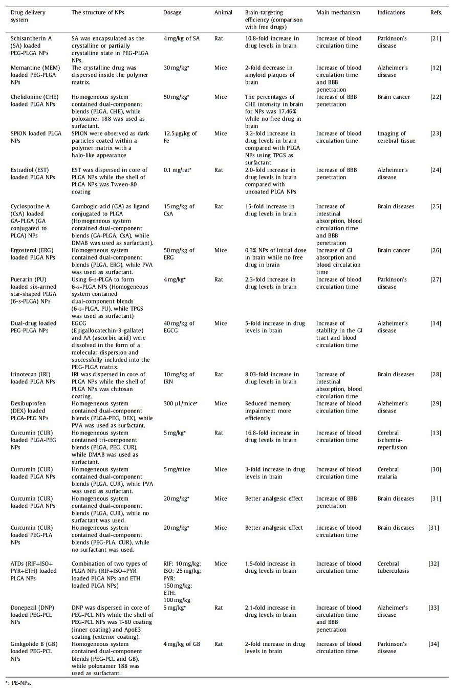
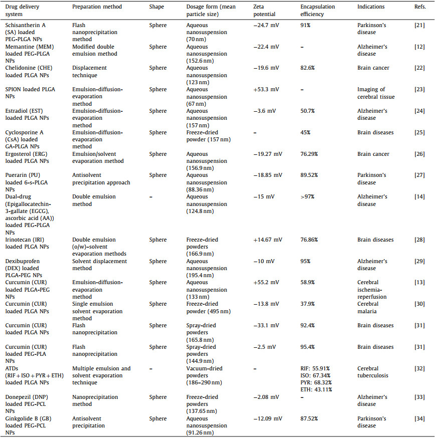
 DownLoad: