Login In
64Cu radiolabeled nanomaterials for positron emission tomography (PET) imaging
-
*Corresponding authors.
E-mail addresses: chmsudd@bjut.edu.cn (D. Su), gaoxy@ihep.ac.cn(X. Gao).
Figures(8)
Citation:
Xueqian Chen, Wenchao Niu, Zhongying Du, Yong Zhang, Dongdong Su, Xueyun Gao. 64Cu radiolabeled nanomaterials for positron emission tomography (PET) imaging[J]. Chinese Chemical Letters,
;2022, 33(7): 3349-3360.
doi:
10.1016/j.cclet.2022.02.070

Figures(8)


Lithium-ion batteries (LIBS) have dominated the power source electronic devices and electric vehicles (EVs) in the modern society. However, the possible safety issues deriving from organic electrolyte and shortage of lithium source inevitably prompt researchers to search for the substitutes for LIBs [1-3]. Recently, aqueous zinc ion batteries (AZIBs) have attracted various research interest due to their high safety, low standard electrode potential (−0.76 V vs. SHE), and high volumetric capacity (5851 mAh/cm3) [4-9]. As the key component for AZIBs, cathode has a prominent influence on the capacity, cycling stability and rate performance for the entire battery. However, the cathodes currently suffer from faces problems of electrode dissolution, low intrinsic conductivity, and sluggish slow mass transfer reaction, thus degrading which limit the further improvement of the application prospect performance of AZIBs [10-14]. Therefore, it's desirable to develop the suitable cathode for AZIBs.
Among various candidates, spinel materials are considered as one of the suitable candidates for AZIBs due to the low price, tunable structure, favorable structural stability, and high open circuit voltage [15-18]. Normally, the chemical formula of spinel materials is denoted as AB2X4, where the A and B ions are tetrahedral coordination and octahedron coordination, respectively, and X can be an anion of O, S, Se or Te [19]. According to the distribution of cations, spinel can be divided into three types: normal, inverse, and complex spinels [20,21]. Benefiting from the unique structure and superior physicochemical property, spinel-based cathode materials have been diffusely applied in the field of AZIBs. For example, Yuan et al. reported a novel Mg2MnO4 active material with inverse-spinel structure and utilized it as cathode for AZIBs [22]. Owing to the presence of rich high-valence-state Mn4+, the as-constructed Zn//Mg2MnO4 battery exhibits superior Zn2+ storage ability. In addition, Zhang et al. reported a non-stoichiometric ZnMn2O4@C cathode which exhibits high specific capacity (~150 mAh/g at 50 mA/g), long-term cyclability and superior rate capability [23].
Despite these advancements, the most development of spinel-based cathode in AZIBs is based on traditional experimental methods. This process inevitably increases the time and cost of developing new electrode, thereby leading to the insufficient efficiency [24-26]. Therefore, it is necessary to search for a more efficient and convenient method to identify suitable candidates for AZIB cathodes from a large number of spinel materials. Recently, the continuous development of computers techniques and theoretical models have provided a high-efficiency avenue for exploring the energy storage mechanism of new electrode materials. Specially, a variety of high-throughput screening methods for new materials have been extensively reported, which can greatly shorten the period and cost of developing new materials [27-30]. As a typical example, Scott et al. combined the high-throughput density functional theory (DFT) calculations and automatic grand canonical linear programming (GCLP) method to acquire 16 high-performance anode materials for lithium-ion batteries by predicting several key performance indicators: open circuit voltage, volume change per lithium addition, gravimetric and volumetric capacities [31]. Similarly, Cai et al. utilized the high-throughput combined with machine learning method to screen spinel containing Mg/Zn elements as cathode materials for organic-based magnesium/zinc ion batteries, and reported six new spinel materials with high conductivity, high ion diffusion rate and low volume expansion rate [32]. However, there are few works reported the application of high-throughput screening method to design the cathode for AZIBs.
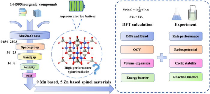
In this paper, we proposed a DFT-based high-throughput screening method combined with experimental validation to enable rapid development of high-performance spinel cathode material for AZIBs. Firstly, 9 Mn-based and 5 Zn-based spinel materials form 12,047 Mn/Zn-O based materials were obtained by checking their structures and whether they satisfy the basic properties of electrodes. Then, five candidates were obtained by further evaluating their electronic structure, open circuit voltage, volume expansion rate, and ionic diffusion coefficient, etc. by first-principles calculations. Meanwhile, taking Mg2MnO4 (one of the five candidates) as an example, we verified the performance of Mg2MnO4-based AZIBs. The cycling stability, reversible capacity, and rate performance of Mg2MnO4-based AZIBs are all excellent. Both electrochemical kinetics characterization and computational results show that the Mg2MnO4 cathode possesses excellent rate capability due to its fast reaction kinetics and low activation energy for ion migration. Therefore, all our results suggest that high-throughput screening based on first-principles calculations can assist us to experimentally obtain promising AZIBs cathode materials and greatly improve the development efficiency.
Fig. 1 shows the basic procedure of high-throughput computing method combined with experiments. Firstly, using the Python-based matminer Discovery Framework tool [33], we extracted all oxides containing Zn/Mn elements from the Materials Projects [34-36] database containing a total of 144,595 inorganic compounds. A total of 9454 Mn-O-based compounds and 2593 Zn-O-based compounds were obtained. The space group determines the internal arrangement of the crystal lattice, and our targets are spinel materials whose space group is Fd-3m. Through the discriminant screening of their space groups, 36 Mn-based and 23 Zn-based spinel oxides were obtained. Then, we obtained a total of 16 Mn-based and 8 Zn-based spinel materials by determining whether their band gaps are zero. Finally, comprehensively considering toxicity, commonness and price of the elements (Eu, Sc, Hy, Dy, Ag, Pd), 9 Mn-based and 5 Zn-based spinel materials were finally determined for further verification. In respond to the 14 selected spinel materials, we further calculated their microscopic properties directly related to their electrochemical performance when used as electrodes, including density of states, band structure, open circuit voltage, volume expansive ratio, ion diffusion coefficient, and energy barrier. To verify the computational results, we also selected a representative material for experimental synthesis and battery device application.
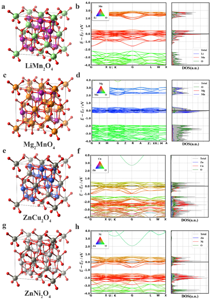
The calculated density of states and bands structures of the 14 spinel materials are shown in Fig. 2 (LiMn2O4, Mg2MnO4, ZnCu2O4 and ZnNi2O4) and Fig. S1 (Supporting information, Mn2AlO4, Mn2CuO4, Mn2NiO4, MnCo2O4, MnV2O4, NaMn2O4, Ti2MnO4, Mn2ZnO4, Ti2ZnO4, V2ZnO4) [37]. It can be seen that the valance and conduction bands of these 14 spinel materials are continuous around the Fermi level, confirming that they have the excellent electrical conductivity, which is consistent with the expectation of high-throughput screening.
As mentioned previously, to validate the high-throughput screening strategy based on first-principles predictions, we need to select a representative material for experimental testing. Considering the simple fabrication process and admirable electrochemical performance [22,38], and also as one of the promising candidates based on our theoretical predictions (Fig. S2 in Supporting information, we comprehensively compared the as-obtained 8 spinel materials from the aspect of OCV, volume expansion, energy barrier, as well as synthesis), Mg2MnO4 was selected as representative to explore its Zn2+ storage ability. We synthesized the Mg2MnO4 material and constructed the coin cell to measure its electrochemical performance, and the preparation process is exhibited in Fig. 3a. Firstly, the X-ray diffraction (XRD) pattern was conducted to characterize the sample's crystal structure. As depicted in Fig. 3b, all the characteristic diffraction peaks can match well with the standard Mg2MnO4 phase (JCPDS No. 19–0773), indicating the successful synthesis of electrode materials. In addition, the element composition and valence states of the Mg2MnO4 sample are analyzed by X-ray photoelectron spectroscopy (XPS). As illustrated in Fig. S3 (Supporting information), XPS spectra affirmed the existence of Mn, Mg, and O elements in the Mg2MnO4 composite. For Mn 2p spectrum (Fig. 3c), two distinct peaks centered at 641.5 eV and 653.2 eV can be assigned to Mn 2p3/2 and Mn 2p1/2, respectively. The spin energy difference of 11.7 eV, indicating that the manganese element mainly exists in the form of Mn4+ in the sample [39-41]. In addition, the high-resolution Mg 1s spectra also displays a distinct peak situated in 1302.6 eV (Fig. 3d) which originates from the Mg2+. All the afore-said results are consistent with the XRD analysis, further suggesting the successful formation of Mg2MnO4 phase.
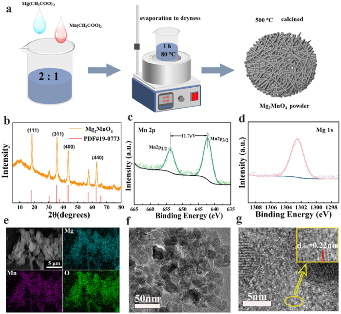
The morphology and microstructure of the as-fabricated samples were characterized by scanning electron microscopy (SEM) and transmission electron microscopy (TEM) [42]. The SEM-EDS pattern in Fig. 3e shows that the elements Mg, Mn and O are uniformly distributed in Mg2MnO4. According to EDS mapping (Fig. S4 and Table S1 in Supporting information), the ratio of Mn to Mg is calculated to be 0.433:1, which is close to the 1:2. In addition, the element content in the Mg2MnO4 sample was also estimated using the inductively coupled plasma-optical emission spectrometer (ICP-OES) technique. As summarized in Table S2 (Supporting information), the molar ratio of Mg and Mn is 0.08 to 0.043, which is consistent with the EDS mapping, further confirming the existence of Mg2MnO4 phase. Subsequently, the micro-structure of the Mg2MnO4 material was characterized by TEM and shown in Fig. 3f and Fig. S5 (Supporting information). Obviously, the as-prepared Mg2MnO4 sample exhibit uniform nanosheets connected to form nanorods. Lattice stripes were observed by high-resolution transmission electron microscopy (HRTEM) and the spacing of the lattice stripes is about 0.22 nm (Fig. 3g), which corresponding to the characteristic crystal plane (400) of Mg2MnO4. All the characterization results confirmed the successful synthesis of Mg2MnO4.
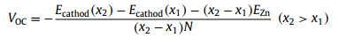
To further explore the performance of these spinel materials as cathodes, as shown in Fig. S6 (Supporting information), we calculated the open circuit voltage (OCV) generated when 2, 4 and 6 Zn2+were intercalated. The definition of OCV is as follows [43]:
|
|
(1) |
where x1 and x2 represent the number of embedded Zn2+, Ecathod represents the energy of the electrode when embedded with different number of Zn2+, EZn represents the energy of a single zinc atom, and N represents the valence of the embedded ions. Firstly, we checked the stability of the embedded sites. Taking Mg2MnO4 as an example, we found that the two embedded Zn2+ were more stable when they were located on opposite surfaces. The sites and their corresponding energies of the Zn2+ embedded in the Mg2MnO4 are summarized in Fig. S7 and Table S2 (Supporting information). Only 8 spinel materials were found to exhibit voltage by OCV calculations and their average voltages are summarized in Fig. 4a. We can see 7 of them have average voltages above 0.5 V, which can be considered as high-voltage electrode materials [44]. Among them, as exhibited in Fig. 4b, ZnCu2O4 and ZnNi2O4 exhibit the best performance with the average OCV reaching 1.775 and 0.963 V, respectively. The voltages of other materials with different numbers of Zn2+ embedded are summarized in Fig. S8 (Supporting information). Need to note that, the average voltage of Mg2MnO4 is 0.6 V, indicating that it is also a high voltage material, which is further confirmed by the cyclic voltammetry (CV) profiles of Mg2MnO4 electrode in Mg2MnO4//Zn coin cells (Fig. 4c). During the first cathodic scan, two obvious reduction peaks situated at 1.34 and 1.18 V can be detected which are ascribed to the insertion of H+ and Zn2+, respectively, whereas the oxidation peak around 1.6 V is corresponding to the ionic extraction behavior. In the subsequent cycles, all the CV curves well overlap, demonstrating the superior reversibility. Furthermore, the galvanostatic charge/discharge (GCD) curves shown in Fig. 4d are consistent with the CV curves, which show an average discharge voltage of about 1.35 V and a discharge capacity of 358.8 mAh/g at 200 mA/g after 50 cycles. The charge-discharge curves at other current densities can be found in Fig. S9 (Supporting information). Obviously, the relevant charge and discharge platform and the redox peaks in the CV curve can be well matched. For example, the discharge platform at 1.5 and 1.3 V is ascribed to the ion insertion behavior, whereas the charge platform of 1.6 V corresponds to the ionic extraction behavior.
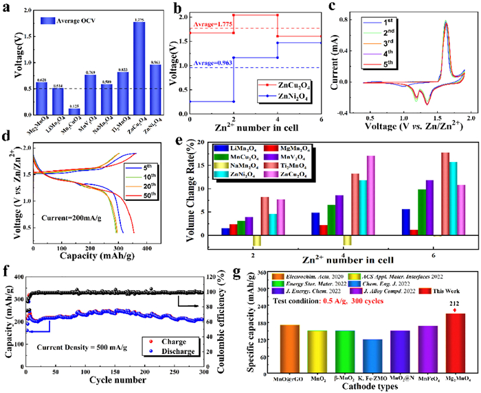
Moreover, we also calculated the volume expansion changes of the spinel materials after embedding Zn2+, which is related to the cycling stability of AZIBs. In normal, the low volume expansion rate indicates the admirable cycling stability. As summarized in Fig. 4e, the volume expansion rate of all 8 materials is less than 20%, indicating that all of them possess outstanding structural stability from the viewpoint of theory [32]. In particular, the Mg2MnO4 and NaMn2O4 model exhibit very low volume expansions (e.g., less than 5%). The experimental cycling stability measurements are shown in Fig. 4f and Fig. S10 (Supporting information), Mg2MnO4 electrode exhibits high cycling stability at 500 mA/g for 300 cycles and 200 mA/g for 100 cycles. Finally, as shown in Fig. 4g, we can see that the Mg2MnO4 cathode does exhibit superior capacity retention and excellent cycling stability (212 mAh/g at 0.5 A/g, 300 cycles) after comparing the cycling performance of Mg2MnO4 with other cathode materials [45-50].
In addition, the rate performance of Mg2MnO4//Zn full cell is also conducted, as illustrated in Fig. 5a. According to the results of the rate performance, we calculated the energy density (cathode only) at power densities of 428.5 W/kg and 2857.1 W/kg, which were 578.1 Wh/kg and 224.1 Wh/kg, respectively. As summarized in Fig. S11 (Supporting information), by comparing the related values of energy and power density, it is found that our Mg2MnO4 cathode has better performance than the previously manganese-based cathodes. With the increasement of current densities, the reversible specific capacity of Mg2MnO4 shows favorable stability. When the current density attains to 4 A/g, the battery system could have a high capacity of 70 mAh/g. When the current density is back to 0.2 A/g, the capacity retention ratio exceeds 100%. These results indicated that it has high conductivity and fast ion diffusion efficiency. In order to check its electrochemical kinetics, we further investigate the diffusion behavior of Zn2+ (Fig. 5b), the diffusion of Zn2+ at the electrode driven by the electric field can be divided into the diffusion between the electrolyte-electrode interface and the diffusion inside the electrode material. Generally, diffusion inside the electrode dominates due to its larger resistance [51,52]. Therefore, we only focus on the migration of Zn2+ inside the spinel materials.
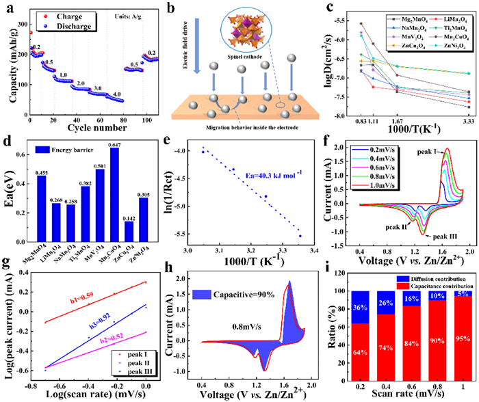
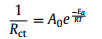
Through ab initio molecular dynamics (AIMD) simulations, we investigated the diffusion behavior of Zn2+ in these 8 spinel materials. The relationship between logD and 1000/T are summarized in Fig. 5c. The calculated diffusion coefficients of Zn2+ in these 8 materials are all greater than 10−9 cm2/s (Table S3 in Supporting information), which indicates that Zn2+ can diffuse rapidly in these electrodes. We can calculate the diffusion barriers of Zn2+ in these eight spinel materials by fitting the Arrhenius formula [53]. As shown in Fig. 5d, their migration energy barriers are all less than 1 eV, which also indicates that Zn2+ can diffuse easily in these electrodes. Moreover, electrochemical impedance spectroscopy (EIS) analysis was performed for Mg2MnO4-based battery. As depicted in Fig. S12 (Supporting information), the Nyquist plot consists of a semicircle in the high-frequency region and a sloping line in the low-frequency region, corresponding to the charge transfer resistance (Rct) and the ion diffusion resistance (Zw), respectively [54]. From the R values at different temperatures (Table S4 in Supporting information), the activation energy of the whole reaction process can also be obtained according to the Arrhenius formula [55,56]:
|
|
(2) |
where A0 is a constant factor, T stands for the temperature (K), R is the gas constant (8.314 J mol−1 K−1), and Ea represents the activation energy, respectively. As shown in Fig. 5e, the Ea value of 40.3 kJ/mol was obtained by linear fitting of the Rct values at different temperatures, which is also in good agreement with the theoretical result (0.455 eV in Fig. 5d, and 1 eV = 96 kJ/mol).
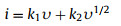
To understand the charge storage process, the electrochemical kinetics was investigated by testing the CV curves at different scan rates in Fig. 5f. Their peak currents i and scan rate υ satisfy the following equation [57,58]:
|
|
(3) |
where a and b are adjustable parameters. When the value of b is close to 0.5, the reaction process depends on the control of the ion diffusion process, while the value of b reaches 1, the corresponding electrochemical behavior is controlled by the capacitance. According to the slopes of the log(i) vs. log(υ) curves for all peaks in Fig. 5g, the b values for peaks Ⅰ, Ⅱ and Ⅲ are 0.59, 0.52 and 0.92, respectively, which implies that the combination of ion diffusion and capacitive behavior synergistically controls the charge storage process. Synergistic effects of diffusion and capacitance are favorable for fast ion diffusion kinetics. In addition, the capacitance contribution can be calculated according to the following equation [42,59,60]:
|
|
(4) |
where i is the current (mA), υ corresponds to the scan rate (mV/s), k1υ corresponds to the current contributed by the capacitor and k2υ1/2 corresponds to the current contributed by diffusion. According to Eq. 4, the capacitance contribution at different sweep speeds can be obtained. The shaded part of Fig. 5h represents the capacitance contribution at 0.8 mV/s. The capacitance contributions at all scan rate are displayed in Fig. 5i. The high capacitance contributions with increasing scan rate indicate fast ion transfer kinetics, corresponding to the good rate performance of the Mg2MnO4 material.
According to the above analysis, the Mg2MnO4 cathode showed excellent electrochemical ability. To further understand the relevant ion storage mechanism, ex situ XRD and XPS were performed on the Mg2MnO4 cathode. The GCD curves of the Mg2MnO4 cathode in different states in the second cycle are shown in Fig. 6a. Combined with the ex situ XRD results (Fig. 6b), it can be seen that the diffraction peak of ZnSO4·6Zn(OH)2·4H2O (JCPDS No. 11–0280) disappears after charging from 0.4 V to 1.6 V, and appears after discharging from 1.9 V to 1.3 V. These results confirm that H+ and Zn2+ are reversibly de-intercalate/intercalate into the Mg2MnO4 electrode [60,61]. It is worth noting that the diffraction peaks at about 18.3°, 35.5°, 43.3°, 62.8°, corresponding to the (111), (311), (400) and (440) crystal planes of Mg2MnO4 respective, did not show significant changes during the discharging/charging process. This means that the Mg2MnO4 cathode has excellent cycle stability. In addition, ex situ XPS of Zn 2p further showed that the insertion of Zn2+ ingress into/egress from the Mg2MnO4 electrode during the discharging/charging process [62]. As shown in Fig. 6c, the peaks at 1045.2 eV and 1022.2 eV correspond to signals of Zn 2p1/2 and Zn 2p3/2, respectively. When discharged to 0.4 V, a strong Zn 2p signal was detected, which weakened in the charged state. As depicted in Fig. 6d, compared with the initial state, the peak corresponding to Mn 2p3/2 in the discharged state shifts to a lower binding energy, indicating that the valence of Mn decreases with the insertion of H+ and Zn2+ into Mg2MnO4 [63]. The increase of the binding energy of Mn 2p3/2 at the charged state indicates the full de-intercalation of H+ and Zn2+. Therefore, the ion intercalation process of the Mg2MnO4 cathode is shown in Fig. 6e, where both H+ and Zn2+ are de-intercalate/intercalate into the Mg2MnO4 electrode. The analysis of ex situ XPS spectra also confirmed the ion storage mechanism of the Mg2MnO4 electrode.
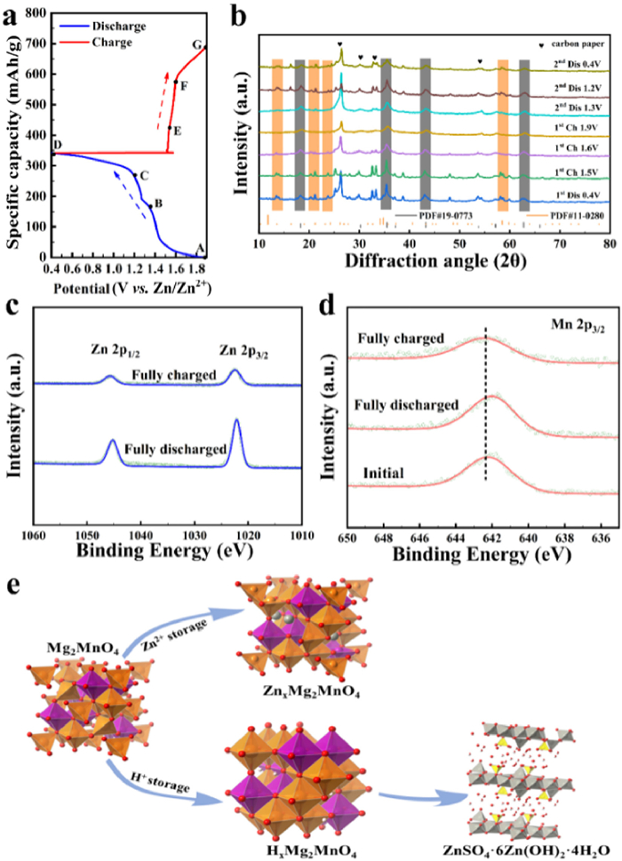
Generally, the electrostatic interaction between ions in the process of Zn2+ insertion and extraction will affect the battery performance. Here, we calculate the Electron Localization Function (ELF) to compare the electrostatic interaction of the cathode material with/without Zn2+ ions. Fig. S13 (Supporting information) shows the electrostatic potential diagrams at 0.6 d on the (001) plane when 0, 2, 4 and 6 Zn ions are inserted. As shown in the figure, when more Zn2+ are in inserted, the variation of electrostatic potential is not obvious, which indicates that the Mg2MnO4 framework has a strong electrostatic screening effect [43,64]. The strong dielectric screening effect of Mg2MnO4 makes the electrostatic interaction between Zn ions very small during Zn storage, which is another reason why Mg2MnO4 shows well performance.
In conclusion, we proposed and demonstrated a high-throughput screening strategy based on first-principles calculations that facilitates the rapid development of high-performance spinel cathode materials for AZIBs. Through high-throughput screening and first-principles calculations, we found that 7 materials (Mg2MnO4, LiMn2O4, NaMn2O4, ZnCu2O4, ZnNi2O4, MnV2O4, Ti2MnO4) exhibited high average open circuit voltage, and 8 materials (Mg2MnO4, LiMn2O4, NaMn2O4, ZnCu2O4, ZnNi2O4, MnV2O4, Ti2MnO4, MnCu2O4) possess high ionic mobility and low volume expansion rate. After taking all the properties into consideration, there are five spinel materials (Mg2MnO4, LiMn2O4, NaMn2O4, ZnCu2O4, ZnNi2O4) that have the potential to be used as cathode materials for high-performance AZIBs. Meanwhile, we experimentally synthesized Mg2MnO4 and applied it to AZIBs cathode. The Mg2MnO4-based battery still has a stable capacity retention of 99% after 300 cycles at a current density of 0.5 A/g, showing good cycling stability, which is consistent with our calculated low volume expansion rate. Both computational and experimental electrochemical kinetics results show that Mg2MnO4 material has high capacitive contribution and a low ion migration activation energy, indicating that the material has fast ion transfer kinetics and thus exhibits good rate performance. The agreement between theoretical calculations and experimental results confirms the effectiveness of our high-throughput material screening strategy, which will significantly reduce the development time and cost of AZIBs cathode materials and make obtaining perfect cathode materials more achievable. We believe that this high-throughput screening strategy based on first-principles calculations should also be applicable to the development of other ionic cathode materials.
The authors declare that they have no known competing financial interests or personal relationships that could have appeared to influence the work reported in this paper.
This work was financially supported by research grants from the Natural Science Foundation of China (Nos. 12004057, 62074022, 52173235), Support plan for Overseas Students to Return to China for Entrepreneurship and Innovation (No. cx2020075), Open Fund of Key Laboratory of Low-grade Energy Utilization Technologies and Systems (No. LLEUTS-2020008), Chongqing Funds for Distinguished Young Scientists (No. cstc2021jcyj-jqX0015), Chongqing Talent Plan (No. CQYC2021059206), Fundamental Research Funds for the Central Universities (No. 2020CDJQY-A055).
Supplementary material associated with this article can be found, in the online version, at doi:
M.F. Ullah, M. Aatif, Cancer Treat. Rev. 35(2009) 193-200.
doi: 10.1016/j.ctrv.2008.10.004
S.K. Arya, S. Bhansali, Chem. Rev. 111(2011) 6783-6809.
doi: 10.1021/cr100420s
M. Gao, F. Yu, C. Lv, J. Choo, L. Chen, Chem. Soc. Rev. 46(2017) 2237-2271.
doi: 10.1039/C6CS00908E
A. Razgulin, N. Ma, J. Rao, Chem. Soc. Rev. 40(2011) 4186-4216.
doi: 10.1039/c1cs15035a
D. Su, L. Gao, F. Gao, X. Zhang, X. Gao, Chem. Sci. 11(2020) 5614-5629.
doi: 10.1039/D0SC01201G
D. Su, X. Chen, Y. Zhang, X. Gao, Trac-Trends Anal. Chem. 133(2020) 116112.
doi: 10.1016/j.trac.2020.116112
Y. Xing, J. Zhao, P.S. Conti, K. Chen, Theranostics 4(2014) 290-306.
doi: 10.7150/thno.7341
K. Heinzmann, L.M. Carter, J.S. Lewis, E.O. Aboagye, Nat. Biomed. Eng. 1(2017) 697-713.
doi: 10.1038/s41551-017-0131-8
H.S. Krishnan, L. Ma, N. Vasdev, S.H. Liang, Chem. Eur. J. 23(2017) 15553-15577.
doi: 10.1002/chem.201701581
J. Pellico, P.J. Gawne, R. T.M. d. R, Chem. Soc. Rev. 50(2021) 3355-3423.
doi: 10.1039/D0CS00384K
S.R. Cherry, T. Jones, J.S. Karp, et al., J. Nucl. Med. 59(2018) 3-12.
doi: 10.2967/jnumed.116.184028
R.D. Badawi, H. Shi, P. Hu, et al., J. Nucl. Med. 60(2019) 299-303.
doi: 10.2967/jnumed.119.226498
K.V. Vimalnath, A. Rajeswari, K.C. Jagadeesan, et al., J. Radioanal. Nucl. Chem. 294(2012) 43-47.
doi: 10.1007/s10967-011-1548-2
R. Chakravarty, S. Chakraborty, A. Dash, Mol. Pharmaceutics 13(2016) 3601-3612.
doi: 10.1021/acs.molpharmaceut.6b00582
L.A. Bass, M. Wang, M.J. Welch, C.J. Anderson, Bioconjugate Chem. 11(2000) 527-532.
doi: 10.1021/bc990167l
T.J. Wadas, E.H. Wong, G.R. Weisman, C.J. Anderson, Curr. Pharm. Des. 13(2007) 3-16.
doi: 10.2174/138161207779313768
P. Szymanski, T. Fraczek, M. Markowicz, E. Mikiciuk-Olasik, BioMetals 25(2012) 1089-1112.
doi: 10.1007/s10534-012-9578-y
M.T. Ma, P.S. Donnelly, Curr. Top. Med. Chem. 11(2011) 500-520.
doi: 10.2174/156802611794785172
S. Li, C.G. England, E.B. Ehlerding, Am. J. Transl. Res. 11(2019) 6007-6015.
D.W. Bartlett, H. Su, I.J. Hildebrandt, W.A. Weber, M.E. Davis, Proc. Natl. Acad. Sci. U. S. A. 104(2007) 15549-15554.
doi: 10.1073/pnas.0707461104
K. Chen, X. Sun, G. Niu, et al., Mol. Imaging Biol. 14(2012) 96-105.
doi: 10.1007/s11307-011-0479-1
A. Krishnan, V. Adhikarla, E.K. Poku, et al., Blood Adv. 4(2020) 5194-5202.
doi: 10.1182/bloodadvances.2020002603
Y. Zhang, C. Zhang, C. Xu, et al., Talanta 200(2019) 432-442.
doi: 10.1016/j.talanta.2019.03.068
J. Ge, Q. Zhang, J. Zeng, Z. Gu, M. Gao, Biomaterials 228(2020) 119553.
doi: 10.1016/j.biomaterials.2019.119553
N. Khlebtsov, L. Dykman, Chem. Soc. Rev. 40(2011) 1647-1671.
doi: 10.1039/C0CS00018C
E. Porret, X. Le Guevel, J.L. Coll, J. Mater. Chem. B 8(2020) 2216-2232.
doi: 10.1039/C9TB02767J
J.E. Mortimer, J.R. Bading, J.M. Park, et al., J. Nucl. Med. 59(2018) 38-43.
doi: 10.2967/jnumed.117.193888
S.K. Woo, S.J. Jang, M.J. Seo, et al., J. Nucl. Med. 60(2019) 26-33.
doi: 10.2967/jnumed.118.210294
A.C. Sedgwick, J.T. Brewster, P. Harvey, et al., Chem. Soc. Rev. 49(2020) 2886-2915.
doi: 10.1039/C8CS00986D
Q. Fan, K. Cheng, X. Hu, et al., J. Am. Chem. Soc. 136(2014) 15185-15194.
doi: 10.1021/ja505412p
R. Zhang, Q. Fan, M. Yang, et al., Adv. Mater. 27(2015) 5063-5069.
doi: 10.1002/adma.201502201
T.M. Shaffer, S. Harmsen, E. Khwaja, et al., Nano Lett. 16(2016) 5601-5604.
doi: 10.1021/acs.nanolett.6b02161
S. Shen, D. Jiang, L. Cheng, et al., ACS Nano 11(2017) 9103-9111.
doi: 10.1021/acsnano.7b03857
C. Perez-Medina, A.J.P. Teunissen, E. Kluza, W.J.M. Mulder, R. van der Meel, Adv. Drug Delivery Rev. 154(2020) 123-141.
Y. Zhao, D. Sultan, L. Detering, et al., Angew. Chem. Int. Ed. 53(2014) 156-159.
doi: 10.1002/anie.201308494
Y. Tao, M. Li, J. Ren, X. Qu, Chem. Soc. Rev. 44(2015) 8636-8663.
doi: 10.1039/C5CS00607D
X. Sun, W. Cai, X. Chen, Acc. Chem. Res. 48(2015) 286-294.
doi: 10.1021/ar500362y
F. Gao, P. Cai, W. Yang, et al., ACS Nano 9(2015) 4976-4986.
doi: 10.1021/nn507130k
X. Sun, X. Huang, X. Yan, et al., ACS Nano 8(2014) 8438-8446.
doi: 10.1021/nn502950t
M. Yang, D. Huo, K.D. Gilroy, et al., ChemNanoMat 3(2017) 44-50.
doi: 10.1002/cnma.201600281
A.F. Frellsen, A.E. Hansen, R.I. Jolck, et al., ACS Nano 10(2016) 9887-9898.
doi: 10.1021/acsnano.6b03144
Y. Zhao, D. Sultan, L. Detering, H. Luehmann, Y. Liu, Nanoscale 6(2014) 13501-13509.
doi: 10.1039/C4NR04569F
A. Niccoli Asabella, G.L. Cascini, C. Altini, et al., BioMed Res. Int. (2014) 7864632014.
M. Shokeen, C.J. Anderson, Acc. Chem. Res. 42(2009) 832-841.
doi: 10.1021/ar800255q
G.S. Heo, Y. Zhao, D. Sultan, et al., ACS Appl. Mater. Interfaces 11(2019) 19669-19678.
doi: 10.1021/acsami.8b22752
X. Zhang, D. Ye, L. Yang, et al., ACS Appl. Nano Mater. 3(2020) 11129-11134.
doi: 10.1021/acsanm.0c02297
M. Zhou, R. Zhang, M. Huang, et al., J. Am. Chem. Soc. 132(2010) 15351-15358.
doi: 10.1021/ja106855m
M. Zhou, J. Li, S. Liang, et al., ACS Nano 9(2015) 7085-7096.
doi: 10.1021/acsnano.5b02635
Z. Wang, P. Huang, O. Jacobson, et al., ACS Nano 10(2016) 3453-3460.
doi: 10.1021/acsnano.5b07521
X. Zhang, L. Detering, D. Sultan, et al., ACS Nano 15(2021) 1186-1198.
doi: 10.1021/acsnano.0c08185
D. Ye, D. Sultan, X. Zhang, et al., J. Control. Release 283(2018) 143-150.
doi: 10.1016/j.jconrel.2018.05.039
D. Ye, X. Zhang, Y. Yue, et al., J. Control. Release 286(2018) 145-153.
doi: 10.1016/j.jconrel.2018.07.020
Y. Zhao, L. Detering, D. Sultan, et al., ACS Nano 10(2016) 5959-5970.
doi: 10.1021/acsnano.6b01326
D. Sultan, W. Li, L. Detering, et al., Nanomed. Nanotechnol. Biol. Med. 36(2021) 102416.
doi: 10.1016/j.nano.2021.102416
H. Hu, P. Huang, O.J. Weiss, et al., Biomaterials 35(2014) 9868-9876.
doi: 10.1016/j.biomaterials.2014.08.038
B. Pang, Y. Zhao, H. Luehmann, et al., ACS Nano 10(2016) 3121-3131.
doi: 10.1021/acsnano.5b07968
R. Madru, M. Budassi, H. Benveniste, et al., Cancer Biother. Radiopharm. 33(2018) 213-220.
doi: 10.1089/cbr.2017.2412
Y. Wang, Y. Liu, H. Luehmann, et al., ACS Nano 6(2012) 5880-5888.
doi: 10.1021/nn300464r
Y. Zhao, B. Pang, L. Detering, et al., Mol. Imaging 17(2018) 1536012118775827.
H. Xie, B. Goins, A. Bao, Z. Wang, W.T. Phillips, Int. J. Nanomed. 7(2012) 2227-2238.
doi: 10.2147/IJN.S30699
M. Nahrendorf, H. Zhang, S. Hembrador, et al., Circulation 117(2008) 379-387.
doi: 10.1161/CIRCULATIONAHA.107.741181
J. Xie, K. Chen, J. Huang, et al., Biomaterials 31(2010) 3016-3022.
doi: 10.1016/j.biomaterials.2010.01.010
H.Y. Lee, Z. Li, K. Chen, et al., J. Nucl. Med. 49(2008) 1371-1379.
doi: 10.2967/jnumed.108.051243
R. Torres Martin de Rosales, R. Tavare, R.L. Paul, et al., Angew. Chem. Int. Ed. 50(2011) 5509-5513.
doi: 10.1002/anie.201007894
X. Yang, H. Hong, J.J. Grailer, et al., Biomaterials 32(2011) 4151-4160.
doi: 10.1016/j.biomaterials.2011.02.006
Z. Liu, W. Cai, L. He, et al., Nat. Nanotechnol. 2(2007) 47-52.
doi: 10.1038/nnano.2006.170
S. Shi, C. Xu, K. Yang, et al., Angew. Chem. Int. Ed. 56(2017) 2889-2892.
doi: 10.1002/anie.201610649
P. Cai, D. Su, W. Yang, et al., ACS Appl. Bio Mater. 3(2019) 611-621.
L. Cheng, A. Kamkaew, H. Sun, et al., ACS Nano 10(2016) 7721-7730.
doi: 10.1021/acsnano.6b03074
L. Feng, L. Cheng, Z. Dong, et al., ACS Nano 11(2017) 927-937.
doi: 10.1021/acsnano.6b07525
B. Yu, H. Wei, Q. He, et al., Angew. Chem. Int. Ed. 57(2018) 218-222.
doi: 10.1002/anie.201710232
D. Luo, S. Goel, H.J. Liu, et al., ACS Nano 11(2017) 12482-12491.
doi: 10.1021/acsnano.7b06578
F. Zhang, Q. Ni, O. Jacobson, et al., Angew. Chem. Int. Ed. 57(2018) 7066-7070.
doi: 10.1002/anie.201801984
M.A. Rajora, L. Ding, M. Valic, et al., Chem. Sci. 8(2017) 5371-5384.
doi: 10.1039/C7SC00732A
Z. He, H. Jia, M. Zheng, et al., ACS Appl. Bio Mater. 4(2021) 5707-5716.
J.L.J. Dearling, A.B. Packard, J. Control. Release 261(2017) 23-30.
doi: 10.1016/j.jconrel.2017.06.011
W.F. Lai, W.T. Wong, A.L. Rogach, Adv. Mater. 32(2020) 1906872.
doi: 10.1002/adma.201906872
P.S. Patrick, L.K. Bogart, T.J. Macdonald, et al., Chem. Sci. 10(2019) 2592-2597.
doi: 10.1039/C8SC04895A
K. Yang, L. Zhu, L. Nie, et al., Theranostics 4(2014) 134-141.
doi: 10.7150/thno.7217
Z. Zha, S. Wang, S. Zhang, et al., Nanoscale 5(2013) 3216-3219.
doi: 10.1039/c3nr00541k
Z. Xiao, Nanomedicine 9(2014) 373-375.
A. Riedinger, T. Avellini, A. Curcio, et al., J. Am. Chem. Soc. 137(2015) 15145-15151.
doi: 10.1021/jacs.5b07973
P.K. Jain, I.H. El-Sayed, M.A. El-Sayed, Nano Today 2(2007) 18-29.
doi: 10.1016/s1748-0132(07)70016-6
J. Xie, L. Gong, S. Zhu, et al., Adv. Mater. 31(2019) 1802244.
doi: 10.1002/adma.201802244
Y. Zhang, X. Chen, Q. Yuan, et al., Chem. Sci. 12(2021) 14855-14862.
doi: 10.1039/D1SC04825B
X. Chen, Y. Zhang, Q. Yuan, et al., J. Mater. Chem. B 9(2021) 6614-6622.
doi: 10.1039/D1TB00836F
M. Nahrendorf, E. Keliher, B. Marinelli, et al., Proc. Natl. Acad. Sci. U. S. A. 107(2010) 7910-7915.
doi: 10.1073/pnas.0915163107
Y. Wang, C. Xu, Y. Chang, et al., ACS Appl. Mater. Interfaces 9(2017) 28959-28966.
doi: 10.1021/acsami.7b10030
R.M. Wong, D.A. Gilbert, K. Liu, A.Y. Louie, ACS Nano 6(2012) 3461-3467.
doi: 10.1021/nn300494k
Y. Hu, S. Huang, X. Zhao, L. Chang, Z. Chen, Chem. Eng. J. 421(2021) 129744.
doi: 10.1016/j.cej.2021.129744
C.M. Cobley, L. Au, J. Chen, Y. Xia, Expert Opin, Drug Deliv. 7(2010) 577-587.
doi: 10.1517/17425240903571614
R.A. Davis, D.A. Rippner, S.H. Hausner, et al., Environ. Sci. Technol. 51(2017) 12537-12546.
doi: 10.1021/acs.est.7b03333
H. Hong, T. Gao, W. Cai, Nano Today 4(2009) 252-261.
doi: 10.1016/j.nantod.2009.04.002
Z. Liu, S. Tabakman, K. Welsher, H. Dai, Nano Res. 2(2009) 85-120.
doi: 10.1007/s12274-009-9009-8
S. Beg, M. Rizwan, A.M. Sheikh, et al., J. Pharm. Pharmacol. 63(2011) 141-163.
doi: 10.1111/j.2042-7158.2010.01167.x
H. Wang, S.T. Yang, A. Cao, Y. Liu, Acc. Chem. Res. 46(2013) 750-760.
doi: 10.1021/ar200335j
H. Hong, K. Yang, Y. Zhang, et al., ACS Nano 6(2012) 2361-2370.
doi: 10.1021/nn204625e
X. Huang, F. Zhang, L. Zhu, et al., ACS Nano 7(2013) 5684-5693.
doi: 10.1021/nn401911k
N. Licciardello, S. Hunoldt, R. Bergmann, et al., Nanoscale 10(2018) 9880-9891.
doi: 10.1039/C8NR01063C
Y. Peng, D. Yang, W. Lu, et al., Acta Biomater 61(2017) 193-203.
doi: 10.1016/j.actbio.2017.08.011
G. Biagiotti, F. Pisaneschi, S.T. Gammon, F, et al., J. Mater. Chem. B 7(2019) 2678-2687.
doi: 10.1039/C8TB03299H
B.T. Cisneros, J.J. Law, M.L. Matson, et al., Nanomedicine 9(2014) 2499-2509.
doi: 10.2217/nnm.14.26
J. Xu, S. Yu, X. Wang, et al., ACS Nano 13(2019) 10242-10260.
doi: 10.1021/acsnano.9b03466
L. Huang, S. Asghar, T. Zhu, et al., Expert Opin. Drug Delivery 18(2021) 1473-1500.
doi: 10.1080/17425247.2021.1950685
S. Kumar, D. Jiang, B. Sun, et al., Adv. NanoBiomed Res. 1(2021) 2000013.
doi: 10.1002/anbr.202000013
B. Liu, G. Qiao, Y. Han, et al., Acta Biomater 117(2020) 361-373.
doi: 10.1016/j.actbio.2020.09.040
W. Fan, B. Yung, P. Huang, X. Chen, Chem. Rev. 117(2017) 13566-13638.
doi: 10.1021/acs.chemrev.7b00258
W. Yu, J. Huang, M. Lin, et al., Anal. Chem. 93(2021) 4647-4656.
doi: 10.1021/acs.analchem.1c00223
G. Yang, S.Z.F. Phua, W.Q. Lim, et al., Adv. Mater. 31(2019) 1901513.
doi: 10.1002/adma.201901513
E. Chang, J. Bu, L. Ding, et al., J. Nanobiotechnol. 19(2021) 154.
doi: 10.1186/s12951-021-00898-1
T.W. Liu, T.D. MacDonald, J. Shi, B.C. Wilson, G. Zheng, Angew. Chem. Int. Ed. 51(2012) 13128-13131.
doi: 10.1002/anie.201206939
J.W. Seo, H. Zhang, D.L. Kukis, C.F. Meares, K.W. Ferrara, Bioconjugate Chem. 19(2008) 2577-2584.
doi: 10.1021/bc8002937
E.E. Paoli, D.E. Kruse, J.W. Seo, et al., J. Control. Release 143(2010) 13-22.
doi: 10.1016/j.jconrel.2009.12.010
C.B. Rygh, S. Qin, J.W. Seo, et al., Clin. Cancer Res. 17(2011) 550-559.
doi: 10.1158/1078-0432.CCR-10-2049
C.M. Kang, H.J. Koo, S. Lee, et al., Nucl. Med. Biol. 40(2013) 1018-1024.
doi: 10.1016/j.nucmedbio.2013.08.003
L.M. Mahakian, D.G. Farwell, H. Zhang, et al., Mol. Imaging Biol. 16(2014) 284-292.
doi: 10.1007/s11307-013-0676-1
S.G. Lee, K. Gangangari, T.M. Kalidindi, et al., Nucl. Med. Biol. 43(2016) 781-787.
doi: 10.1016/j.nucmedbio.2016.08.011
Y. Du, X. Liang, Y. Li, et al., Mol. Pharmaceutics 14(2017) 3978-3986.
doi: 10.1021/acs.molpharmaceut.7b00649
A.I. Jensen, G.W. Severin, A.E. Hansen, et al., J. Control. Release 269(2018) 100-109.
doi: 10.1016/j.jconrel.2017.11.006
A.W. Wong, E. Ormsby, H. Zhang, et al., Am. J. Nucl. Med. Mol. Imaging 3(2013) 32-43.
J.W. Seo, L.M. Mahakian, A. Kheirolomoom, et al., Bioconjugate Chem. 21(2010) 1206-1215.
doi: 10.1021/bc100018n
J.W. Seo, S. Qin, L.M. Mahakian, et al., J. Control. Release 151(2011) 28-34.
doi: 10.1016/j.jconrel.2011.01.008
M. Jaymand, Y. Davatgaran Taghipour, A. Rezaei, et al., Coord. Chem. Rev. 440(2021) 213974.
doi: 10.1016/j.ccr.2021.213974
Y. Liu, M.J. Welch, Bioconjugate Chem. 23(2012) 671-682.
doi: 10.1021/bc200264c
A.E. Hansen, A.L. Petersen, J.R. Henriksen, et al., ACS Nano 9(2015) 6985-6995.
doi: 10.1021/acsnano.5b01324
A.L. Petersen, T. Binderup, P. Rasmussen, et al., Biomaterials 32(2011) 2334-2341.
doi: 10.1016/j.biomaterials.2010.11.059
L.W. Locke, M.W. Mayo, A.D. Yoo, M.B. Williams, S.S. Berr, Biomaterials 33(2012) 7785-7793.
doi: 10.1016/j.biomaterials.2012.07.022
J.R. Henriksen, A.L. Petersen, A.E. Hansen, et al., ACS Appl. Mater. Interfaces 7(2015) 22796-22806.
doi: 10.1021/acsami.5b04612
B. Borresen, J.R. Henriksen, G. Clergeaud, et al., ACS Nano 12(2018) 11386-11398.
doi: 10.1021/acsnano.8b06266
H. Lee, J. Zheng, D. Gaddy, et al., Nanomedicine 11(2015) 155-165.
doi: 10.1016/j.nano.2014.08.011
S.J. Blocker, K.A. Douglas, L.A. Polin, et al., Theranostics 7(2017) 4229-4239.
doi: 10.7150/thno.21688
H. Lee, A.F. Shields, B.A. Siegel, et al., Clin. Cancer Res. 23(2017) 4190-4202.
doi: 10.1158/1078-0432.CCR-16-3193
H. Lee, D. Gaddy, M. Ventura, et al., Theranostics 8(2018) 2300-2312.
doi: 10.7150/thno.21670
S. Edmonds, A. Volpe, H. Shmeeda, et al., ACS Nano 10(2016) 10294-10307.
doi: 10.1021/acsnano.6b05935
M.D. Majmudar, J. Yoo, E.J. Keliher, et al., Circ. Res. 112(2013) 755-761.
doi: 10.1161/CIRCRESAHA.111.300576
M.L. Senders, A.E. Meerwaldt, M.M.T. van Leent, et al., Nat. Nanotechnol. 15(2020) 398-405.
doi: 10.1038/s41565-020-0642-4
J. Llop, P. Jiang, M. Marradi, et al., J. Mater. Chem. B 3(2015) 6293-6300.
doi: 10.1039/C5TB01157D
M. Lameijer, T. Binderup, M.M.T. van Leent, et al., Nat. Biomed. Eng. 2(2018) 279-292.
doi: 10.1038/s41551-018-0221-2
J.L.J. Dearling, A.B. Packard, J. Control. Release 261(2017) 23-30.
doi: 10.1016/j.jconrel.2017.06.011
D.L. Bailey, K.P. Willowson, Eur. J. Nucl. Med. Mol. Imaging 41(2014) S17-S25.
doi: 10.1007/s00259-013-2542-4
A. Hosny, C. Parmar, J. Quackenbush, L.H. Schwartz, H.J.W.L. Aerts, Nat. Rev. Cancer 18(2018) 500-510.
doi: 10.1038/s41568-018-0016-5
G. Wang, J.C. Ye, K. Mueller, J.A. Fessler, IEEE Trans. Med. Imaging 37(2018) 1289-1296.
doi: 10.1109/TMI.2018.2833635
P. Zou, H. Chen, H.J. Paholak, D. Sun, Mol. Pharmaceutics 10(2013) 4185-4194.
doi: 10.1021/mp4002393
L. Wang, L. Yan, J. Liu, C. Chen, Y. Zhao, Anal. Chem. 90(2018) 589-614.
doi: 10.1021/acs.analchem.7b04765
J. Guo, K. Feng, W. Wu, et al., Angew. Chem. Int. Ed. 60(2021) 21884-21889.
doi: 10.1002/anie.202107231

Jing-Jing Zhang , Lujun Lou , Rui Lv , Jiahui Chen , Yinlong Li , Guangwei Wu , Lingchao Cai , Steven H. Liang , Zhen Chen . Recent advances in photochemistry for positron emission tomography imaging. Chinese Chemical Letters, 2024, 35(8): 109342-. doi: 10.1016/j.cclet.2023.109342
Yiling Li , Zekun Gao , Xiuxiu Yue , Minhuan Lan , Xiuli Zheng , Benhua Wang , Shuang Zhao , Xiangzhi Song . FRET-based two-photon benzo[a] phenothiazinium photosensitizer for fluorescence imaging-guided photodynamic therapy. Chinese Chemical Letters, 2024, 35(7): 109133-. doi: 10.1016/j.cclet.2023.109133
Leichen Wang , Anqing Mei , Na Li , Xiaohong Ruan , Xu Sun , Yu Cai , Jinjun Shao , Xiaochen Dong . Aza-BODIPY dye with unexpected bromination and high singlet oxygen quantum yield for photoacoustic imaging-guided synergetic photodynamic/photothermal therapy. Chinese Chemical Letters, 2024, 35(6): 108974-. doi: 10.1016/j.cclet.2023.108974
Xuejian Xing , Pan Zhu , E Pang , Shaojing Zhao , Yu Tang , Zheyu Hu , Quchang Ouyang , Minhuan Lan . D-A-D-structured boron-dipyrromethene with aggregation-induced enhanced phototherapeutic efficiency for near-infrared fluorescent and photoacoustic imaging-guided synergistic photodynamic and photothermal cancer therapy. Chinese Chemical Letters, 2024, 35(10): 109452-. doi: 10.1016/j.cclet.2023.109452
Xiaofang Luo , Ye Wu , Xiaokun Zhang , Min Tang , Feiye Ju , Zuodong Qin , Gregory J Duns , Wei-Dong Zhang , Jiang-Jiang Qin , Xin Luan . Peptide-based strategies for overcoming multidrug-resistance in cancer therapy. Chinese Chemical Letters, 2025, 36(1): 109724-. doi: 10.1016/j.cclet.2024.109724
Tao LIU , Yuting TIAN , Ke GAO , Xuwei HAN , Ru'nan MIN , Wenjing ZHAO , Xueyi SUN , Caixia YIN . A photothermal agent with high photothermal conversion efficiency and high stability for tumor therapy. Chinese Journal of Inorganic Chemistry, 2024, 40(8): 1622-1632. doi: 10.11862/CJIC.20240107
Biao Huang , Tao Tang , Fushou Liu , Shi-Hui Chen , Zhi-Ling Zhang , Mingxi Zhang , Ran Cui . Quantum dots boost large-view NIR-Ⅱ imaging with high fidelity for fluorescence-guided tumor surgery. Chinese Chemical Letters, 2024, 35(12): 109694-. doi: 10.1016/j.cclet.2024.109694
Boran Cheng , Lei Cao , Chen Li , Fang-Yi Huo , Qian-Fang Meng , Ganglin Tong , Xuan Wu , Lin-Lin Bu , Lang Rao , Shubin Wang . Fluorine-doped carbon quantum dots with deep-red emission for hypochlorite determination and cancer cell imaging. Chinese Chemical Letters, 2024, 35(6): 108969-. doi: 10.1016/j.cclet.2023.108969
Gongcheng Ma , Qihang Ding , Yuding Zhang , Yue Wang , Jingjing Xiang , Mingle Li , Qi Zhao , Saipeng Huang , Ping Gong , Jong Seung Kim . Palladium-free chemoselective probe for in vivo fluorescence imaging of carbon monoxide. Chinese Chemical Letters, 2024, 35(9): 109293-. doi: 10.1016/j.cclet.2023.109293
Makhloufi Zoulikha , Zhongjian Chen , Jun Wu , Wei He . Approved delivery strategies for biopharmaceuticals. Chinese Chemical Letters, 2025, 36(2): 110225-. doi: 10.1016/j.cclet.2024.110225
Pengfei Li , Chulin Qu , Fan Wu , Hu Gao , Chengyan Zhao , Yue Zhao , Zhen Shen . Robust free-base and metalated corrole radicals with reduction-induced emission. Chinese Chemical Letters, 2025, 36(2): 110292-. doi: 10.1016/j.cclet.2024.110292
Ling-Ling Wu , Xiangchuan Meng , Qingyang Zhang , Xiaowan Han , Feiya Yang , Qinghua Wang , Hai-Yu Hu , Nianzeng Xing . Heavy-atom engineered hypoxia-responsive probes for precisive photoacoustic imaging and cancer therapy. Chinese Chemical Letters, 2024, 35(4): 108663-. doi: 10.1016/j.cclet.2023.108663
Jinyu Guo , Yandai Lin , Shaohua He , Yueqing Chen , Fenglu Li , Renjie Ruan , Gaoxing Pan , Hexin Nan , Jibin Song , Jin Zhang . Utilizing dual-responsive iridium(Ⅲ) complex for hepatocellular carcinoma: Integrating photoacoustic imaging with chemotherapy and photodynamic therapy. Chinese Chemical Letters, 2024, 35(9): 109537-. doi: 10.1016/j.cclet.2024.109537
Du Liu , Yuyan Li , Hankun Zhang , Benhua Wang , Chaoyi Yao , Minhuan Lan , Zhanhong Yang , Xiangzhi Song . Three-in-one erlotinib-modified NIR photosensitizer for fluorescence imaging and synergistic chemo-photodynamic therapy. Chinese Chemical Letters, 2025, 36(2): 109910-. doi: 10.1016/j.cclet.2024.109910
Baoli Yin , Xinlin Liu , Zhe Li , Zhifei Ye , Youjuan Wang , Xia Yin , Sulai Liu , Guosheng Song , Shuangyan Huan , Xiao-Bing Zhang . Ratiometric NIR-Ⅱ fluorescent organic nanoprobe for imaging and monitoring tumor-activated photodynamic therapy. Chinese Chemical Letters, 2025, 36(5): 110119-. doi: 10.1016/j.cclet.2024.110119
Zhaomin Tang , Qian He , Jianren Zhou , Shuang Yan , Li Jiang , Yudong Wang , Chenxing Yao , Huangzhao Wei , Keda Yang , Jiajia Wang . Active-transporting of charge-reversal Cu(Ⅱ)-doped mesoporous silica nanoagents for antitumor chemo/chemodynamic therapy. Chinese Chemical Letters, 2024, 35(7): 109742-. doi: 10.1016/j.cclet.2024.109742
Xiaoyi Meng , Xinyue Sun , Zhaogang Sun , Yue Cheng , Yong Wang , Jun Ye , Yin Xiao , Hongqian Chu . Supramolecular-orchestrated carrier-free chemodynamic synergists with augmented oxidative damage for potentiated cancer therapy. Chinese Chemical Letters, 2025, 36(5): 110765-. doi: 10.1016/j.cclet.2024.110765
Qinyu Zhao , Yunchao Zhao , Songjing Zhong , Zhaoyang Yue , Zhuoheng Jiang , Shaobo Wang , Quanhong Hu , Shuncheng Yao , Kaikai Wen , Linlin Li . Urchin-like piezoelectric ZnSnO3/Cu3P p-n heterojunction for enhanced cancer sonodynamic therapy. Chinese Chemical Letters, 2024, 35(12): 109644-. doi: 10.1016/j.cclet.2024.109644
Shuo Zhang , Haitao Liao , Zhi-Qun Liu , Chong Yan , Jia-Qi Huang . Re-evaluating the nano-sized inorganic protective layer on Cu current collector for anode free lithium metal batteries. Chinese Chemical Letters, 2024, 35(7): 109284-. doi: 10.1016/j.cclet.2023.109284
Ze Wang , Hao Liang , Annan Liu , Xingchen Li , Lin Guan , Lei Li , Liang He , Andrew K. Whittaker , Bai Yang , Quan Lin . Strength through unity: Alkaline phosphatase-responsive AIEgen nanoprobe for aggregation-enhanced multi-mode imaging and photothermal therapy of metastatic prostate cancer. Chinese Chemical Letters, 2025, 36(2): 109765-. doi: 10.1016/j.cclet.2024.109765
 DownLoad:

 DownLoad: