State Key Laboratory of Bioelectronics, National Demonstration Centre for Experimental Biomedical Engineering Education, School of Biological Science and Medical Engineering, Southeast University, Nanjing 210096, China
b.
Southeast University Jiangbei New Area Innovation Institute, Nanjing 210096, China
c.
Nanjing Jinling Hosp, Dept Orthoped, Nanjing 210002, China
d.
School of Chemical Engineering, Nanjing University of Science and Technology, Nanjing 210094, China
* Corresponding authors. E-mail addresses: tingwang@seu.edu.cn (T. Wang), yuant1205@163.com (T. Yuan). 1 These authors contributed equally to this work
Received Date:
26 July 2021 Revised Date:
13 November 2021 Accepted Date:
15 November 2021 Available Online:
19 November 2021
In clinical settings the wound-dressing was required easy to use and can match the wound area immediately, at the same time they need to have the properties of hemostats, anti-inflammation and promoting wound healing. To get an ideal wound dressing, we developed a type of gel-like wound adhesive patch from spraying double-network hydrogel, which own the properties of self-antibacterial and can promote wound healing. By spraying, the gel-like wound adhesive patch can match the wound area immediately and form a gel-film in 10 s. Sodium carboxymethyl cellulose as pH sensitive materials accelerated the speed to form the gel-film and enhanced ductility of the wound adhesive patch. In vitro experiments show that, this gel-like wound adhesive patch can promote cell proliferation and reduce cell apoptosis. In vivo studies show that, compared with commercialized wound dressings in clinic using, the spraying gel-like wound adhesive patch from our work has a better effect on wound healing. In conclusion, the spraying gel-like wound patch in our work is easy to use and can form a gel-film match on wound area in a short time, also it has the properties of hemostats, anti-inflammation and promoting wound healing. Its feasibility for mass production shows a good potential for commercial use.
Adenosine triphosphate (ATP), known as "the energy currency of the cell", plays an important role in living cells, including protein synthesis, intracellular signal transduction and cell division [1,2]. The abnormal levels of intracellular ATP have been proved to be closely associated with a variety of diseases, such as cancer and Parkinson [3]. On the other hand, as a signal molecule, hydrogen sulfide (H2S) widely exists in heart, brain, liver and other major organs, and plays an irreplaceable role in physiological functions including neural regulation, apoptosis, insulin signal inhibition and blood pressure [4]. The normal concentration of H2S in cells is about 0.01–3 µmol/L [5]. Abnormal H2S level can lead to the disfunction of cell, which is related to many diseases, like cirrhosis and Alzheimer's disease [6]. Therefore, the detection of ATP and H2S level has important physiological and pathological significance.
Recently, fluorescent sensors of ATP and H2S within cellular environments are well developed for the fast response, excellent selectivity, high sensitivity and simple operation of the fluorescent technique [7]. The ATP sensors are mainly on the basis of the different host-guest interaction mechanism, for instance, metal ion complexation, hydrogen bonding, π-π interaction and the electrostatic interaction [8–12], while the recognition of H2S is based on the different types of chemical reaction, including copper sulfide precipitation, nucleophilic addition and reductive reaction [13,14]. Nevertheless, the traditional sensors are mainly limited to detect single species, fluorescent sensors for the simultaneous recognition of ATP and H2S are still rare.
Currently, fluorescent sensors for simultaneous detection dual or multiple chemical species in biology have attracted much attention [15–17]. Some multifunctional fluorescent sensors for ATP and another analyte, including H2O2, H2S, ONOO− and nitroreductase (NTR), were also documented [18–23]. Typically, the multi-analyte chemosensors were elegantly prepared by covalently linked multiple analyte recognition sites, including complexation and chemical reaction, with one or two flurophores in a single molecule. This strategy requires cumbersome chemical synthesis methods and is time-consuming and labor-intensive.
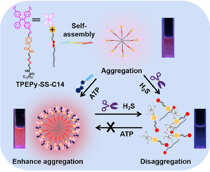
Aggregation-induced emission (AIE) fluorogens, also named as AIEgens, which have attracted much attention for their unique fluorescence emission properties [24]. The AIEgens show non- or weak emission in dilution solutions, but emit strongly at the aggregation state. Previously, we have reported imidazolium-functionalized tetraphenylethylenes, which have a good fluorescence turn-on sensing toward ATP for the complexation enhanced aggregation [25]. We envisioned that if the second recognition site such as the disulfide bond is rationally incorporated into this system, a dual functional probe which can detect ATP and another analyte will be obtained. In this context, herein, we design and synthesize a novel AIE bifunctional probe TPEPy-SS-C14 (Scheme 1), which can simultaneously detect ATP and H2S based on the aggregation/disaggregation mechanism. The probe TPEPy-SS-C14 is rationally designed as following: (1) The TPEPy unit makes probe a red AIE-based emission, (2) the pyridinium and amide groups are used as the ATP binding site via the electrostatic interactions and hydrogen bonding, (3) the disulfide bond is cleavable by H2S, and (4) the long alkyl chain endows probe a good amphiphilic property in aqueous solution. In addition, the probe locates mitochondria, and can detect ATP and hydrogen sulfide levels in living cells.
Scheme 1
Scheme 1.
Chemical structure of TPEPy-SS-C14 and its proposed response mechanism to ATP and H2S.
The detailed synthesis route of TPEPy-SS-C14 is shown in Scheme S1 (Supporting information). And the chemical structure of TPEPy-SS-C14 was verified by high-resolution mass spectrometry (HRMS), 1H nuclear magnetic resonance (NMR) and 13C NMR spectra (Figs. S1-S5 in Supporting information).
Firstly, the ultraviolet–visible (UV–vis) absorption and fluorescence spectra of TPEPy-SS-C14 (10 µmol/L) was investigated in aqueous solution. As shown in Fig. 1a, TPEPy-SS-C14 exhibits a red emission with the maximum peak at 630 nm and a low-energy absorption peak at 428 nm. The Stokes shift is 202 nm, which can prevent the interference effectively caused by self-absorption in the excitation process of biological imaging.
Figure 1
Figure 1.
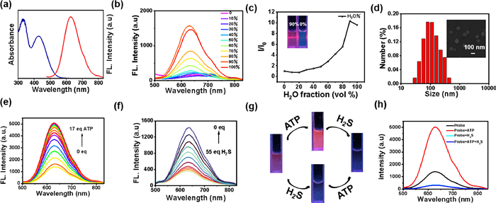
(a) UV–vis absorption and emission spectra of TPEPy-SS-C14 (10 µmol/L) in HEPES (10 mmol/L, pH 7.4) buffer solution. (b) The fluorescence spectra of TPEPy-SS-C14 (10 µmol/L) in water/dimethyl sulfoxide (DMSO) mixtures with different H2O fractions. (c) The plot of the relative emission intensity (I/I0) of TPEPy-SS-C14versus the H2O fractions in H2O/DMSO mixtures. Inset: Tyndall experiment of TPEPy-SS-C14 (10 µmol/L) in the DMSO solution and in H2O/DMSO mixtures with 90% H2O fractions under 365 nm UV irradiation. (d) Size distribution of TPEPy-SS-C14 (10 µmol/L). Inset: SEM image of TPEPy-SS-C14 (10 µmol/L). The fluorescence emission spectrum of TPEPy-SS-C14 (10 µmol/L) after adding different amounts of ATP (e) and H2S (f) in HEPES (10 mmol/L, pH 7.4) buffer solution. (g) Photographs of TPEPy-SS-C14 (10 µmol/L) for ATP (0.17 mmol/L) and H2S (55 mmol/L) under 365 nm UV irradiation. (h) The fluorescence spectra of TPEPy-SS-C14 (10 µmol/L) after addition of ATP (0.17 mmol/L) or H2S (55 mmol/L). The excitation wavelength is 420 nm.
Next, the AIE properties of TPEPy-SS-C14 were studied by emission spectra in water/DMSO mixture (Figs. 1b and c). In DMSO, the soluble TPEPy-SS-C14 molecule shows very weak emission at 630 nm. The fluorescence intensity of TPEPy-SS-C14 increased gradually with the increasing fraction of water and attained the maximum intensity in 90% aqueous solution, which may be due to the formation of nano aggregates. Additionally, the Tyndall experiment, scanning electron microscopy (SEM) and dynamic light scattering (DLS) measurements well confirmed the formation of nona-aggregation. The TPEPy-SS-C14 aggregations show a sphere morphology with a diameter of 160–180 nm (Fig. 1d). Besides, the critical micelle concentration (CMC) of TPEPy-SS-C14 was also investigated by concentration-dependent fluorescence experiment in aqueous solution, with the calculated value of 7.78 µmol/L, lower than the concentration used for testing (10 µmol/L, Fig. S6 in Supporting information).
We evaluated the sensing properties of TPEPy-SS-C14 toward ATP in 4-(2-hydroxyethyl)–1-piperazineethanesulfonic acid (HEPES) aqueous buffer solution (pH 7.4). With the addition of ATP, the fluorescence of TPEPy-SS-C14 gradually increased and got saturated when 17 equiv. ATP was added (Fig. 1e), with the fluorescence emission intensity at 630 nm about 3.6-fold enhancement. In addition, according to fluorescence titration data, the binding constant of TPEPy-SS-C14 and ATP is calculated to be 2.57 × 10⁵ mol/L and the stoichiometric binding ratio is 1:1 (Fig. S7a in Supporting information). The detection limit of TPEPy-SS-C14 towards ATP is 12.3 nmol/L (Fig. S7b in Supporting information), indicating that the probe can recognize ATP quantitatively and effectively in physiological conditions.
Next, the fluorescence responding of TPEPy-SS-C14 toward H2S alone was also investigated. In Fig. 1f, upon the increasing concentration of sodium sulfide, the fluorescence of TPEPy-SS-C14 will gradually decrease, with the quenching ratio about 80% when 55 equiv. sodium sulfide was added. This change can be well observed by the naked eye under the irradiation of a 365 nm ultraviolet lamp, along with the fluorescence color varying from red fluorescence to dark luminescence. The fluorescence intensity at 630 nm versus the concentration of hydrogen sulfide exhibits a great linear relation from 0 to 200 µmol/L, and the calculated limit of detection (LOD) is 5.0 × 10⁻⁷ mol/L (Fig. S8 in Supporting information). Then the time-dependent fluorescence spectra of TPEPy-SS-C14 added with 40 equiv. H2S were investigated. The fluorescence of probe decreases rapidly in a few seconds and basically reaches equilibrium in three minutes, which indicates that TPEPy-SS-C14 can be used for rapid detection of H2S (Fig. S9 in Supporting information).
Compared to other reported probes for simultaneous detection of ATP and H2S (Table S1 in Supporting information), TPEPy-SS-C14 has a lower detection limit and shorter detection time [18–23]. These listed probes are all using rhodamine linked 1,8-naphthalimide as fluorescence signal matrix, which can recognize ATP through the spirolactam ring-opening mechanism of rhodamine and recognize H2S by the reduction of -N3 on 1,8-naphthalimide. These probes have complex synthesis process and single recognition mechanism, but here we design and synthesize a novel AIE bifunctional probe TPEPy-SS-C14, which can simultaneously detect ATP and H2S based on the aggregation/disaggregation mechanism. TPEPy-SS-C14 can detect ATP with a fluorescence enhancement response due to complexation enhanced aggregation. And H2S can quench the fluorescence due to the disaggregation of the cleavable disulfide bond. In addition, the probe locates mitochondria, and can detect ATP and hydrogen sulfide levels in living cells.
The specific guest induced aggregation-disaggregation mechanism of TPEPy-SS-C14 to detect ATP and H2S are shown in Scheme 1. Upon the addition of ATP, strong intermolecular interactions will occur between pyridinium and amide donors of TPEPy-SS-C14 and the negative phosphate groups of ATP. This proposed binding model can be confirmed by 1H NMR titration of TPEPy-SS-C14 with ATP in DMSO-d6/D2O (8:2, v/v) solution, therein a large chemical shift was observed in the pyridinium and amide protons (Fig. S10 in Supporting information). After binding with ATP, the nano-aggregation of TPEPy-SS-C14 became larger, with the DLS data changing from 180 nm to 360 nm in aqueous solution, and the transmission electron microscope (TEM) increasing up to 340 nm in solid state (Fig. S11 in Supporting information). Thus, the complexation of ATP can enhance the aggregation of TPEPy-SS-C14, which makes the fluorescence a turn-on response. We also investigated the reaction mechanism of TPEPy-SS-C14 toward H2S by high-resolution mass spectrum. In Fig. S12 (Supporting information), after treated with H2S, TPEPy-SS-C14 is cleaved into TPEPy-SSH, C14-SSH and C14-SS-C14 species, indication that the addition of H2S leads to disaggregation.
Next, we studied the sensing performance of TPEPy-SS-C14 in the simultaneous detection of ATP and H2S in HEPES buffer. TPEPy-SS-C14 still can detect H2S effectively in the presence of ATP, but can not recognize ATP with the coexistence in H2S (Figs. 1g and h). We further tested the concentration dependent change of H2S added with ATP. From Fig. S13 (Supporting information), we can see that the fluorescence of TPEPy-SS-C14 at 630 nm first increase upon addition of ATP for complexation. The fluorescence intensity gradually decreases when further addition of H2S, with the quenching ratio as high as 94%. Interesting, the calculated LOD of TPEPy-SS-C14 /ATP toward H2S is 1.57 × 10⁻⁷ mol/L, which is lower than that of TPEPy-SS-C14 (5.0 × 10⁻⁷ mol/L). This result is reasonable because the H2S trigged disulfide bond cleavage can happen in both TPEPy-SS-C14 and TPEPy-SS-C14 /ATP nano-aggregation. The addition of ATP will not affect the quenching effect of hydrogen sulfide on TPEPy-SS-C14 probe.
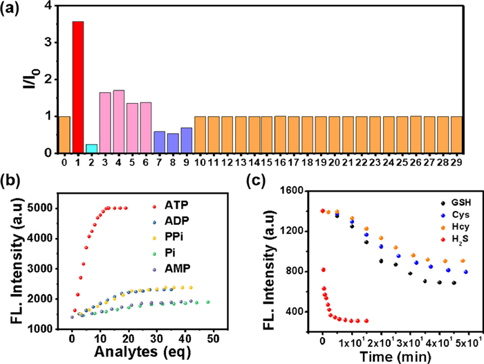
To further study the specificity of TPEPy-SS-C14 for ATP and H2S, the responses of the probe to other interfering species, including metal cations, various anions and amino acids were also studied (Fig. 2a). As shown in Fig. 2b and Fig. S14 (Supporting information), a few other phosphate anions, for example, adenosine diphosphate (ADP), adenosine monophosphate (AMP), pyrophosphate (PPi) and inorganic phosphate (Pi), can also slightly enhance the fluorescence. A weak response to ADP, PPi, Pi and AMP, with a partial enhance of the emission spectra of TPEPy-SS-C14 was recorded in Figs. S15 and S16 (Supporting information). However, this interference can be ignored because TPEPy-SS-C14 exhibits far better binding affinity of ATP than these similar structured polyphostates. The detection limits and binding constants of TPEPy-SS-C14 to these anions were also shown in Table S2 (Supporting information). Particularly, the KATP/KADP ratio is about 230.
Figure 2
Figure 2.
(a) Relative fluorescence histogram chart of TPEPy-SS-C14 (10 µmol/L) at 630 nm upon the addition of 0.55 mmol/L various analytes, 0-free, 1-ATP, 2-H2S, 3-ADP, 4-PPi, 5-AMP, 6-Pi, 7-GSH, 8-Cys, 9-Hcy, 10-CH3COO−, 11-NO3−, 12-Cl−, 13-Br−, 14-HSO4−, 15-SO32−, 16-Glu, 17-Arg, 18-His, 19-Met, 20-Thr, 21-Ser, 22-K+, 23-Na+, 24-Ca2+, 25-Mg2+, 26-Fe2+, 27-Fe3+, 28-Zn2+, 29-Al3+. (b) Fluorescence intensities of TPEPy-SS-C14 at 630 nm versus the number of equivalents of several phosphate anions. (c) Time-dependent fluorescence intensity at 630 nm of TPEPy-SS-C14 (10 µmol/L) upon addition of 55 equivalents of biothiols in HEPES buffer solution (pH 7.4).
On the other hand, we find the addition of some thiols-containing amide acid, including glutathione (GSH), cysteine (Cys) and homocysteine (Hcy), can also leads to a little decrease of the fluorescence. However, the quench ratio by the thiols is much lower than that of H2S, indicating that TPEPy-SS-C14 can discriminate H2S from thiols and other analytes. The difference fluorescence response of TPEPy-SS-C14 toward H2S and thiols may be attributed their different reaction mechanism. For H2S, which mainly exists as HS− in physiological conditions, can effectively cleave the disulfide bond to release the fluorescent active species TPEPy-SSH [8–12]. However, the thiols reaction may give both the cleavage and the disulfide exchange products. We also studied the reaction mechanism of GSH with the probe (Fig. S17 in Supporting information), which captures not only the cleavage product TPEPy-SH, but also the disulfide exchange species TPEPy-SG. Regarding that TPEPy-SG is also amphiphily for bearing the hydrophilic glutathione group, it may exhibit self-assembly property with high emission. Therefore, the fluorescence is only slightly reduced after adding GSH and other thiols [26]. Furthermore, H2S shows stronger nucleophilicity than bio-thiols. We compared the time kinetics of H2S and other thiols with TPEPy-SS-C14 (Fig. 2c), and found that the fluorescence of hydrogen sulfide added decreased rapidly in a few seconds, and basically reached equilibrium in 5 min, while other thiols took nearly 50 min. So probe TPEPy-SS-C14 reacting with H2S is dominant under the complexed physiological conditions.
For really application, a good multifunctional probe should also behaves an excellent selectively in many competitive ions. Thus, the competitive experiments of TPEPy-SS-C14 toward ATP/H2S under the coexistence of many interference species were carried out (Fig. S18 in Supporting information). It was revealed that TPEPy-SS-C14 still displays a excellent fluorescence enhancement ability toward ATP in the presence of these analytes, except H2S. However, it can recognize H2S in the presence of the following 27 analytes, containing biothiols GSH, Cys and Hcy (Fig. S19 in Supporting information). In addition, the influences of pH experiments reveals that this probe can work in wide pH range of 6‒10, indicating the physiological applicability of the TPEPy-SS-C14 probe (Fig. S20 in Supporting information).
Based on the good selective competition and anti-interference ability of the probe in vitro, TPEPy-SS-C14 was used to recognize ATP and H2S in living cells. Firstly, the toxicity of TPEPy-SS-C14 on SMMC cells was conducted by standard MTT assay. As shown in Fig. S21 (Supporting information), TPEPy-SS-C14 possessed a low cytotoxicity at the test concentration and is appropriate for intracellular imaging. The subcellular location of TPEPy-SS-C14 was investigated by co localization experiment with Mito tracker green. TPEPy-SS-C14 overlaps well with Mito tracker green dye, with an overlap rate of 0.91 (Fig. S22 in Supporting information). These results show that TPEPy-SS-C14 has a good localization effect on mitochondria and provides the imaging ability of ATP and hydrogen sulfide in mitochondria.
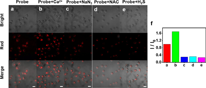
In order to investigate the ability of TPEPy-SS-C14 to recognize ATP in cells, we divided the cells into three groups: control group (row a), calcium ion treated (row b) and sodium azide treated (row c) (Fig. 3). Ca2+ increases ATP concentration by activating mitochondrial dehydrogenase and the fluorescence intensity of corresponding cell imaging is 120% of that of the control group. On the contrary, sodium azide will inhibit enzyme activity, leading to the decrease of ATP concentration and the fluorescence intensity of corresponding cell imaging is only 29% of that of the control group. This indicates that the probe can successfully detect the level of ATP in cells.
Figure 3
Figure 3.
Confocal laser scanning microscopy (CLSM) images of SMMC cells that were incubated with TPEPy-SS-C14 (10 µmol/L) for 45 min (a). Cells were pretreated with Ca2+ (1.5 mmol/L; 1 h) (b), NaN3 (1.5 mmol/L; 1 h) (c), N-acetyl-L-cysteine (NAC) (1 mmol/L; 1 h) (d), Na2S (0.6 mmol/L; 1 h) (e), followed incubation with TPEPy-SS-C14 (10 µmol/L) for another 45 min, respectively. (f) Relative fluorescence intensity of a, b, c, d and e. Scale bar: 20 µm. λex = 488 nm, λem = 570–700 nm.
Then, we evaluated the imaging ability of TPEPy-SS-C14 for endogenous and exogenous H2S. In Fig. 3, after incubating the cells pretreated with NAC (1 mmol/L) with 10 µmol/L probes for 1 h, the red fluorescence decreased significantly (row d), indicating that NAC induced the production of endogenous hydrogen sulfide concentration, and leaded to cleavage TPEPy-SS-C14 in living cells. Similarly, the fluorescence of cells treated with exogenous H2S was also reduced obviously as expected (row e). These results demonstrate that TPEPy-SS-C14 can effectively evaluate the level of endogenous and exogenous H2S in cells. Probe TPEPy-SS-C14 can be applied as a tool to explore the level of ATP and hydrogen sulfide in living cell mitochondria.
To sum up, we constructed a dual site AIE fluorescence probe TPEPy-SS-C14 and proposed a new strategy of assembly/disassembly for simultaneous recognition of ATP and H2S. As a proof of concept, the assembly/disassembly mechanism was applied to modulate the fluorescence of the amphiphilic AIEgen. After interaction ATP or H2S with probe, the aggregation state of the probe in the aqueous solution changes, showing that the fluorescence increases or decreases. It is worth mentioning that TPEPy-SS-C14 has good sensitivity and selectivity to ATP and hydrogen sulfide in SMMC cells under physiological conditions. This system has superiority over previously reported work, such as easier synthesis, lower detection limit, shorter detection time and simple operation. The sensor possesses the potential to become a effective tool to research the relationship between ATP and H2S in mitochondria of living cells.
Declaration of competing interest
The authors declare that they have no known competing financial interests or personal relationships that could have appeared to influence the work reported in this paper.
Acknowledgments
This work was supported by the National Nature Science Foundation of China (No. 22061028) and Jiangxi Provincial Natural Science Foundation (No. 20224ACB203012).
Supplementary materials
Supplementary material associated with this article can be found, in the online version, at doi:10.1016/j.cclet.2023.108925.
References
[1]
A. Moeini, P. Pedram, P. Makvandi, M. Malinconico, G.G. d'ayala, Carbohyd. Polym. 233 (2020) 16.
Haiying Lu
, Weijie Li
. The electrolyte solvation and interfacial chemistry for anode-free sodium metal batteries. Chinese Journal of Structural Chemistry,
2024, 43(11): 100334-100334.
doi: 10.1016/j.cjsc.2024.100334
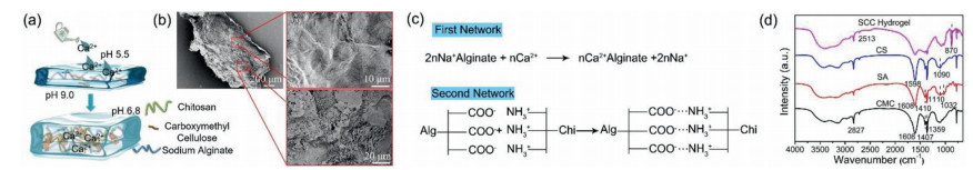
Figure 1. (a) Schematic illustration of cartoon for hydrogel structure and formation process; (b) SEM of SCC hydrogel membrane and microstructure of hydrogel surface film; (c) Schematic illustration of formula for forming hydrogel network; (d) The FT-IR spectra of CMC, SA, CS and SCC hydrogel.
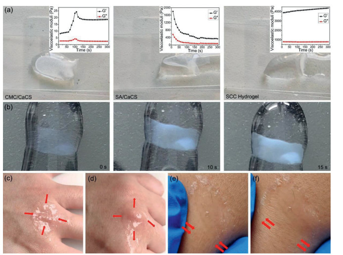
Figure 2. (a) the rheology moduli of CMC/CaCS, SA/CaCS and SCC hydrogel from left to right, in 5min (The left, middle and right inset represent the hydrogel rheological property of CMC/CaCS, SA/CaCS and SCC hydrogel.); (b) The film forming speed test of SCC hydrogel; (c) the SCC hydrogel contract under moisture condition; (d) the SCC hydrogel stretches under moisture condition; (e) the SCC hydrogel contract under dry condition; (f) the SCC hydrogel stretches under dry condition.
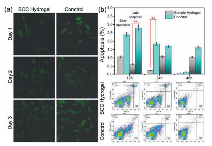
Figure 3. (a) Cell (cultured on SCC hydrogel or in six-well dishes) morphology. (b) Cells flow cytometry analysis for apoptosis detection (**P < 0.01) among the indicated groups.
Figure 4. Wound healing progress: (a) Schematic diagram of hydrogel for wound healing in mice; (b) Gross observation of the heal in vivo during different periods, quantitative statistical analysis of wounds closure for market group and SCC hydrogel; (c) Haemotoxylin and eosin (H & E) staining of wounds at day 3 and day 21 (scale bar: 20 μm).

 Login In


 DownLoad:

 DownLoad: