Citation:
Tianfang Yang, Ye Chen, Yang Liu, Xupo Liu, Shuyan Gao. Self-sacrificial template synthesis of Fe, N co-doped porous carbon as efficient oxygen reduction electrocatalysts towards Zn-air battery application[J]. Chinese Chemical Letters,
;2022, 33(4): 2171-2177.
doi:
10.1016/j.cclet.2021.09.014
Self-sacrificial template synthesis of Fe, N co-doped porous carbon as efficient oxygen reduction electrocatalysts towards Zn-air battery application
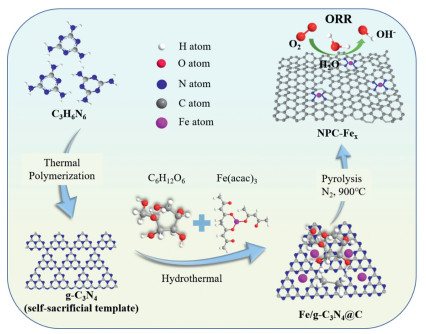
Designing highly efficient non-precious based electrocatalysts for oxygen reduction reaction (ORR) is of significance for the rapid development of metal-air batteries. Herein, a hydrothermal-pyrolysis method is employed to fabricate Fe, N co-doped porous carbon materials as effective ORR electrocatalyst through adopting graphitic carbon nitride (g-C3N4) as both the self-sacrificial templates and N sources. The g-C3N4 provides a high concentration of unsaturated pyridine-type N to coordinate with iron to form Fe-N active sites. Through adjusting the Fe doping amounts, it is proved that appropriate Fe doping content is conducive to the construction of abundant defects and active sites of Fe-N. The as-prepared catalyst exhibits superior electrocatalytic ORR performance in alkaline media with half-wave potential (E1/2 = 0.82 V) and onset potential (Eonset = 0.95 V), equivalent to the commercial Pt/C catalyst. Moreover, there is almost no activity loss after 10 k continuous cyclic voltammetry cycles and methanol tolerance, indicating the excellent durability and superior methanol tolerance. Remarkably, when assembled as the cathode in a Zn-air battery, the device displays a power density of 99 mW/cm2, an open-circuit potential of 1.48 V and long-term discharge-charge cycling stability, indicating the promising potential to substitute the Pt catalyst for practical application.
The interaction between dyes and surrounding molecules and the process of photoexcitation lead to changes in the conjugated system or molecular configuration of dye molecules, resulting in fluorescence changes, which create the signal transduction mechanism of fluorescent probes [1-3]. With the coordinated development of labeling techniques and dye chemistry, fluorescent probes have been continuously updated, from general environment-sensitive fluorescence changes to selective fluorescence responses after molecular recognition for specific target molecules [4,5]. The most typical fluorophore with molecular recognition fluorescence response performance is the fluorescent protein [6]. The molecular recognition between its endogenous chromophore and the fluorescent protein cavity effectively inhibits the rotation of the chromophore in the protein cavity and produces a unique phenolic hydroxyl dehydrogenation reaction. The hydroxyl dehydrogenation reaction ensures that the chromophore can only have fluorescence emission ability in the GFP cavity [7]. Such chromophores that activate fluorescence through molecular recognition are called fluorogens. Drawing on the pattern of recognition and binding of such endogenous chromophores and biomolecules to emit light, in recent years, relying on the screening of substrate molecules by antibodies or aptamers, non-covalently bound fluorescent antibodies and aptamers activated against exogenous chromophores has been developed [8-11]. Depending on genetic coding and enzymatic reactions, covalently linked protein tags, such as the widely used SNAP-tag [11,12] and Halo-tag [13], have been developed. This strategy of combining and activating fluorogen emission has stimulated the study of fluorescent probes that recognize biological macromolecules (including proteins, nucleic acids and carbohydrates) with low background fluorescence, but how to realize molecular recognition and activate fluorescence is a challenge in the research of such fluorescent probes [14-18].
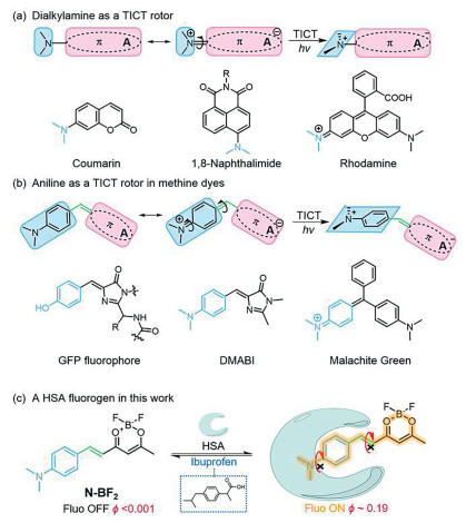
Inhibition of twisted intramolecular charge transfer (TICT) is one of the main mechanisms by which molecular recognition activates fluorescence, with dialiphatic substituted amino groups being the most commonly used rotors (Fig. 1a) [19-21]. The structure of such molecules is characterized by the direct conjugation of tertiary amine groups in planar rigid chromophores, common fluorophores such as coumarin, naphthalimide and rhodamine. The lone pair of electrons of the tertiary amine is delocalized to the aromatic system of the chromophore due to the effect of electron donating and acceptance, forming a partial C=N double bond. After being excited by light, an electron is completely intramolecularly transferred to the acceptor, and the tertiary amine group rotates to form a vertical configuration with the chromophore to stabilize the zwitterion with intramolecular charge separation. However, such molecules typically have high background fluorescence and lack strong binding to biomolecules due to their large size and structural rigidity.
Figure 1
Figure 1.
(a) The fluorophores with dialkylamine as a TICT rotator. (b) Aniline as a TICT rotor in methine dyes. (c) HSA probe with aniline as a TICT rotator in this work.
Reducing the size of the chromophore, and increasing the flexibility of the molecule is a conventional improvement idea (Fig. 1b). The fluorophore of the fluorescent protein is connected a methine group to a single benzene ring conjugated a system as the electron donor and acceptor, respectively. The existence of the methine group increases the flexibility when it is combined with biomolecules, and the size of the conjugated structure of the single benzene ring is moderate. This type of methionine chromophore has the structural characteristics of strong binding force with biological macromolecules. Inspired by fluorescent protein chromophores, aniline-substituted methine chromophores, with typical structures such as DMABI and Malachite Green, have been shown to have strong binding to both proteins and nucleic acids [22,23]. More importantly, aniline acts as an electron-donating group to form a quinoid structure, and the tertiary amine group and the aniline as a whole can co-rotate during the intramolecular charge transfer process in the excited state, which can more effectively quench the fluorescence. The background fluorescence of these chromophores is very low, and the fluorescence intensity increases by 2–3 orders of magnitude after binding to proteins or nucleic acids. In one of our recent works, the importance of the methine structure in binding to biomacromolecules and activating fluorescence was also demonstrated through the modification of the methine structure in the fluorophore by linking electron-donating and electron-withdrawing groups.
In this paper, we introduced aniline into the difluoroboron β-diketonate chromophore via a methine bridge, resulting in N-BF2 that is almost non-fluorescent in aqueous solution (Fig. 1c). N-BF2 has fast and strong binding to HSA or BSA, accompanied by the activation of fluorescence (fluorescence enhancement 90 (HSA)/112 (BSA)-fold). This intermolecular binding was found to be rapid and reversible in solution and intracellularly. Since N-BF2 also has lipid droplet-targeted ability, the complex of N-BF2HSA realizes the regulation of reversible lipid droplets staining in cells.
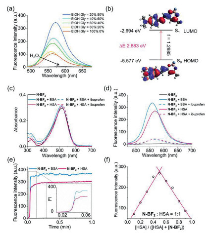
Firstly, we inspected the quenching effect of aniline-rotor in N-BF2, through the analysis of fluorescence spectra depending on different solvents and viscosity. Similar with the most traditional d-π-A dyes, N-BF2 shown solvochromic effect with obvious red-shift in fluorescence emission from 541 nm in dioxane to 591 nm in DMSO (Fig. S1 in Supporting information). And it should be noted that N-BF2 exhibited negligible fluorescence in strong polar solvent (ϕ < 0.01) which indicated the facile formation of TICT and its low background (Fig. 2a and Table S1 in Supporting information). This was further illustrated through the electron density changing in LUMO and HOMO (Fig. 2b). Once excited, the electron density of aniline decreased and flowed toward difluoroboron β-diketonate thus enhancing the charge separation which favored to form TICT. While with the increase of viscosity, the corresponding emission intensity of N-BF2 significantly enhanced (Fig. 2a). Such viscosity-dependent fluorescence intensification is also a typical characteristic of TICT emission. These results indicated potential applications of N-BF2 in developing fluorogenic probes based on restricted rotation.
Figure 2
Figure 2.
(a) Fluorescence spectra of N-BF2 in different proportions of glycerol/EtOH mixture and H2O. (b) Molecular orbital amplitude plots of HOMO and LUMO energy levels of N-BF2. (c) The absorption and (d) fluorescence spectra of N-BF2 upon addition of HSA/BSA and pretreated with ibuprofen (200 µmol/L). (e) Time course of fluorescence intensity of 5 µmol/L N-BF2 in the presence of 5 µmol/L HSA and BSA. (f) The Job's plot of N-BF2 and HSA at difffferent ratios, total concentration of N-BF2 and HSA was 10 µmol/L. λex = 460 nm.
Surprisingly, the rotation of aniline-rotor in N-BF2 can be inhibited upon binding HSA and BSA thus inducing great fluorescence enhancement. After the addition of HSA or BSA, the maximum absorption of N-BF2 at 513 nm slightly blue-shifted to 506 nm and 507 nm, respectively (Fig. 2c). Similarly, the emission at 583 nm blue-shifted to 568 nm and 559 nm, respectively, which indicated the polar microenvironment of cavity binding the chromophore (Fig. 2d). In the meanwhile, the fluorescence intensity increased more than 90-fold (HSA) and 112-fold (BSA), respectively (Table S2 in Supporting information). We also found N-BF2 could sense HSA and BSA in ~1.2 s much faster than those reported HSA/BSA probes (Fig. 2e and Table S3 in Supporting information). Furthermore, the Job's plot was conducted through monitoring fluorescence intensity at 568/559 nm, and indicated the N-BF2/HSA and N-BF2/BSA complexes all had 1:1 stoichiometry (Fig. 2f and Fig. S2 in Supporting information).
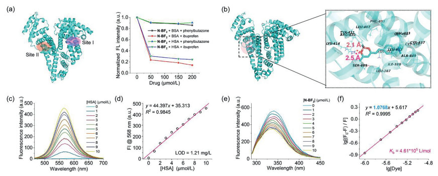
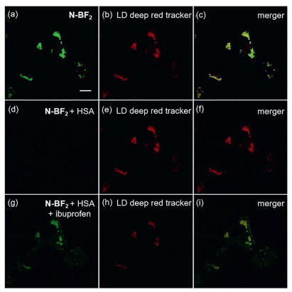
To further understand the binding between HSA and N-BF2, drug competition and molecular docking experiments were next performed. Phenylbutazone and ibuprofen, which have been known to bind HSA/BSA at site Ⅰ and site Ⅱ, respectively, are used to inspect the binding model. Both N-BF2/HSA and N-BF2/BSA complexes shown slight decrease in presence of 50–200 µmol/L phenylbutazone. However, the addition of ibuprofen could induce the fluorescence intensity of N-BF2 sharply decreased to 30% (Fig. 3a). It indicated that N-BF2 most likely to bind HSA/BSA at narrow site Ⅱ which considered as the common drug sites. The following docking study between N-BF2 and HSA/BSA also demonstrated this result (Fig. 3b and Fig. S3 in Supporting information). It revealed that oxygen in difluoroboron β-diketonate could formed two stable hydrogen bonds of 2.1 Å and 2.5 Å with the basic amino acid Arg-410 and Lys-414, which made N-BF2 specifically bind site Ⅱ. On the other side, hydrophobic interactions of aniline unit with surrounding neutral amino acids, Phe-403, Val-433, Leu-453, Ala-449, Cys-438, Cys-392 and Ile-388, were found thus inhibiting the rotation of aniline and enhancing fluorescence intensity of N-BF2. Inspired by the stable binding model, titration experiment and double logarithmic equation was further conducted to examine the limit of detection (LOD) and binding constant (Kb). With the addition of HSA, the fluorescence intensity gradually enhanced and the emission wavelength shifted from 583 nm to 568 nm (Fig. 3c). A linear relationship (R2 = 0.9845) was found between fluorescence intensity of N-BF2 and HSA concentration in the range of 0–10 µmol/L (Fig. 3d). Based on 3σ/k, the corresponding limit of detection (LOD) for HSA was calculated to be 1.21 mg/mL (Fig. 3d). Besides, with the addition of N-BF2, the fluorescence intensity at 340 nm gradually decreased and the concentration-dependent fluorescence changes were utilized to find the binding constant, the Kb was 4.61 × 10⁵ L/mol (Figs. 3e and f). LOD and Kb of N-BF2 toward BSA was also inspected to be 0.93 mg/mL and 4.67 × 10⁵ L/mol which indicated the strong chelation between HSA/BSAand N-BF2 (Figs. S4 and S5 in Supporting information). As an endogenous substance in vivo, HSA not only has high stability but also acts as a drug carrier which can increase the water solubility of drugs and regulate drug release. Therefore, we used N-BF2/HSA complexes as a trigger to control the entry and exit of N-BF2 in living cells. As shown in Figs. 4a–c, N-BF2 could specifically light up lipid droplets in HeLa cells. With the addition of 30 µmol/L HSA, the fluorescence of N-BF2 in lipid droplets disappeared, indicating that N-BF2 gradually dissociated from lipid droplets and flowed out of cells to bind HSA (Figs. 4d–f). Ibuprofen could bind HSA with higher affinity. Then the addition of ibuprofen replaced N-BF2 from HSA and the released N-BF2 could re-cross the cell membrane and go back into lipid droplets which had been confirmed by the recovery of fluorescence in lipid droplets (Figs. 4g–i). So, it could be concluded that the complex of N-BF2/HSA indeed realized the regulation of reversible lipid droplet staining in cells.
Figure 3
Figure 3.
(a) Normalized fluorescence intensity of N-BF2 (5 µmol/L) in presence of HSA (5 µmol/L) or BSA (5 µmol/L) which pretreated with different concentrations of site-specific marker (0–200 µmol/L ibuprofen or phenylbutazone). (b) Binding modes of N-BF2 with contacting residues in binding site Ⅱ of HSA. (c) Fluorescence spectra of N-BF2 (5 µmol/L) in presence of different concentration of HSA (0–10 µmol/L). (d) The linear relationship of fluorescence intensity at 568 nm with concentration of HSA (0–10 µmol/L). λex = 460 nm. (e) Fluorescence emission spectra of HSA (5 µmol/L) in presence of increasing concentration of N-BF2 (0–10 µmol/L) and (f) double logarithmic relationship of fluorescence intensity at 340 nm with the concentration of N-BF2 (0–10 µmol/L). λex = 280 nm.
Figure 4.
Confocal cellular imaging of 1 µmol/L N-BF2 in HeLa cells in presence or absence of HSA and ibuprofen. [HSA] = 30 µmol/L. [Ibuprofen] = 30 µmol/L. Scale bar = 10 µm.
In conclusion, we developed a fluorogenic HSA/BSA probe through introducing aniline and a methine bridge into the difluoroboron β-diketonate chromophore. Due to the inhibition of aniline rotation, N-BF2 could light up HSA/BSA in 1.2 s with 90/112-fold fluorescence enhancements. Furthermore, the stable hydrogen bonds made N-BF2 exhibit high sensitivity with HSA and Kb towards site Ⅱ of HSA/BSA, with LOD 1.21/0.93 mg/mL, Kb 4.61 × 10⁵/4.67 × 105 L/mol, which had been confirmed by molecular docking experiments. Finally, we realized the regulation of reversible lipid droplets staining through binding and dissociation between N-BF2 and HSA in living cells, which may be applied in drug site-specific delivery.
Declaration of competing interest
The authors declare that they have no known competing financial interests or personal relationships that could have appeared to influence the work reported in this paper.
Acknowledgment
This work is supported by the National Natural Science Foundation of China (Nos. 22078314, 21878286, 21908216, 22078201, U1908202).
Supplementary materials
Supplementary material associated with this article can be found, in the online version, at doi:10.1016/j.cclet.2022.04.070.
References
[1]
S. Chu, Y. Cui, N. Liu, Nat. Mater. 16 (2016) 16-22.
doi: 10.1038/nmat4834
[2]
G. Ren, S. Chen, J. Zhang, J. Mater. Chem. A 9 (2021) 5751-5758.
doi: 10.1039/D0TA11493F
[3]
J. Song, S. Qiu, F. Hu, Adv. Funct. Mater. 31 (2021) 2100618.
doi: 10.1002/adfm.202100618
Scheme 1. Schematic illustration of preparing NPC-Fex catalysts through a facile hydrothermal-pyrolysis process.
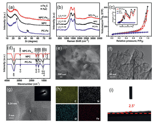
Figure 1. (a) XRD patterns. (b) Raman spectra. (c) nitrogen gas adsorption-desorption isotherms (the insert is pore-size distribution). (d) FT-IR spectra of NPC-Fe0.3, NPC and PC-Fe. (e) FE-SEM image. (f) TEM image. (g) HR-TEM image (insert is SAED pattern). (h) EDS mapping images of C, N, O and Fe elements. (i) contact angle of NPC-Fe0.3.
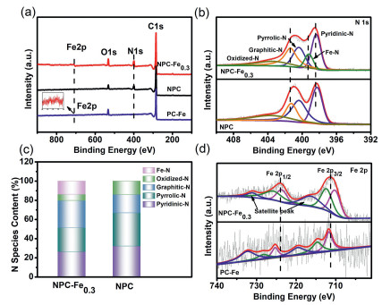
Figure 2. (a) XPS survey spectra of NPC-Fe0.3, NPC and PC-Fe. (b) N 1s spectra of NPCFe0.3 and NPC. (c) The contents of various N configurations for NPC-Fe0.3 and NPC. (d) Fe 2p spectra of NPC-Fe0.3 and PC-Fe.
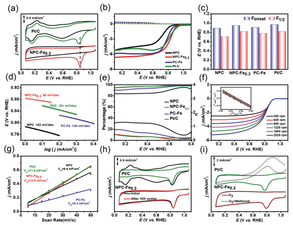
Figure 3. Electrocatalytic behavior in 0.1 mol/L KOH: (a) CV curves of NPC-Fe0.3 and Pt/C. (b) LSV curves. (c) comparison of E1/2 and Eonset. (d) Tafel plots. (e) H2O2 yields and electron numbers of NPC, NPC-Fe0.3, PC-Fe and Pt/C. (f) LSV curves of NPC-Fe0.3 with a sweep rate of 10 mV/s at different rotating speeds ranging from 400 rpm to 2025 rpm (the insert is K-L plots in different potentials). (g) plots extraction. (h) stability tests. (i) resistance of methanol crossover of NPC-Fe0.3 and Pt/C.
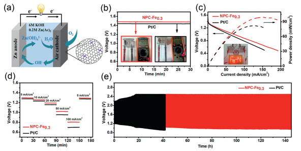
Figure 4. (a) Schematic diagram of the Zn-air battery. (b) The open-circuit voltage. (c) The polarization curves and corresponding power density plots (inset is the photograph of 69 red LED powered by two ZABs in series with NPC-Fe0.3 as the air cathode). (d) Galvanostatic discharge curves with different current densities from 0 to 100 mA/cm2. (e) The voltage-capacity curves of ZABs at 5 mA/cm2 assembled with NPC-Fe0.3 and commercial Pt/C.

 Login In


 DownLoad:

 DownLoad: