Citation: Yujie Dai, Yiming Ding, Linlin Li. Nanozymes for regulation of reactive oxygen species and disease therapy[J]. Chinese Chemical Letters, ;2021, 32(9): 2715-2728. doi: 10.1016/j.cclet.2021.03.036 shu

Nanozymes for regulation of reactive oxygen species and disease therapy

    * Corresponding author at: Beijing Institute of Nanoenergy and Nanosystems, Chinese Academy of Sciences, Beijing 101400, China.
    E-mail address: lilinlin@binn.cas.cn (L. Li).
  • Received Date: 10 January 2021
    Revised Date: 12 March 2021
    Accepted Date: 15 March 2021
    Available Online: 16 March 2021

Figures(10)

  • With high catalytic activity and stability, nanozymes have huge advantage in generating or eliminating the reactive oxygen species (ROS) due to their intrinsic enzyme-mimicking abilities, therefore attracting wide attention in ROS-related disease therapy. To better design nanozyme-based platforms for ROS-related biological application, we firstly illustrate the catalytic mechanism of different activities, and then introduce different strategies for using nanozymes to augment or reduce ROS level for the applications in cancer therapy, pathogen infection, neurodegeneration, etc. Finally, the challenges and future opportunities are proposed for the development and application of nanozymes.
  • Adenosine triphosphate (ATP), known as "the energy currency of the cell", plays an important role in living cells, including protein synthesis, intracellular signal transduction and cell division [1,2]. The abnormal levels of intracellular ATP have been proved to be closely associated with a variety of diseases, such as cancer and Parkinson [3]. On the other hand, as a signal molecule, hydrogen sulfide (H2S) widely exists in heart, brain, liver and other major organs, and plays an irreplaceable role in physiological functions including neural regulation, apoptosis, insulin signal inhibition and blood pressure [4]. The normal concentration of H2S in cells is about 0.01–3 µmol/L [5]. Abnormal H2S level can lead to the disfunction of cell, which is related to many diseases, like cirrhosis and Alzheimer's disease [6]. Therefore, the detection of ATP and H2S level has important physiological and pathological significance.

    Recently, fluorescent sensors of ATP and H2S within cellular environments are well developed for the fast response, excellent selectivity, high sensitivity and simple operation of the fluorescent technique [7]. The ATP sensors are mainly on the basis of the different host-guest interaction mechanism, for instance, metal ion complexation, hydrogen bonding, π-π interaction and the electrostatic interaction [812], while the recognition of H2S is based on the different types of chemical reaction, including copper sulfide precipitation, nucleophilic addition and reductive reaction [13,14]. Nevertheless, the traditional sensors are mainly limited to detect single species, fluorescent sensors for the simultaneous recognition of ATP and H2S are still rare.

    Currently, fluorescent sensors for simultaneous detection dual or multiple chemical species in biology have attracted much attention [1517]. Some multifunctional fluorescent sensors for ATP and another analyte, including H2O2, H2S, ONOO and nitroreductase (NTR), were also documented [1823]. Typically, the multi-analyte chemosensors were elegantly prepared by covalently linked multiple analyte recognition sites, including complexation and chemical reaction, with one or two flurophores in a single molecule. This strategy requires cumbersome chemical synthesis methods and is time-consuming and labor-intensive.

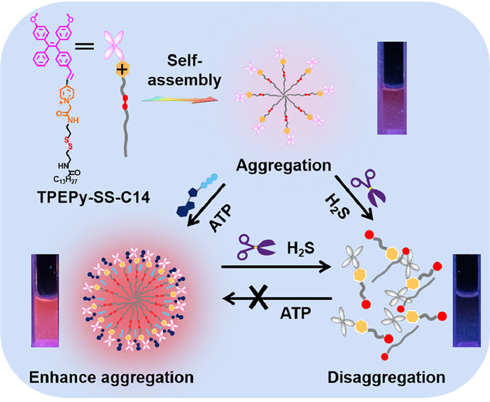
    Aggregation-induced emission (AIE) fluorogens, also named as AIEgens, which have attracted much attention for their unique fluorescence emission properties [24]. The AIEgens show non- or weak emission in dilution solutions, but emit strongly at the aggregation state. Previously, we have reported imidazolium-functionalized tetraphenylethylenes, which have a good fluorescence turn-on sensing toward ATP for the complexation enhanced aggregation [25]. We envisioned that if the second recognition site such as the disulfide bond is rationally incorporated into this system, a dual functional probe which can detect ATP and another analyte will be obtained. In this context, herein, we design and synthesize a novel AIE bifunctional probe TPEPy-SS-C14 (Scheme 1), which can simultaneously detect ATP and H2S based on the aggregation/disaggregation mechanism. The probe TPEPy-SS-C14 is rationally designed as following: (1) The TPEPy unit makes probe a red AIE-based emission, (2) the pyridinium and amide groups are used as the ATP binding site via the electrostatic interactions and hydrogen bonding, (3) the disulfide bond is cleavable by H2S, and (4) the long alkyl chain endows probe a good amphiphilic property in aqueous solution. In addition, the probe locates mitochondria, and can detect ATP and hydrogen sulfide levels in living cells.

    Scheme 1

    Scheme 1.  Chemical structure of TPEPy-SS-C14 and its proposed response mechanism to ATP and H2S.

    The detailed synthesis route of TPEPy-SS-C14 is shown in Scheme S1 (Supporting information). And the chemical structure of TPEPy-SS-C14 was verified by high-resolution mass spectrometry (HRMS), 1H nuclear magnetic resonance (NMR) and 13C NMR spectra (Figs. S1-S5 in Supporting information).

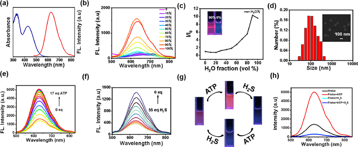
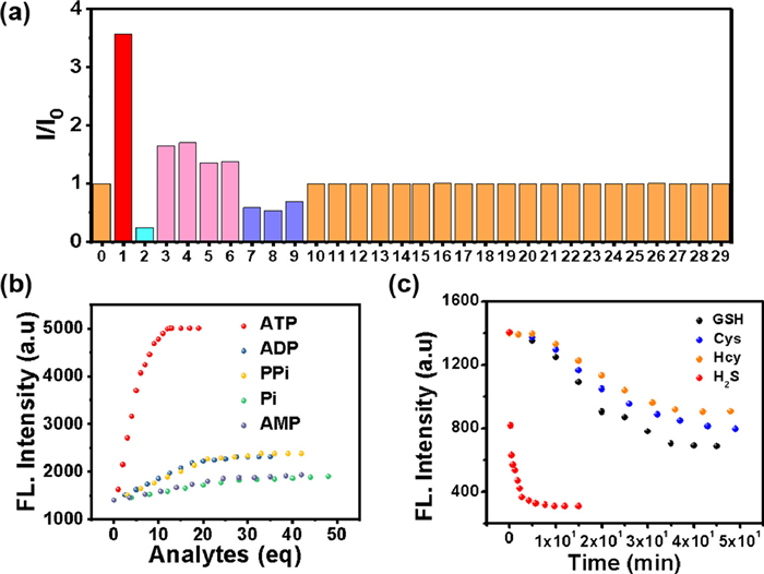
    Firstly, the ultraviolet–visible (UV–vis) absorption and fluorescence spectra of TPEPy-SS-C14 (10 µmol/L) was investigated in aqueous solution. As shown in Fig. 1a, TPEPy-SS-C14 exhibits a red emission with the maximum peak at 630 nm and a low-energy absorption peak at 428 nm. The Stokes shift is 202 nm, which can prevent the interference effectively caused by self-absorption in the excitation process of biological imaging.

    Figure 1

    Figure 1.  (a) UV–vis absorption and emission spectra of TPEPy-SS-C14 (10 µmol/L) in HEPES (10 mmol/L, pH 7.4) buffer solution. (b) The fluorescence spectra of TPEPy-SS-C14 (10 µmol/L) in water/dimethyl sulfoxide (DMSO) mixtures with different H2O fractions. (c) The plot of the relative emission intensity (I/I0) of TPEPy-SS-C14 versus the H2O fractions in H2O/DMSO mixtures. Inset: Tyndall experiment of TPEPy-SS-C14 (10 µmol/L) in the DMSO solution and in H2O/DMSO mixtures with 90% H2O fractions under 365 nm UV irradiation. (d) Size distribution of TPEPy-SS-C14 (10 µmol/L). Inset: SEM image of TPEPy-SS-C14 (10 µmol/L). The fluorescence emission spectrum of TPEPy-SS-C14 (10 µmol/L) after adding different amounts of ATP (e) and H2S (f) in HEPES (10 mmol/L, pH 7.4) buffer solution. (g) Photographs of TPEPy-SS-C14 (10 µmol/L) for ATP (0.17 mmol/L) and H2S (55 mmol/L) under 365 nm UV irradiation. (h) The fluorescence spectra of TPEPy-SS-C14 (10 µmol/L) after addition of ATP (0.17 mmol/L) or H2S (55 mmol/L). The excitation wavelength is 420 nm.

    Next, the AIE properties of TPEPy-SS-C14 were studied by emission spectra in water/DMSO mixture (Figs. 1b and c). In DMSO, the soluble TPEPy-SS-C14 molecule shows very weak emission at 630 nm. The fluorescence intensity of TPEPy-SS-C14 increased gradually with the increasing fraction of water and attained the maximum intensity in 90% aqueous solution, which may be due to the formation of nano aggregates. Additionally, the Tyndall experiment, scanning electron microscopy (SEM) and dynamic light scattering (DLS) measurements well confirmed the formation of nona-aggregation. The TPEPy-SS-C14 aggregations show a sphere morphology with a diameter of 160–180 nm (Fig. 1d). Besides, the critical micelle concentration (CMC) of TPEPy-SS-C14 was also investigated by concentration-dependent fluorescence experiment in aqueous solution, with the calculated value of 7.78 µmol/L, lower than the concentration used for testing (10 µmol/L, Fig. S6 in Supporting information).

    We evaluated the sensing properties of TPEPy-SS-C14 toward ATP in 4-(2-hydroxyethyl)–1-piperazineethanesulfonic acid (HEPES) aqueous buffer solution (pH 7.4). With the addition of ATP, the fluorescence of TPEPy-SS-C14 gradually increased and got saturated when 17 equiv. ATP was added (Fig. 1e), with the fluorescence emission intensity at 630 nm about 3.6-fold enhancement. In addition, according to fluorescence titration data, the binding constant of TPEPy-SS-C14 and ATP is calculated to be 2.57 × 10⁵ mol/L and the stoichiometric binding ratio is 1:1 (Fig. S7a in Supporting information). The detection limit of TPEPy-SS-C14 towards ATP is 12.3 nmol/L (Fig. S7b in Supporting information), indicating that the probe can recognize ATP quantitatively and effectively in physiological conditions.

    Next, the fluorescence responding of TPEPy-SS-C14 toward H2S alone was also investigated. In Fig. 1f, upon the increasing concentration of sodium sulfide, the fluorescence of TPEPy-SS-C14 will gradually decrease, with the quenching ratio about 80% when 55 equiv. sodium sulfide was added. This change can be well observed by the naked eye under the irradiation of a 365 nm ultraviolet lamp, along with the fluorescence color varying from red fluorescence to dark luminescence. The fluorescence intensity at 630 nm versus the concentration of hydrogen sulfide exhibits a great linear relation from 0 to 200 µmol/L, and the calculated limit of detection (LOD) is 5.0 × 10⁻⁷ mol/L (Fig. S8 in Supporting information). Then the time-dependent fluorescence spectra of TPEPy-SS-C14 added with 40 equiv. H2S were investigated. The fluorescence of probe decreases rapidly in a few seconds and basically reaches equilibrium in three minutes, which indicates that TPEPy-SS-C14 can be used for rapid detection of H2S (Fig. S9 in Supporting information).

    Compared to other reported probes for simultaneous detection of ATP and H2S (Table S1 in Supporting information), TPEPy-SS-C14 has a lower detection limit and shorter detection time [1823]. These listed probes are all using rhodamine linked 1,8-naphthalimide as fluorescence signal matrix, which can recognize ATP through the spirolactam ring-opening mechanism of rhodamine and recognize H2S by the reduction of -N3 on 1,8-naphthalimide. These probes have complex synthesis process and single recognition mechanism, but here we design and synthesize a novel AIE bifunctional probe TPEPy-SS-C14, which can simultaneously detect ATP and H2S based on the aggregation/disaggregation mechanism. TPEPy-SS-C14 can detect ATP with a fluorescence enhancement response due to complexation enhanced aggregation. And H2S can quench the fluorescence due to the disaggregation of the cleavable disulfide bond. In addition, the probe locates mitochondria, and can detect ATP and hydrogen sulfide levels in living cells.

    The specific guest induced aggregation-disaggregation mechanism of TPEPy-SS-C14 to detect ATP and H2S are shown in Scheme 1. Upon the addition of ATP, strong intermolecular interactions will occur between pyridinium and amide donors of TPEPy-SS-C14 and the negative phosphate groups of ATP. This proposed binding model can be confirmed by 1H NMR titration of TPEPy-SS-C14 with ATP in DMSO-d6/D2O (8:2, v/v) solution, therein a large chemical shift was observed in the pyridinium and amide protons (Fig. S10 in Supporting information). After binding with ATP, the nano-aggregation of TPEPy-SS-C14 became larger, with the DLS data changing from 180 nm to 360 nm in aqueous solution, and the transmission electron microscope (TEM) increasing up to 340 nm in solid state (Fig. S11 in Supporting information). Thus, the complexation of ATP can enhance the aggregation of TPEPy-SS-C14, which makes the fluorescence a turn-on response. We also investigated the reaction mechanism of TPEPy-SS-C14 toward H2S by high-resolution mass spectrum. In Fig. S12 (Supporting information), after treated with H2S, TPEPy-SS-C14 is cleaved into TPEPy-SSH, C14-SSH and C14-SS-C14 species, indication that the addition of H2S leads to disaggregation.

    Next, we studied the sensing performance of TPEPy-SS-C14 in the simultaneous detection of ATP and H2S in HEPES buffer. TPEPy-SS-C14 still can detect H2S effectively in the presence of ATP, but can not recognize ATP with the coexistence in H2S (Figs. 1g and h). We further tested the concentration dependent change of H2S added with ATP. From Fig. S13 (Supporting information), we can see that the fluorescence of TPEPy-SS-C14 at 630 nm first increase upon addition of ATP for complexation. The fluorescence intensity gradually decreases when further addition of H2S, with the quenching ratio as high as 94%. Interesting, the calculated LOD of TPEPy-SS-C14 /ATP toward H2S is 1.57 × 10⁻⁷ mol/L, which is lower than that of TPEPy-SS-C14 (5.0 × 10⁻⁷ mol/L). This result is reasonable because the H2S trigged disulfide bond cleavage can happen in both TPEPy-SS-C14 and TPEPy-SS-C14 /ATP nano-aggregation. The addition of ATP will not affect the quenching effect of hydrogen sulfide on TPEPy-SS-C14 probe.

    To further study the specificity of TPEPy-SS-C14 for ATP and H2S, the responses of the probe to other interfering species, including metal cations, various anions and amino acids were also studied (Fig. 2a). As shown in Fig. 2b and Fig. S14 (Supporting information), a few other phosphate anions, for example, adenosine diphosphate (ADP), adenosine monophosphate (AMP), pyrophosphate (PPi) and inorganic phosphate (Pi), can also slightly enhance the fluorescence. A weak response to ADP, PPi, Pi and AMP, with a partial enhance of the emission spectra of TPEPy-SS-C14 was recorded in Figs. S15 and S16 (Supporting information). However, this interference can be ignored because TPEPy-SS-C14 exhibits far better binding affinity of ATP than these similar structured polyphostates. The detection limits and binding constants of TPEPy-SS-C14 to these anions were also shown in Table S2 (Supporting information). Particularly, the KATP/KADP ratio is about 230.

    Figure 2

    Figure 2.  (a) Relative fluorescence histogram chart of TPEPy-SS-C14 (10 µmol/L) at 630 nm upon the addition of 0.55 mmol/L various analytes, 0-free, 1-ATP, 2-H2S, 3-ADP, 4-PPi, 5-AMP, 6-Pi, 7-GSH, 8-Cys, 9-Hcy, 10-CH3COO, 11-NO3, 12-Cl, 13-Br, 14-HSO4, 15-SO32−, 16-Glu, 17-Arg, 18-His, 19-Met, 20-Thr, 21-Ser, 22-K+, 23-Na+, 24-Ca2+, 25-Mg2+, 26-Fe2+, 27-Fe3+, 28-Zn2+, 29-Al3+. (b) Fluorescence intensities of TPEPy-SS-C14 at 630 nm versus the number of equivalents of several phosphate anions. (c) Time-dependent fluorescence intensity at 630 nm of TPEPy-SS-C14 (10 µmol/L) upon addition of 55 equivalents of biothiols in HEPES buffer solution (pH 7.4).

    On the other hand, we find the addition of some thiols-containing amide acid, including glutathione (GSH), cysteine (Cys) and homocysteine (Hcy), can also leads to a little decrease of the fluorescence. However, the quench ratio by the thiols is much lower than that of H2S, indicating that TPEPy-SS-C14 can discriminate H2S from thiols and other analytes. The difference fluorescence response of TPEPy-SS-C14 toward H2S and thiols may be attributed their different reaction mechanism. For H2S, which mainly exists as HS in physiological conditions, can effectively cleave the disulfide bond to release the fluorescent active species TPEPy-SSH [812]. However, the thiols reaction may give both the cleavage and the disulfide exchange products. We also studied the reaction mechanism of GSH with the probe (Fig. S17 in Supporting information), which captures not only the cleavage product TPEPy-SH, but also the disulfide exchange species TPEPy-SG. Regarding that TPEPy-SG is also amphiphily for bearing the hydrophilic glutathione group, it may exhibit self-assembly property with high emission. Therefore, the fluorescence is only slightly reduced after adding GSH and other thiols [26]. Furthermore, H2S shows stronger nucleophilicity than bio-thiols. We compared the time kinetics of H2S and other thiols with TPEPy-SS-C14 (Fig. 2c), and found that the fluorescence of hydrogen sulfide added decreased rapidly in a few seconds, and basically reached equilibrium in 5 min, while other thiols took nearly 50 min. So probe TPEPy-SS-C14 reacting with H2S is dominant under the complexed physiological conditions.

    For really application, a good multifunctional probe should also behaves an excellent selectively in many competitive ions. Thus, the competitive experiments of TPEPy-SS-C14 toward ATP/H2S under the coexistence of many interference species were carried out (Fig. S18 in Supporting information). It was revealed that TPEPy-SS-C14 still displays a excellent fluorescence enhancement ability toward ATP in the presence of these analytes, except H2S. However, it can recognize H2S in the presence of the following 27 analytes, containing biothiols GSH, Cys and Hcy (Fig. S19 in Supporting information). In addition, the influences of pH experiments reveals that this probe can work in wide pH range of 6‒10, indicating the physiological applicability of the TPEPy-SS-C14 probe (Fig. S20 in Supporting information).

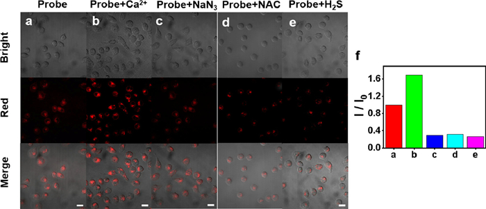
    Based on the good selective competition and anti-interference ability of the probe in vitro, TPEPy-SS-C14 was used to recognize ATP and H2S in living cells. Firstly, the toxicity of TPEPy-SS-C14 on SMMC cells was conducted by standard MTT assay. As shown in Fig. S21 (Supporting information), TPEPy-SS-C14 possessed a low cytotoxicity at the test concentration and is appropriate for intracellular imaging. The subcellular location of TPEPy-SS-C14 was investigated by co localization experiment with Mito tracker green. TPEPy-SS-C14 overlaps well with Mito tracker green dye, with an overlap rate of 0.91 (Fig. S22 in Supporting information). These results show that TPEPy-SS-C14 has a good localization effect on mitochondria and provides the imaging ability of ATP and hydrogen sulfide in mitochondria.

    In order to investigate the ability of TPEPy-SS-C14 to recognize ATP in cells, we divided the cells into three groups: control group (row a), calcium ion treated (row b) and sodium azide treated (row c) (Fig. 3). Ca2+ increases ATP concentration by activating mitochondrial dehydrogenase and the fluorescence intensity of corresponding cell imaging is 120% of that of the control group. On the contrary, sodium azide will inhibit enzyme activity, leading to the decrease of ATP concentration and the fluorescence intensity of corresponding cell imaging is only 29% of that of the control group. This indicates that the probe can successfully detect the level of ATP in cells.

    Figure 3

    Figure 3.  Confocal laser scanning microscopy (CLSM) images of SMMC cells that were incubated with TPEPy-SS-C14 (10 µmol/L) for 45 min (a). Cells were pretreated with Ca2+ (1.5 mmol/L; 1 h) (b), NaN3 (1.5 mmol/L; 1 h) (c), N-acetyl-L-cysteine (NAC) (1 mmol/L; 1 h) (d), Na2S (0.6 mmol/L; 1 h) (e), followed incubation with TPEPy-SS-C14 (10 µmol/L) for another 45 min, respectively. (f) Relative fluorescence intensity of a, b, c, d and e. Scale bar: 20 µm. λex = 488 nm, λem = 570–700 nm.

    Then, we evaluated the imaging ability of TPEPy-SS-C14 for endogenous and exogenous H2S. In Fig. 3, after incubating the cells pretreated with NAC (1 mmol/L) with 10 µmol/L probes for 1 h, the red fluorescence decreased significantly (row d), indicating that NAC induced the production of endogenous hydrogen sulfide concentration, and leaded to cleavage TPEPy-SS-C14 in living cells. Similarly, the fluorescence of cells treated with exogenous H2S was also reduced obviously as expected (row e). These results demonstrate that TPEPy-SS-C14 can effectively evaluate the level of endogenous and exogenous H2S in cells. Probe TPEPy-SS-C14 can be applied as a tool to explore the level of ATP and hydrogen sulfide in living cell mitochondria.

    To sum up, we constructed a dual site AIE fluorescence probe TPEPy-SS-C14 and proposed a new strategy of assembly/disassembly for simultaneous recognition of ATP and H2S. As a proof of concept, the assembly/disassembly mechanism was applied to modulate the fluorescence of the amphiphilic AIEgen. After interaction ATP or H2S with probe, the aggregation state of the probe in the aqueous solution changes, showing that the fluorescence increases or decreases. It is worth mentioning that TPEPy-SS-C14 has good sensitivity and selectivity to ATP and hydrogen sulfide in SMMC cells under physiological conditions. This system has superiority over previously reported work, such as easier synthesis, lower detection limit, shorter detection time and simple operation. The sensor possesses the potential to become a effective tool to research the relationship between ATP and H2S in mitochondria of living cells.

    The authors declare that they have no known competing financial interests or personal relationships that could have appeared to influence the work reported in this paper.

    This work was supported by the National Nature Science Foundation of China (No. 22061028) and Jiangxi Provincial Natural Science Foundation (No. 20224ACB203012).

    Supplementary material associated with this article can be found, in the online version, at doi:10.1016/j.cclet.2023.108925.


    1. [1]

      A.W. Dobson, Y. Xu, M.R. Kelley, et al., J. Biol. Chem. 275(2000) 37518-37523.  doi: 10.1074/jbc.M000831200

    2. [2]

      H. Beinert, D.E. Green, P. Hele, et al., Science 124(1956) 614-615.  doi: 10.1126/science.124.3223.614

    3. [3]

      C.N. Serhan, A. Jain, S. Marleau, et al., J. Immunol. 171(2003) 6856-6865.  doi: 10.4049/jimmunol.171.12.6856

    4. [4]

      S.C. Lu, Mol. Aspects Med. 30(2009) 42-59.  doi: 10.1016/j.mam.2008.05.005

    5. [5]

      D.M. Shih, L.J. Gu, Y.R. Xia, et al., Nature 394(1998) 284-287.  doi: 10.1038/28406

    6. [6]

      C. Mateo, J.M. Palomo, G.F. Lorente, et al., Enzyme Microb. Technol. 40(2007) 1451-1463.  doi: 10.1016/j.enzmictec.2007.01.018

    7. [7]

      M. Liang, X. Yan, Acc. Chem. Res. 52(2019) 2190-2200.  doi: 10.1021/acs.accounts.9b00140

    8. [8]

      F. Manea, F.B. Houillon, L. Pasquato, P. Scrimin, Angew. Chem. Int. Ed. 43(2004) 6165-6169.  doi: 10.1002/anie.200460649

    9. [9]

      L. Gao, J. Zhuang, L. Nie, et al., Nat. Nanotechnol. 2(2007) 577-583.  doi: 10.1038/nnano.2007.260

    10. [10]

      B. Liu, Z. Sun, P.J. Huang, J. Liu, J. Am. Chem. Soc. 137(2015) 1290-1295.  doi: 10.1021/ja511444e

    11. [11]

      F. Wang, Y. Zhang, Z. Liu, et al., Nanoscale 12(2020) 14465-14471.  doi: 10.1039/D0NR03217D

    12. [12]

      W. Yin, J. Yu, F. Lv, et al., ACS Nano 10(2016) 11000-11011.  doi: 10.1021/acsnano.6b05810

    13. [13]

      K. Zhang, M. Tu, W. Gao, et al., Nano Lett. 19(2019) 2812-2823.  doi: 10.1021/acs.nanolett.8b04729

    14. [14]

      C. Liu, J. Xing, O.U. Akakuru, et al., Nano Lett. 19(2019) 5674-5682.  doi: 10.1021/acs.nanolett.9b02253

    15. [15]

      R. Yan, S. Sun, J. Yang, et al., ACS Nano 13(2019) 11552-11560.  doi: 10.1021/acsnano.9b05075

    16. [16]

      M.P. Murphy, Biochem. J. 417(2009) 1-13.  doi: 10.1042/BJ20081386

    17. [17]

      C.C. Winterbourn, Nat. Chem. Biol. 4(2008) 278-286.  doi: 10.1038/nchembio.85

    18. [18]

      V.J. Thannickal, B.L. Fanburg, Am. J. Physiol. Lung Cell Mol. Physiol. 279(2000) L1005-L1028.  doi: 10.1152/ajplung.2000.279.6.L1005

    19. [19]

      S.S. Gill, N. Tuteja, Plant Physiol. Biochem. 48(2010) 909-930.  doi: 10.1016/j.plaphy.2010.08.016

    20. [20]

      H. Pelicano, D. Carney, P. Huang, Drug Resist. Updat. 7(2004) 97-110.  doi: 10.1016/j.drup.2004.01.004

    21. [21]

      E.R. Stadtman, Free Radical Res. 40(2006) 1250-1258.  doi: 10.1080/10715760600918142

    22. [22]

      K. Ito, A. Hirao, F. Arai, et al., Nat. Med. 12(2006) 446-451.  doi: 10.1038/nm1388

    23. [23]

      M. Mittal, M.R. Siddiqui, K. Tran, et al., Antioxid. Redox Signal. 20(2014) 1126-1167.  doi: 10.1089/ars.2012.5149

    24. [24]

      F. Giacco, M. Brownlee, Circul. Res. 107(2010) 1058-1070.  doi: 10.1161/CIRCRESAHA.110.223545

    25. [25]

      M. Brownlee, Diabetes 54(2005) 1615-1625.  doi: 10.2337/diabetes.54.6.1615

    26. [26]

      W.R. Markesbery, Free Radical Biol. Med. 23(1997) 134-147.  doi: 10.1016/S0891-5849(96)00629-6

    27. [27]

      K. Apel, H. Hirt, Annu. Rev. Plant Biol. 55(2004) 373-399.  doi: 10.1146/annurev.arplant.55.031903.141701

    28. [28]

      Z. Zhou, J. Song, L. Nie, X. Chen, Chem. Soc. Rev. 45(2016) 6597-6626.  doi: 10.1039/C6CS00271D

    29. [29]

      X. Qian, Y. Zheng, Y. Chen, Adv. Mater. 28(2016) 8097-8129.  doi: 10.1002/adma.201602012

    30. [30]

      S. Wang, G. Yu, Z. Wang, et al., Angew. Chem. Int. Ed. 58(2019) 14758-14763.  doi: 10.1002/anie.201908997

    31. [31]

      P. Sonveaux, Oncotarget 8(2017) 35482-35483.  doi: 10.18632/oncotarget.16613

    32. [32]

      M. Hu, K. Korschelt, P. Daniel, et al., ACS Appl. Mater. Interfaces 9(2017) 38024-38031.  doi: 10.1021/acsami.7b12212

    33. [33]

      X. Mu, J. Wang, Y. Li, et al., ACS Nano 13(2019) 1870-1884.

    34. [34]

      Y. Huang, C. Liu, F. Pu, et al., Chem. Commun. 53(2017) 3082-3085.  doi: 10.1039/C7CC00045F

    35. [35]

      D. Li, B. Liu, P.J.J. Huang, et al., Chem. Commun. 54(2018) 12519-12522.  doi: 10.1039/C8CC07062H

    36. [36]

      K. Fan, H. Wang, J. Xi, et al., Chem. Commun. 53(2017) 424-427.  doi: 10.1039/C6CC08542C

    37. [37]

      D. Jiang, D. Ni, Z.T. Rosenkrans, et al., Chem. Soc. Rev. 48(2019) 3683-3704.  doi: 10.1039/C8CS00718G

    38. [38]

      M. Liang, X. Yan, Acc. Chem. Res. 52(2019) 2190-2200.  doi: 10.1021/acs.accounts.9b00140

    39. [39]

      X. Shen, Z. Wang, X. Gao, Y. Zhao, ACS Catal. 10(2020) 12657-12665.  doi: 10.1021/acscatal.0c03426

    40. [40]

      Y. Ding, G. Wang, F. Sun, Y. Lin, ACS Appl. Mater. Interfaces 10(2018) 32567-32578.  doi: 10.1021/acsami.8b10560

    41. [41]

      Z. Wang, X. Shen, X. Gao, Y. Zhao, Nanoscale 11(2019) 13289-13299.  doi: 10.1039/C9NR03473K

    42. [42]

      S. Guo, L. Guo, J. Phys. Chem. C 123(2019) 30318-30334.  doi: 10.1021/acs.jpcc.9b07802

    43. [43]

      D. Wang, X. Song, P. Li, et al., J. Mater. Chem. B 39(2020) 9028-9034.

    44. [44]

      X. Gonze, B. Amadon, P.M. Anglade, et al., Comput. Phys. Commun. 180(2009) 2582-2615.  doi: 10.1016/j.cpc.2009.07.007

    45. [45]

      X. Wang, X.J. Gao, L. Qin, et al., Nat. Commun. 10(2019) 704.  doi: 10.1038/s41467-019-08657-5

    46. [46]

      S. Guo, L. Guo, J. Phys. Chem. C 123(2019) 30318-30334.  doi: 10.1021/acs.jpcc.9b07802

    47. [47]

      A.C. Moreno Maldonado, E.L. Winkler, M. Raineri, et al., J. Phys. Chem. C 123(2019) 20617-20627.  doi: 10.1021/acs.jpcc.9b05371

    48. [48]

      B. Jiang, D. Duan, L. Gao, et al., Nat. Protoc. 13(2018) 1506-1520.  doi: 10.1038/s41596-018-0001-1

    49. [49]

      Z. Zhang, X. Zhang, B. Liu, J. Liu, J. Am. Chem. Soc. 139(2017) 5412-5419.  doi: 10.1021/jacs.7b00601

    50. [50]

      S.B. Bankar, M.V. Bule, R.S. Singhal, L. Ananthanarayan, Biotechnol. Adv. 27(2009) 489-501.  doi: 10.1016/j.biotechadv.2009.04.003

    51. [51]

      R. Ragg, F. Natalio, M.N. Tahir, et al., ACS Nano 8(2014) 5182-5189.  doi: 10.1021/nn501235j

    52. [52]

      S.G. Rhee, Science 312(2006) 1882-1883.  doi: 10.1126/science.1130481

    53. [53]

      X. Mei, T. Hu, H. Wang, et al., Biomaterials 258(2020) 120257.  doi: 10.1016/j.biomaterials.2020.120257

    54. [54]

      M. Comotti, C. Della Pina, R. Matarrese, M. Rossi, Angew. Chem. Int. Ed. 43(2004) 5812-5815.  doi: 10.1002/anie.200460446

    55. [55]

      M. Comotti, C. Della Pina, E. Falletta, M. Rossi, Adv. Synth. Catal. 348(2006) 313-316.  doi: 10.1002/adsc.200505389

    56. [56]

      X. Shen, W. Liu, X. Gao, et al., J. Am. Chem. Soc. 137(2015) 15882-15891.  doi: 10.1021/jacs.5b10346

    57. [57]

      Q. Wang, G. Hong, Y. Liu, et al., RSC Adv. 10(2020) 25209-25213.  doi: 10.1039/D0RA05342B

    58. [58]

      Y. Yang, D. Zhu, Y. Liu, et al., Nanoscale 12(2020) 13548-13557.  doi: 10.1039/D0NR02800B

    59. [59]

      F. Charbgoo, M. Bin Ahmad, M. Darroudi, Int. J. Nanomedicine12(2017)1401-1413.  doi: 10.2147/IJN.S124855

    60. [60]

      J. Yao, Y. Cheng, M. Zhou, et al., Chem. Sci. 9(2018) 2927-2933.  doi: 10.1039/C7SC05476A

    61. [61]

      S.M. Hirst, A.S. Karakoti, R.D. Tyler, et al., Small 5(2009) 2848-2856.  doi: 10.1002/smll.200901048

    62. [62]

      T. Pirmohamed, J.M. Dowding, S. Singh, et al., Chem. Commun. (Camb. ) 46(2010) 2736-2738.  doi: 10.1039/b922024k

    63. [63]

      S. Singh, Biointerphases 11(2016) 04B202.  doi: 10.1116/1.4966535

    64. [64]

      I. Celardo, J.Z. Pedersen, E. Traversa, L. Ghibelli, Nanoscale 3(2011) 1411-1420.  doi: 10.1039/c0nr00875c

    65. [65]

      P. Ji, L. Wang, F. Chen, J. Zhang, ChemCatChem 2(2010) 1552-1554.  doi: 10.1002/cctc.201000191

    66. [66]

      D. Damatov, J.M. Mayer, Chem. Commun. (Camb. ) 52(2016) 10281-10284.  doi: 10.1039/C6CC03790A

    67. [67]

      V. Nicolini, E. Gambuzzi, G. Malavasi, et al., J. Phys. Chem. B 119(2015) 4009-4019.

    68. [68]

      I.N. Zelko, T.J. Mariani, R.J. Folz, Free Radical Biol. Med. 33(2002) 337-349.  doi: 10.1016/S0891-5849(02)00905-X

    69. [69]

      A. Okado-Matsumoto, I. Fridovich, J. Biol. Chem. 276(2001) 38388-38393.  doi: 10.1074/jbc.M105395200

    70. [70]

      J.M. McCord, M.A. Edeas, Biomed. Pharmacother. 59(2005) 139-142.  doi: 10.1016/j.biopha.2005.03.005

    71. [71]

      S. Liu, R. Tian, J. Xu, et al., Chem. Commun. 55(2019) 13820-13823.  doi: 10.1039/C9CC07085K

    72. [72]

      Y. Guan, M. Li, K. Dong, et al., Biomaterials 98(2016) 92-102.  doi: 10.1016/j.biomaterials.2016.05.005

    73. [73]

      Y. Sheng, I.A. Abreu, D.E. Cabelli, et al., Chem. Rev. 114(2014) 3854-3918.  doi: 10.1021/cr4005296

    74. [74]

      E.G. Heckert, A.S. Karakoti, S. Seal, W.T. Self, Biomaterials 29(2008) 2705-2709.  doi: 10.1016/j.biomaterials.2008.03.014

    75. [75]

      G. Calabrese, B. Morgan, J. Riemer, Antioxid. Redox Signal. 27(2017) 1162-1177.  doi: 10.1089/ars.2017.7121

    76. [76]

      E.V. Kalinina, N.N. Chernov, M.D. Novichkova, Biochemistry (Mosc. ) 79(2014) 1562-1583.  doi: 10.1134/S0006297914130082

    77. [77]

      N. Couto, J. Wood, J. Barber, Free Radical Biol. Med. 95(2016) 27-42.  doi: 10.1016/j.freeradbiomed.2016.02.028

    78. [78]

      O.M. Ighodaro, O.A. Akinloye, Alex. J. Med. 54(2018) 287-293.

    79. [79]

      R. Brigelius-Flohe, M. Maiorino, Biochim. Biophys. Acta 1830(2013) 3289-3303.  doi: 10.1016/j.bbagen.2012.11.020

    80. [80]

      M.M. Gaschler, A.A. Andia, H. Liu, et al., Nat. Chem. Biol. 14(2018) 507-515.  doi: 10.1038/s41589-018-0031-6

    81. [81]

      M. Jia, D. Qin, C. Zhao, et al., Nat. Immunol. 21(2020) 727.  doi: 10.1038/s41590-020-0699-0

    82. [82]

      L.J. Yant, Q.T. Ran, L. Rao, et al., Free Radical Biol. Med. 34(2003) 496-502.  doi: 10.1016/S0891-5849(02)01360-6

    83. [83]

      Y. Huang, Z. Liu, C. Liu, et al., Chem. Eur. J. 24(2018) 10224-10230.  doi: 10.1002/chem.201801725

    84. [84]

      T. Wirth, Angew. Chem. Int. Ed. 54(2015) 10074-10076.  doi: 10.1002/anie.201505056

    85. [85]

      Y. Huang, C. Liu, F. Pu, et al., Chem. Commun. (Camb. ) 53(2017) 3082-3085.  doi: 10.1039/C7CC00045F

    86. [86]

      N. Singh, M.A. Savanur, S. Srivastava, et al., Angew. Chem. Int. Ed. 56(2017) 14267-14271.  doi: 10.1002/anie.201708573

    87. [87]

      A.A. Vernekar, D. Sinha, S. Srivastava, et al., Nat. Commun. 5(2014) 5301.  doi: 10.1038/ncomms6301

    88. [88]

      S. Ghosh, P. Roy, N. Karmodak, et al., Angew. Chem. Int. Ed. 57(2018) 4510-4515.  doi: 10.1002/anie.201800681

    89. [89]

      B. Yang, Y. Chen, J. Shi, Chem. Rev. 119(2019) 4881-4985.  doi: 10.1021/acs.chemrev.8b00626

    90. [90]

      V.D. Petrov, F. van Breusegem, AoB Plants (2012) pls014.

    91. [91]

      H. Sies, Redox Biol. 11(2017) 613-619.  doi: 10.1016/j.redox.2016.12.035

    92. [92]

      G.P. Bienert, A.L.B. Moller, K.A. Kristiansen, et al., J. Biol. Chem. 282(2007) 1183-1192.  doi: 10.1074/jbc.M603761200

    93. [93]

      L. Huang, J.X. Chen, L.F. Gan, et al., Sci. Adv. 5(2019) 9.

    94. [94]

      J. Chen, H. Gao, Z. Li, et al., Chin. Chem. Lett. 31(2020) 1398-1401.  doi: 10.1016/j.cclet.2020.03.052

    95. [95]

      B. Xu, H. Wang, W. Wang, et al., Angew. Chem. Int. Ed. 58(2019) 4911-4916.  doi: 10.1002/anie.201813994

    96. [96]

      P. Wang, S. Liu, M. Hu, et al., Adv. Funct. Mater. 30(2020) 2000647.  doi: 10.1002/adfm.202000647

    97. [97]

      F. Cao, L. Zhang, H. Wang, et al., Angew. Chem. Int. Ed. 58(2019) 16236-16242.  doi: 10.1002/anie.201908289

    98. [98]

      Y. Tao, E. Ju, J. Ren, X. Qu, Adv. Mater. 27(2015) 1097-1104.  doi: 10.1002/adma.201405105

    99. [99]

      A. Liu, M. Li, J. Wang, et al., Chin. Chem. Lett. 31(2020) 1133-1136.  doi: 10.1016/j.cclet.2019.10.011

    100. [100]

      F. Cao, L. Zhang, Y. You, et al., Angew. Chem. Int. Ed. 59(2020) 5108-5115.  doi: 10.1002/anie.201912182

    101. [101]

      K. Fan, J. Xi, L. Fan, et al., Nat. Commun. 9(2018) 1440.  doi: 10.1038/s41467-018-03903-8

    102. [102]

      Y. Huang, Z. Liu, C. Liu, et al., Angew. Chem. Int. Ed. 55(2016) 6646-6650.  doi: 10.1002/anie.201600868

    103. [103]

      J. Shan, X. Li, K. Yang, et al., ACS Nano 13(2019) 13797-13808.  doi: 10.1021/acsnano.9b03868

    104. [104]

      X. Liu, Z. Yan, Y. Zhang, et al., ACS Nano 13(2019) 5222-5230.  doi: 10.1021/acsnano.8b09501

    105. [105]

      Z. Wang, Y. Zhang, E. Ju, et al., Nat. Commun. 9(2018) 3334.  doi: 10.1038/s41467-018-05798-x

    106. [106]

      Q. Wu, Z. He, X. Wang, et al., Nat. Commun. 10(2019) 240.  doi: 10.1038/s41467-018-08234-2

    107. [107]

      M. Qi, H. Pan, H. Shen, et al., Angew. Chem. Int. Ed. 59(2020) 11748-11753.  doi: 10.1002/anie.202002331

    108. [108]

      C. Wei, Y. Liu, X. Zhu, et al., Biomaterials 238(2020) 119848.  doi: 10.1016/j.biomaterials.2020.119848

    109. [109]

      W. Zhen, Y. Liu, W. Wang, et al., Angew. Chem. Int. Ed. 59(2020) 9491-9497.  doi: 10.1002/anie.201916142

    110. [110]

      Y. Liu, P. Bhattarai, Z. Dai, X. Chen, Chem. Soc. Rev. 48(2019) 2053-2108.  doi: 10.1039/C8CS00618K

    111. [111]

      L. Minai, D.Y. Hayon, D. Yelin, Sci. Rep. 3(2013) 2146.  doi: 10.1038/srep02146

    112. [112]

      I.B. Slimen, T. Najar, A. Ghram, et al., Int. J. Hyperthermia 30(2014) 513-523.  doi: 10.3109/02656736.2014.971446

    113. [113]

      L. Fan, X. Xu, C. Zhu, et al., ACS Appl. Mater. Interfaces 10(2018) 4502-4511.  doi: 10.1021/acsami.7b17916

    114. [114]

      M. Aioub, S.R. Panikkanvalappil, M.A. El-Sayed, ACS Nano 11(2017) 579-586.  doi: 10.1021/acsnano.6b06651

    115. [115]

      S.P. Sun, C.J. Li, J.H. Sun, et al., J. Hazard. Mater. 161(2009) 1052-1057.  doi: 10.1016/j.jhazmat.2008.04.080

    116. [116]

      M. Wang, M. Chang, Q. Chen, et al., Biomaterials 252(2020) 120093.  doi: 10.1016/j.biomaterials.2020.120093

    117. [117]

      M. Zhou, S. Song, J. Zhao, et al., J. Mater. Chem. B 3(2015) 8939-8948.  doi: 10.1039/C5TB01866H

    118. [118]

      S. Shen, S. Wang, R. Zheng, et al., Biomaterials 39(2015) 67-74.  doi: 10.1016/j.biomaterials.2014.10.064

    119. [119]

      S. Li, L. Shang, B. Xu, et al., Angew. Chem. Int. Ed. 58(2019) 12624-12631.  doi: 10.1002/anie.201904751

    120. [120]

      Y. Jiang, X. Zhao, J. Huang, et al., Nat. Commun. 11(2020) 1857.  doi: 10.1038/s41467-020-15730-x

    121. [121]

      S. Dong, Y. Dong, T. Jia, et al., Adv. Mater. 32(2020) 2002439.  doi: 10.1002/adma.202002439

    122. [122]

      A.P. Castano, P. Mroz, M.R. Hamblin, Nat. Rev. Cancer 6(2006) 535-545.  doi: 10.1038/nrc1894

    123. [123]

      J.P. Celli, B.Q. Spring, I. Rizvi, et al., Chem. Rev. 110(2010) 2795-2838.  doi: 10.1021/cr900300p

    124. [124]

      M. Wu, Y. Ding, L. Li, Nanoscale 11(2019) 19658-19683.  doi: 10.1039/C9NR06651A

    125. [125]

      Y. Zhang, F. Wang, C. Liu, et al., ACS Nano 12(2018) 651-661.  doi: 10.1021/acsnano.7b07746

    126. [126]

      D. Wang, H. Wu, S.Z.F. Phua, et al., Nat. Commun. 11(2020) 357.  doi: 10.1038/s41467-019-14199-7

    127. [127]

      W. Hiraoka, H. Honda, L.B. Feril Jr., et al., Ultrason. Sonochem. 13(2006) 535-542.  doi: 10.1016/j.ultsonch.2005.10.001

    128. [128]

      H. Chen, X. Zhou, Y. Gao, et al., Drug Discov. Today 19(2014) 502-509.  doi: 10.1016/j.drudis.2014.01.010

    129. [129]

      J. Chen, H. Luo, Y. Liu, et al., ACS Nano 11(2017) 12849-12862.  doi: 10.1021/acsnano.7b08225

    130. [130]

      X. Wang, X. Zhong, F. Gong, et al., Mater. Horizons 7(2020) 2028-2046.  doi: 10.1039/D0MH00613K

    131. [131]

      F. Gong, L. Cheng, N. Yang, et al., Adv. Mater. 31(2019) 1900730.  doi: 10.1002/adma.201900730

    132. [132]

      P. Zhu, Y. Chen, J. Shi, ACS Nano 12(2018) 3780-3795.  doi: 10.1021/acsnano.8b00999

    133. [133]

      X. Zhong, X. Wang, L. Cheng, et al., Adv. Funct. Mater. 30(2019) 1907954.

    134. [134]

      D. Sun, X. Pang, Y. Cheng, et al., ACS Nano 14(2020) 2063-2076.  doi: 10.1021/acsnano.9b08667

    135. [135]

      M. Feng, Y. Pan, R. Kong, S. Shu, Innovation (New York, N.Y. ) 1(2020) 100032-100032.

    136. [136]

      Z. Tang, Y. Liu, M. He, W. Bu, Angew. Chem. Int. Ed. 58(2019) 946-956.  doi: 10.1002/anie.201805664

    137. [137]

      Z. Tang, P. Zhao, H. Wang, et al., Chem. Rev. 121(2021) 1981-2019.  doi: 10.1021/acs.chemrev.0c00977

    138. [138]

      Y. Zhu, R. Zhu, Y. Xi, et al., Appl. Catal. B: Environ. 255(2019) 117739.  doi: 10.1016/j.apcatb.2019.05.041

    139. [139]

      Y.S. Jung, W.T. Lim, J.Y. Park, Y.H. Kim, Environ. Technol. 30(2009) 183-190.  doi: 10.1080/09593330802468848

    140. [140]

      Y. Sang, F. Cao, W. Li, et al., J. Am. Chem. Soc. 142(2020) 5177-5183.  doi: 10.1021/jacs.9b12873

    141. [141]

      M. Huo, L. Wang, Y. Chen, J. Shi, Nat. Commun. 8(2017) 357.  doi: 10.1038/s41467-017-00424-8

    142. [142]

      C. Fang, Z. Deng, G. Cao, et al., Adv. Funct. Mater. 30(2020) 1910085.  doi: 10.1002/adfm.201910085

    143. [143]

      H. Zhang, X. Liang, L. Han, F. Li, Small 14(2018) 1803256.  doi: 10.1002/smll.201803256

    144. [144]

      M. Chang, M. Wang, M. Wang, et al., Adv. Mater. 31(2019) e1905271.  doi: 10.1002/adma.201905271

    145. [145]

      M. Wang, M. Chang, Q. Chen, et al., Biomaterials 252(2020) 120093.  doi: 10.1016/j.biomaterials.2020.120093

    146. [146]

      B. D'Autreaux, M.B. Toledano, Nat. Rev. Mol. Cell Biol. 8(2007) 813-824.

    147. [147]

      M.L. Circu, T.Y. Aw, Free Radical Biol. Med. 48(2010) 749-762.  doi: 10.1016/j.freeradbiomed.2009.12.022

    148. [148]

      N. Singh, M.A. Savanur, S. Srivastava, et al., Nanoscale 11(2019) 3855-3863.  doi: 10.1039/C8NR09397K

    149. [149]

      Y. Huang, Z. Liu, C. Liu, et al., Angew. Chem. Int. Ed. 55(2016) 6646-6650.  doi: 10.1002/anie.201600868

    150. [150]

      J. Wu, Y. Yu, Y. Cheng, et al., Angew. Chem. Int. Ed. 60(2020) 1227-1234.

    151. [151]

      N. Singh, S.K. NaveenKumar, M. Geethika, G. Mugesh, Angew. Chem. Int. Ed. 60(2020) 3121-3130.

    152. [152]

      S.I. Han, S.W. Lee, M.G. Cho, et al., Adv. Mater. 32(2020) 2001566.  doi: 10.1002/adma.202001566

    153. [153]

      Y. Liu, Y. Cheng, H. Zhang, et al., Sci. Adv. 6(2020) eabb2695.  doi: 10.1126/sciadv.abb2695

    154. [154]

      T. Liu, B. Xiao, F. Xiang, et al., Nat. Commun. 11(2020) 2788.  doi: 10.1038/s41467-020-16544-7

    155. [155]

      X. Mu, J. Wang, Y. Li, et al., ACS Nano 13(2019) 1870-1884.

    156. [156]

      K. Zhang, M. Tu, W. Gao, et al., Nano Lett. 19(2019) 2812-2823.  doi: 10.1021/acs.nanolett.8b04729

    157. [157]

      D. Duan, K. Fan, D. Zhang, et al., Biosens. Bioelectron. 74(2015) 134-141.  doi: 10.1016/j.bios.2015.05.025

    158. [158]

      J. Park, J. Chu, A. Tsou, et al., Biomaterials 32(2011) 3921-3930.  doi: 10.1016/j.biomaterials.2011.02.019

  • 加载中
    1. [1]

      Xiaoshuai WuBailei WangYichen LiXiaoxuan GuanMingjing YinWenquan LvYin ChenFei LuTao QinHuyang GaoWeiqian JinYifu HuangCuiping LiMing GaoJunyu Lu . NIR driven catalytic enhanced acute lung injury therapy by using polydopamine@Co nanozyme via scavenging ROS. Chinese Chemical Letters, 2025, 36(2): 110211-. doi: 10.1016/j.cclet.2024.110211

    2. [2]

      Leichen WangAnqing MeiNa LiXiaohong RuanXu SunYu CaiJinjun ShaoXiaochen Dong . Aza-BODIPY dye with unexpected bromination and high singlet oxygen quantum yield for photoacoustic imaging-guided synergetic photodynamic/photothermal therapy. Chinese Chemical Letters, 2024, 35(6): 108974-. doi: 10.1016/j.cclet.2023.108974

    3. [3]

      Yuanyi ZhouKe MaJinfeng LiuZirun ZhengBo HuYu MengZhizhong LiMingshan Zhu . Is reactive oxygen species the only way for cancer inhibition over single atom nanomedicine? Autophagy regulation also works. Chinese Chemical Letters, 2024, 35(6): 109056-. doi: 10.1016/j.cclet.2023.109056

    4. [4]

      Wei SuXiaoyan LuoPeiyuan LiYing ZhangChenxiang LinKang WangJianzhuang Jiang . Phthalocyanine self-assembled nanoparticles for type Ⅰ photodynamic antibacterial therapy. Chinese Chemical Letters, 2024, 35(12): 109522-. doi: 10.1016/j.cclet.2024.109522

    5. [5]

      Yihao ZhangYang JiaoXianchao JiaQiaojia GuoChunying Duan . Highly effective self-assembled porphyrin MOCs nanomaterials for enhanced photodynamic therapy in tumor. Chinese Chemical Letters, 2024, 35(5): 108748-. doi: 10.1016/j.cclet.2023.108748

    6. [6]

      Yu QinMingyang HuangChenlu HuangHannah L. PerryLinhua ZhangDunwan Zhu . O2-generating multifunctional polymeric micelles for highly efficient and selective photodynamic-photothermal therapy in melanoma. Chinese Chemical Letters, 2024, 35(7): 109171-. doi: 10.1016/j.cclet.2023.109171

    7. [7]

      Yiling LiZekun GaoXiuxiu YueMinhuan LanXiuli ZhengBenhua WangShuang ZhaoXiangzhi Song . FRET-based two-photon benzo[a] phenothiazinium photosensitizer for fluorescence imaging-guided photodynamic therapy. Chinese Chemical Letters, 2024, 35(7): 109133-. doi: 10.1016/j.cclet.2023.109133

    8. [8]

      Hao CaiXiaoyan WuLei JiangFeng YuYuxiang YangYan LiXian ZhangJian LiuZijian LiHong Bi . Lysosome-targeted carbon dots with a light-controlled nitric oxide releasing property for enhanced photodynamic therapy. Chinese Chemical Letters, 2024, 35(4): 108946-. doi: 10.1016/j.cclet.2023.108946

    9. [9]

      Wenkai LiuYanxian HouWeijian LiuRan WangShan HeXiang XiaChengyuan LvHua GuQichao YaoQingze PanZehou SuDanhong ZhouWen SunJiangli FanXiaojun Peng . Se-substituted pentamethine cyanine for anticancer photodynamic therapy mediated using the hot band absorption process. Chinese Chemical Letters, 2024, 35(12): 109631-. doi: 10.1016/j.cclet.2024.109631

    10. [10]

      Du LiuYuyan LiHankun ZhangBenhua WangChaoyi YaoMinhuan LanZhanhong YangXiangzhi Song . Three-in-one erlotinib-modified NIR photosensitizer for fluorescence imaging and synergistic chemo-photodynamic therapy. Chinese Chemical Letters, 2025, 36(2): 109910-. doi: 10.1016/j.cclet.2024.109910

    11. [11]

      Liangliang JiaYe HongXinyu HeYing ZhouLiujiao RenHongjun DuBin ZhaoBin QinZhe YangDi Gao . Fighting hypoxia to improve photodynamic therapy-driven immunotherapy: Alleviating, exploiting and disregarding. Chinese Chemical Letters, 2025, 36(2): 109957-. doi: 10.1016/j.cclet.2024.109957

    12. [12]

      Baoli YinXinlin LiuZhe LiZhifei YeYoujuan WangXia YinSulai LiuGuosheng SongShuangyan HuanXiao-Bing Zhang . Ratiometric NIR-Ⅱ fluorescent organic nanoprobe for imaging and monitoring tumor-activated photodynamic therapy. Chinese Chemical Letters, 2025, 36(5): 110119-. doi: 10.1016/j.cclet.2024.110119

    13. [13]

      Xuejian XingPan ZhuE PangShaojing ZhaoYu TangZheyu HuQuchang OuyangMinhuan Lan . D-A-D-structured boron-dipyrromethene with aggregation-induced enhanced phototherapeutic efficiency for near-infrared fluorescent and photoacoustic imaging-guided synergistic photodynamic and photothermal cancer therapy. Chinese Chemical Letters, 2024, 35(10): 109452-. doi: 10.1016/j.cclet.2023.109452

    14. [14]

      Haoran HouSiwen WeiYutong ShaoYingnan WuGaobo HongJing AnJiarui TianJianjun DuFengling SongXiaojun Peng . A 690-nm-excitable type Ⅰ & Ⅱ photosensitizer based on biotinylation of verteporfin for photodynamic therapy of deep-seated orthotopic breast tumors. Chinese Chemical Letters, 2025, 36(6): 110315-. doi: 10.1016/j.cclet.2024.110315

    15. [15]

      Yuequan WangCongtian WuChengcheng FengQin ChenZhonggui HeShenwu ZhangCong LuoJin Sun . Spatiotemporally-controlled supramolecular hybrid nanoassembly enabling ferroptosis-augmented photodynamic immunotherapy of cancer. Chinese Chemical Letters, 2025, 36(3): 109902-. doi: 10.1016/j.cclet.2024.109902

    16. [16]

      Shaobin HeXiaoyun GuoQionghua ZhengHuanran ShenYuan XuFenglin LinJincheng ChenHaohua DengYiming ZengWei Chen . Engineering nickel-supported osmium bimetallic nanozymes with specifically improved peroxidase-like activity for immunoassay. Chinese Chemical Letters, 2025, 36(4): 110096-. doi: 10.1016/j.cclet.2024.110096

    17. [17]

      Zhongyu WangLijun WangHuaixin Zhao . DNA-based nanosystems to generate reactive oxygen species for nanomedicine. Chinese Chemical Letters, 2024, 35(11): 109637-. doi: 10.1016/j.cclet.2024.109637

    18. [18]

      Caixia ZhuQing HongKaiyuan WangYanfei ShenSongqin LiuYuanjian Zhang . Single nanozyme-based colorimetric biosensor for dopamine with enhanced selectivity via reactivity of oxidation intermediates. Chinese Chemical Letters, 2024, 35(10): 109560-. doi: 10.1016/j.cclet.2024.109560

    19. [19]

      Ziqin LiKai HaoLongwei XiangHuayu Tian . Cationic covalent organic framework nanocarriers integrating both efficient gene silencing and real-time gene detection. Chinese Chemical Letters, 2025, 36(4): 109943-. doi: 10.1016/j.cclet.2024.109943

    20. [20]

      Yixin SunKeke YuXiuchun GuoLanlan ZongZhonggui HeXiaohui Pu . Three-in-one reduction and acid-ignited micelles amplify antitumor efficacy via precise synergistic delivery of paclitaxel and naringenin. Chinese Chemical Letters, 2025, 36(6): 110393-. doi: 10.1016/j.cclet.2024.110393

Metrics
  • PDF Downloads(44)
  • Abstract views(1914)
  • HTML views(89)

通讯作者: 陈斌, bchen63@163.com
  • 1. 

    沈阳化工大学材料科学与工程学院 沈阳 110142

  1. 本站搜索
  2. 百度学术搜索
  3. 万方数据库搜索
  4. CNKI搜索
Address:Zhongguancun North First Street 2,100190 Beijing, PR China Tel: +86-010-82449177-888
Powered By info@rhhz.net

/

DownLoad:  Full-Size Img  PowerPoint
Return