
 Login In
Photo-responsive nanoparticles for β-lapachone delivery in vitro
- Corresponding author: Huo Guoyan, huoguoyan@yahoo.com
Figures(4)
Citation:
Li Huibo, Chen Changmai, An Qin, Huo Guoyan, Run Mingtao. Photo-responsive nanoparticles for β-lapachone delivery in vitro[J]. Chinese Chemical Letters,
;2018, 29(9): 1347-1349.
doi:
10.1016/j.cclet.2017.11.025

Figures(4)


Drug delivery system can improve the pharmacological and therapeutic properties of drugs. Researchers have used many strategies to prepare stimuli responsive vectors for drug delivery, such as micelles [1], liposomes, dendrimers, hydrogels as stimuli responsive drug delivery system. Triggers that have been applied include pH [2, 3], temperature [4, 5], light [6-9], enzymes [10, 11] and so on. However, these polymers also have some problems, such as poor biocompatibility, low drug loading in these systems, we cannot control the drug delivery in spatial and temporal dimension, thus stimuli responsive drug delivery system have attracted much attention.
Some anticancer drugs are toxic with serious side effects [12]. To reduce these side effects, we designed drug delivery system to encapsulate the drugs. Compared with traditional method like chemotherapy, nanoparticles encapsulated with drugs can increase the efficiency of drugs, prevent the drugs from being degraded in the transport process, prolong the time of circulation in blood, increase the bioavailability of the drugs of poor soluble in water, and deliver the drugs to the appropriate site with high efficiency.
In our previous study, we have studied the photo-degradable polyesters and photo-responsive polymeric nanoparticles for triggered release. However, after UV irradiation, the particle will expand several folds larger, which may result in cell toxicity. In this paper, we used a large molecular weight cross-link agent, which would prevent the particle size increasing after UV irradiation. We used β-lapachone (β-Lapa) as an antiproliferative agent, which selectively induces apoptosis-related cell death in a variety of human cancer cells [13]. We used two compounds containing photo-labile groups, 2-nitrophenylethylene glycol and 7-diethylamino-4-hydroxymethylcoumarin as monomers to form two kinds of polymers. After UV irradiation, the two groups will cleave from the polymers and release the drug β-Lapa at the same time.
All the solvents and reagents we used were commercial and without further purification except as noted below. Dichloromethane (DCM), trimethylamine (TEA) were distilled over calcium hydride and store in dry flasks. 1, 2-Eethanediamine, 1, 6-hexanediamine, 2, 20-(ethylenedioxy) bis(ethylamine), 2-nitrophenylactone, were purchased from Alfa Aesar. β-Lapa was purchased from Biovision. Poly(ethylene glycol) dimethacrylate was purchased from Energy Chemical, China.
The size of the nanoparticles was determined using a Zatasizer Nano-ZS from Malvern Instruments. UV-vis spectra were performed on DU800 UV-vis spectrophotometer. Uptake of the nanoparticles was visualized with an inverted fluorescence microscope.
We prepared the nanoparticles by emulsion polymerization. Firstly, 5 mg SDS was dissolved in 5 mL distilled water, then 50 mg M1 or M2 were dissolved in anhydrous DCM, then added 4 μL hexadecane, 1.5 μL cross-linker, 10 μL AIBN (1 wt%, in DCM), and 3 mg β-Lapa. They are mixed together, and treated by ultrasound wave in the ice bath, under 80 W for 5 min and then stirred at 80 ℃ for 18 h. Then the polymerization was dialysis in distilled water for 3 days, and lyophilized for further use.
Nanoparticles size distribution was determined by DLS, particle was dispersed in water and results were presented as an intensity distribution. Nanoparticles release was characterized by UV-vis absorbance spectra, the particles were dispersed in water, and detected by the instrument before and after 365 nm UV irradiation.
We used MTT assay to study the toxicity of the particles with RAW 264.7 cells. Seed 5000 cells/well, after incubated at 37 ℃ humidified atmosphere with 5% CO2 for 24 h, we treated the cells with various concentration of nanoparticles and incubated for 24 h. Added 10 μL MTT solution (10 mg/mL) and incubated for another 4 h, the solution was changed with 100 μL DMSO. The cell viability was determined by comparing the absorbance of particle-treated cells to that of control ones.
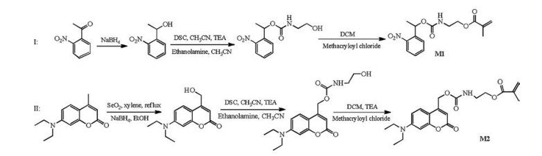
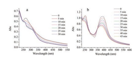
The two photoresponsive monomers were synthesized based on the literature reported else [14, 15] and they were 2-(((1-(2-nitrophenyl) ethoxy) carbonyl)amino)ethyl methacrylate (M1) and 2-((((7-(diethylamino)-2-oxo-2H-chromen-4-yl)methoxy)carbonyl)amino)ethyl methacrylate (M2), the process of synthesis was shown in Scheme 1. We used the two monomers to synthesize polymers (NP1 and NP2) through free emulsion polymerization [16], the particles without encapsulated drugs we defined them as NP1-blk, NP2-blk respectively, and the nanoparticles containing β-Lapa we defined them as NP1-β-Lapa and NP2-β-Lapa respectively. In this polymerization, we used poly(ethylene glycol) dimethacrylate (CL2), with a long chain and large molecular weight (550 Da) as a new cross-linker in the free emulsion polymerization. After UV irradiation, the particles size will increase a little larger. The polymers are not soluble in organic system, so we cannot characterize them with gel permeation chromatography (GPC), we characterized the two nanoparticles by dynamic light scattering (DLS), UV-vis absorbance spectra. We measured the absorbance spectra of the two nanoparticles under different UV irradiation time, and shown in Fig. 1. At λ = 325 nm with different times, with the increase of irradiation time, there was a new absorbance shoulder peak at 305 nm in the spectra of Fig. 1a, which indicated a clean conversion from esters to carbonyl moieties [14]. There was no new peaks appear in the spectra of NP2-β-Lapa (see Fig. 1b), but with the increase of UV irradiation time, absorbance strength at 380 nm and 250 nm will decrease, suggesting that the structure of the nanoparticle changed after irradiation of UV.
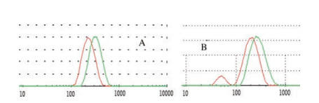
We also measured the size of the nanoparticles by DLS, which can show the size distribution of the particles before and after irradiation of UV, th increase in the particles size, arising from the formation of carbon dioxide, and an amine group, it was hydrophilic and results in the expand of the particle. Compared with our previous studies [14], the particles size increased less. But it also helps the e irradiation time was 15 min. As shown in Fig. 2, after 15 min UV irradiation, there was an obvious release of the drug.
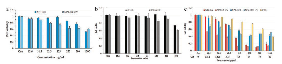
To evaluate the uptake efficacy of the β-Lapa encausulated into nanoparticles of cells, we studied uptake and release of the nanoparticles in macrophages cells. We also compared β-Lapa with curcumin to cellular uptake efficiency. In this study, we prepared two groups of cells, the first one was normally incubated with nanoparticles, and the other one was treated with 15 min UV irradiation. In this experiment, we choose NP2, owning to its fluorescence. In Figs. 3a and b were about the toxicity of NP1 and NP2, before and after UV irradiation. At low concentration, there was no obvious toxicity, suggesting that the particle itself has low toxicity before and after irradiation. As shown in Fig. 3c, we focused on the NP2-β-Lapa, after 15 min irradiation at 365 nm, cell toxicity was lower, it was because the β-Lapa was released from the particle under UV irradiation. Meantime we evaluated the cell toxicity of the NP2-β-Lapa and NP2-cur (nanoparticle2 encapsulated with curcumin), after UV, the cell toxicity of NP2-β-Lapa was much lower than that of NP2-cur, indicating that the efficacy to cure of the β-Lapa was higher than that of NP2-cur, indicating that the efficacy to cure of the β-Lapa was higher than curcumin.
In this paper, we synthesized two photo-responsive nanoparticles NP1, NP2, and characterized them by DLS, UV-vis absorbance spectra. The average size of the NP1 changed from 220 nm to 340 nm after UV irradiation, and NP2 changed from 220 nm to 360 nm. It was benefit for the release of the drug. The photoresponsive polymers formed nanoparticles with the encapsulation of curcumin and β-Lapachone. The DLS results showed that the size of the particles will swell big after UV irradiation but much smaller compared with those reported in the literature mentioned in this paper. It may be caused by the using of the large molecule weight crosslinker. Furthermore, cell toxicity of these particles also evaluated at the concentrations up to 1000 mg/mL, after UV irradiation, the cell toxicity was much lower than before. Compared with NP2-cur, the cell toxicity of the NP2-β-Lapa was lower, it showed that the nanoparticles were photoresponsive and it can release encapsulates to achieve treatment of the cancers. These studies may show a new promising strategy for photocontrolled drug delivery system.
This work is supported by SKL Professor Tang' Laboratory. The machine we used for nanoparticles characterization are available free of charge via the internet at http://pubs.acs.org.
K. Ding, L. Shi, L. Zhang, et al., Polym. Chem. 7(2016) 899-904.
doi: 10.1039/C5PY01690H
K.C. Remant Bahadur, B. Thapa, P. Xu, Mol. Pharm. 9(2012) 2719-2729.
doi: 10.1021/mp300274g
K. Ulbrich, V. Subr, Adv. Drug Deliv. Rev. 56(2004) 1023-1050.
doi: 10.1016/j.addr.2003.10.040
D.E. Meyer, B.C. Shin, G.A. Kong, M.W. Dewhirst, A. Chilkoti, J. Control Release 74(2001) 213-224.
doi: 10.1016/S0168-3659(01)00319-4
M.R. Chilkoti, D.E. Dreher, D. Meyer, D. Raucher, Adv. Drug Deliver. Rev. 54(2002) 613-630.
doi: 10.1016/S0169-409X(02)00041-8
Y. Bansal, Y. Zhang, Acc. Chem. Res. 47(2014) 3052-3060.
doi: 10.1021/ar500217w
S. Barman, S.K. Mukhopadhyay, K.K. Behara, S. Dey, N.D. Singh, ACS Appl. Mater. Interfaces 6(2014) 7045-7054.
doi: 10.1021/am500965n
S. Karthik, N. Puvvada, B.N. Kumar, et al., ACS Appl. Mater. Interfaces 5(2013) 5232-5238.
doi: 10.1021/am401059k
A.Y. Rwei, W. Wang, D.S. Kohane, Nano Today 10(2015) 451-467.
doi: 10.1016/j.nantod.2015.06.004
J. Nunthanid, K. Huanbutta, M. Luangtana-anan, et al., Eur. J. Pharm. Biopharm. 68(2008) 253-259.
doi: 10.1016/j.ejpb.2007.05.017
M.R. Dzamukova, E.A. Naumenko, Y.M. Lvov, R.F. Fakhrullin, Sci. Rep. 5(2015) 10560.
doi: 10.1038/srep10560
W. Cheng, L. Gu, W. Ren, Y. Liu, Mater. Sci. Eng. C 45(2014) 600-608.
doi: 10.1016/j.msec.2014.05.050
Y.J. Jeon, W. Bang, Y.H. Choi, J.H. Shim, J.I. Chae, Biol. Pharm. Bull. 38(2015) 1302-1308.
doi: 10.1248/bpb.b15-00159
Z. Lv, P. Wang, X. Wang, X. Tang, Int. J. Mol. Sci. 13(2012) 16387-16399.
doi: 10.3390/ijms131216387
Z. Lv, P. Wang, X. Wang, X. Tang, Langmuir 28(2012) 9387-9394.
doi: 10.1021/la301534h
K. Landfester, Angew. Chem. Int. Ed. 48(2009) 4488-4507.
doi: 10.1002/anie.v48:25

Shuang Liang , Jianjun Yao , Dan Liu , Mengli Zhou , Yong Cui , Zhaohui Wang . Tumor-responsive covalent organic polymeric nanoparticles enhancing STING activation for cancer immunotherapy. Chinese Chemical Letters, 2025, 36(3): 109856-. doi: 10.1016/j.cclet.2024.109856
Zhiwei Zhong , Yanbin Huang , Wantai Yang . A simple photochemical method for surface fluorination using perfluoroketones. Chinese Chemical Letters, 2024, 35(5): 109339-. doi: 10.1016/j.cclet.2023.109339
Chengde Wang , Liping Huang , Shanshan Wang , Lihao Wu , Yi Wang , Jun Dong . A distinction of gliomas at cellular and tissue level by surface-enhanced Raman scattering spectroscopy. Chinese Chemical Letters, 2024, 35(5): 109383-. doi: 10.1016/j.cclet.2023.109383
Wenlong Li , Feishi Shan , Qingdong Bao , Qinghua Li , Hua Gao , Leyong Wang . Supramolecular assembly nanoparticle for trans-epithelial treatment of keratoconus. Chinese Chemical Letters, 2024, 35(10): 110060-. doi: 10.1016/j.cclet.2024.110060
Zijian Jiang , Yuang Liu , Yijian Zong , Yong Fan , Wanchun Zhu , Yupeng Guo . Preparation of Nano Zinc Oxide by Microemulsion Method and Study on Its Photocatalytic Activity. University Chemistry, 2024, 39(5): 266-273. doi: 10.3866/PKU.DXHX202311101
Haijing Cui , Weihao Zhu , Chuning Yue , Ming Yang , Wenzhi Ren , Aiguo Wu . Recent progress of ultrasound-responsive titanium dioxide sonosensitizers in cancer treatment. Chinese Chemical Letters, 2024, 35(10): 109727-. doi: 10.1016/j.cclet.2024.109727
Xueying Shi , Xiaoxuan Zhou , Bing Xiao , Hongxia Xu , Wei Zhang , Hongjie Hu , Shiqun Shao , Zhuxian Zhou , Youqing Shen , Xiaodan Xu , Jianbin Tang . A β-lapachone-loaded iron-polyphenol nanocomplex enhances chemodynamic therapy through cascade amplification of ROS in tumor. Chinese Chemical Letters, 2025, 36(5): 110178-. doi: 10.1016/j.cclet.2024.110178
Hongxia Li , Xiyang Wang , Du Qiao , Jiahao Li , Weiping Zhu , Honglin Li . Mechanism of nanoparticle aggregation in gas-liquid microfluidic mixing. Chinese Chemical Letters, 2024, 35(4): 108747-. doi: 10.1016/j.cclet.2023.108747
Yixin Zhang , Ting Wang , Jixiang Zhang , Pengyu Lu , Neng Shi , Liqiang Zhang , Weiran Zhu , Nongyue He . Formation mechanism for stable system of nanoparticle/protein corona and phospholipid membrane. Chinese Chemical Letters, 2024, 35(4): 108619-. doi: 10.1016/j.cclet.2023.108619
Yiran Tao , Chunlei Dai , Zhaoxiang Xie , Xinru You , Kaiwen Li , Jun Wu , Hai Huang . Redox responsive polymeric nanoparticles enhance the efficacy of cyclin dependent kinase 7 inhibitor for enhanced treatment of prostate cancer. Chinese Chemical Letters, 2024, 35(8): 109170-. doi: 10.1016/j.cclet.2023.109170
Tong Tong , Lezong Chen , Siying Wu , Zhong Cao , Yuanbin Song , Jun Wu . Establishment of a leucine-based poly(ester amide)s library with self-anticancer effect as nano-drug carrier for colorectal cancer treatment. Chinese Chemical Letters, 2024, 35(12): 109689-. doi: 10.1016/j.cclet.2024.109689
Boyuan Liu , Zixu Liu , Ping Wang , Yu Zhang , Haibing He , Tian Yin , Jingxin Gou , Xing Tang . Nanomedicine-based targeting delivery systems for peritoneal cavity localized therapy: A promising treatment of ovarian cancer and its peritoneal metastasis. Chinese Chemical Letters, 2025, 36(6): 110229-. doi: 10.1016/j.cclet.2024.110229
Botao QU , Qian WANG , Xiaogang NING , Yuxin ZHOU , Ruiping ZHANG . Deeply penetrating photoacoustic imaging in tumor tissues based on dual-targeted melanin nanoparticle. Chinese Journal of Inorganic Chemistry, 2024, 40(5): 1025-1032. doi: 10.11862/CJIC.20230416
Shenglan Zhou , Haijian Li , Hongyi Gao , Ang Li , Tian Li , Shanshan Cheng , Jingjing Wang , Jitti Kasemchainan , Jianhua Yi , Fengqi Zhao , Wengang Qu . Recent advances in metal-loaded MOFs photocatalysts: From single atom, cluster to nanoparticle. Chinese Chemical Letters, 2025, 36(1): 110142-. doi: 10.1016/j.cclet.2024.110142
Xueqi Zhang , Han Gao , Jianan Xu , Min Zhou . Polyelectrolyte-functionalized carbon nanocones enable rapid and accurate analysis of Ag nanoparticle colloids. Chinese Chemical Letters, 2025, 36(4): 110148-. doi: 10.1016/j.cclet.2024.110148
Yan Gao , Zi-Lin Song , Shuang Yu , Xiu-Li Zhao , Da-Wei Chen , Ming-Xi Qiao . Enhanced ferroptosis by a nanoparticle mimicking hemoglobin coordinate pattern with self-supplying hydrogen peroxide. Chinese Chemical Letters, 2025, 36(5): 110097-. doi: 10.1016/j.cclet.2024.110097
Jijoe Samuel Prabagar , Kumbam Lingeshwar Reddy , Dong-Kwon Lim . Visible-light responsive gold nanoparticle and nano-sized Bi2O3-x sheet heterozygote structure for efficient photocatalytic conversion of N2 to NH3. Chinese Journal of Structural Chemistry, 2025, 44(4): 100564-100564. doi: 10.1016/j.cjsc.2025.100564
Tingting Liu , Pengfei Sun , Wei Zhao , Yingshuang Li , Lujun Cheng , Jiahai Fan , Xiaohui Bi , Xiaoping Dong . Magnesium doping to improve the light to heat conversion of OMS-2 for formaldehyde oxidation under visible light irradiation. Chinese Chemical Letters, 2024, 35(4): 108813-. doi: 10.1016/j.cclet.2023.108813
Yan-Kai Zhang , Yong-Zheng Zhang , Chun-Xiao Jia , Fang Wang , Xiuling Zhang , Yuhang Wu , Zhongmin Liu , Hui Hu , Da-Shuai Zhang , Longlong Geng , Jing Xu , Hongliang Huang . A stable Zn-MOF with anthracene-based linker for Cr(VI) photocatalytic reduction under sunlight irradiation. Chinese Chemical Letters, 2024, 35(12): 109756-. doi: 10.1016/j.cclet.2024.109756
Qian Wu , Mengda Xu , Tianjiao Ma , Shuzhen Yan , Jin Li , Xuesong Jiang . Chalcone-derived oxime esters with efficient photoinitiation properties under LED irradiation. Chinese Chemical Letters, 2025, 36(3): 110427-. doi: 10.1016/j.cclet.2024.110427
 DownLoad:
 DownLoad: