Login In
Proteins as functional interlayer in organic field-effect transistor
- Corresponding author: Peng Yang, yangpeng@snnu.edu.cn
Figures(5)
Citation:
Wei-Hong Zhang, Bo-Jing Jiang, Peng Yang. Proteins as functional interlayer in organic field-effect transistor[J]. Chinese Chemical Letters,
;2016, 27(8): 1339-1344.
doi:
10.1016/j.cclet.2016.06.044

Figures(5)


Tetracyclines (TC) represent a prominent class of antibiotics with global significance, and their presence in surface and groundwater is noteworthy due to their chemical stability and resistance to biodegradation [1,2]. Notably, TC concentrations in hospital and pharmaceutical wastewater can reach exceptionally high levels, exceeding 100 mg/L in certain instances [3]. In response to the pressing need for effective removal of these persistent organics, advanced oxidation processes (AOPs) have been demonstrated as a highly efficient and green technologies by generating strong oxidizing free radicals [4–7]. Among the various AOPs, H2O2-based heterogeneous Fenton process using solid catalysts instead of Fe2+ have been fully developed [8]. Solid catalysts extend the pH operational range, while also minimizing chemical usage and sludge formation, effectively addressing the limitations of homogeneous Fenton systems [9–12]. Furthermore, the facile recovery and recyclability of these catalysts markedly diminish associated costs [13–17]. However, the performance of these heterogeneous Fenton catalysts depends on their specific surface area, active sites, and structural properties [18,19]. They still face some inherent problems of limited active sites, low H2O2 activation and difficulty in meeting the industrial requirements compared to the homogeneous Fenton process [20].
Single-atom catalysts (SACs), which combine the high activity of homogeneous catalysts with the easy recovery of heterogeneous catalysts, have emerged as a promising research area in the field of environmental catalysis [21,22]. By maximizing atom utilization, the catalyst activity and selectivity have also been greatly exploited [23]. Currently, single-atom transition metal-nitrogen-carbon (M-N-C, M = Fe, Ni, Mn, etc.) has been widely studied in oxygen reduction reaction (ORR) [24], hydrogen evolution reaction (HER) [25], and CO2 reduction reaction (CO2RR) [26], due to its distinctive catalytic activity. Previous studies have shown that M-Nx is the main active site affecting the catalytic reaction rate [27–30]. In recent years, it has been shown that the electron spin state can be effectively optimized by changing the coordination number of Fe-Nx, which in turn affects the catalytic activity [31]. It was also reported that carbon nanotube-based catalysts with five Co-N coordination numbers performed significantly better than the four-nitrogen coordination case prepared by the same method [32]. Based on the interaction of light and adsorbed O2, g-C3N4 anchored with Ti single atoms could be converted from Ti-N6 to Ti-N4, prompting a rapid generation of clean active substances [33]. Nevertheless, research on the effect of coordination number of Fe-N-C and its degradation mechanism are still unclear [34–36].
In this study, a series of heterogeneous Fenton-like catalysts with Fe-N-C structures were prepared by a two-step pyrolysis method (Fig. S1 in supporting information). The Fe-N coordination number could be conveniently tuned by adjusting the amount of nitrogen source using this method. The differences in the performance of Fe-N-C catalysts affected by Fe-N coordination number in H2O2-based Fenton systems were investigated using TC as the target contaminant. Subsequently, the adsorption, major radicals, possible degradation mechanisms and pathways within the studied systems were explored. This study unveils the coordination-dependent activity behavior and mechanism of heterogeneous Fenton-like catalyst, which lays the foundation for the design of non-precious Fe-N-C catalyst.
The prepared catalysts were labeled as Fe-N-C-1, Fe-N-C-2, Fe-N-C-3 and Fe-C by the difference in the amount of Fe added (Text S1 in Supporting information). The scanning electron microscopy (SEM) images (Figs. S2a-h in Supporting information) reveal that the amounts of irregular particles adhered to the catalyst surface elevated with an increase in the Fe ratio. It can also be observed from the Fig. S3 (Supporting information) that the particles were mainly composed of Fe and O, while the C and N elements were uniformly distributed on the surface of the catalyst. The transmission electron microscope (TEM) image (Fig. 1a) shows that obvious nanoparticles were evenly distributed on the catalyst. Moreover, high resolution transmission electron microscope (HRTEM) characterizations (Fig. 1b) demonstrate that the lattice fringe spacing of the nanoparticle was 0.252 nm, which could be correspond well to (311) crystallographic plane of Fe2O3 [37]. In addition, the TEM-energy-dispersive X-ray spectroscopy (TEM-EDS) scan in Fig. S4 (Supporting information) reveals that the ratio of Fe to O in the nanoparticles was 2:3, which partly confirmed the presence of Fe2O3. Conversely, the EDS scans of the region without nanoparticles (Fig. 1c) demonstrate that the four elements (C, N, O, and Fe) were uniformly distributed in Fe-N-C-2. The Fe content of Fe-N-C-2 was determined to be 16.4 wt% by Inductively Coupled Plasma Optical Emission Spectrometer (ICP-OES). Table S1 (Supporting information) shows weight percentage of the elements in different regions. In the region without nanoparticles, the weight percentages of Fe and N were 1.46% and 6.83%, respectively. This Fe mass fraction of 1.46% was close to the Fe content reported in other studies involving Fe-SACs [38,39]. These results indicate that N, and Fe atoms were successfully integrated into the C framework. Therefore, it can be speculated that Fe-N-C catalysts comprises both Fe2O3 nanoparticles and atomically dispersed Fe.
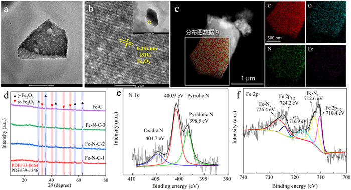
The crystal structures of the catalysts were further investigated using X-ray Diffractometer (XRD). As depicted in Fig. 1d, all studied samples exhibited characteristic peaks corresponding to the cubic γ-Fe2O3 crystalline phases. The presence of N contributed to a higher degree of crystallinity of γ-Fe2O3. It is noteworthy that the crystallinity increased gradually with the increase of Fe injection, and the addition of Fe beyond a certain point led to the formation of α-Fe2O3. These Fe2O3 provide the magnetism for the catalyst to be recovered (Text S2 and Fig. S5 in Supporting information). X-ray Photoelectron Spectroscopy (XPS) was carried out to analyze the valence states and the chemical bond among the elements of the catalyst. As shown in Fig. S6 (Supporting information), the existence of C-N and O-Fe in the Fe-N-C-2 catalyst were confirmed [40]. The corresponding fine-scan N 1s XPS spectrum (Fig. 1e) could be nicely deconvoluted into three component peaks, which were pyridinic N (398.5 eV), pyrrolic N (400.9 eV), and oxidized N (404.7 eV) [41,42]. Furthermore, Fe 2p3/2 and Fe 2p1/2 exhibited two peaks at 710.4 eV and 724.2 eV, respectively, indicating the +3-valence of Fe (Fig. 1f). The peaks at 712.6 eV and 726.4 eV belonged to the Fe-Nx configuration.
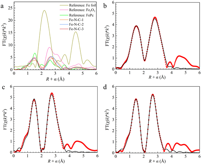
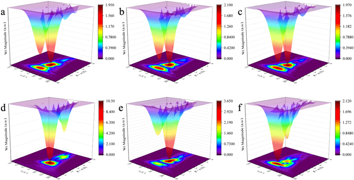
The presence of Fe-N bonds was further confirmed by the Fourier transform (FT) k3-weighted extended X-ray absorption fine structure (EXAFS) in Fig. 2a. The distinct peaks of Fe-N-C samples at 1.5 Å corresponded to the Fe-N and Fe-O bonds, which markedly different from Fe foil. The curve fit and fitting results reveal that the first shell Fe-N coordination numbers of Fe-N-C-1, Fe-N-C-2 and Fe-N-C-3 were determined as 3.3, 3.8 and 4.1, respectively (Figs. 2b–d, Fig. S7 and Tables S2-S4 in Supporting information). The Wavelet Transform (WT) of EXAFS could visualize the types of coordination atoms while demonstrating the radial distances. The WT spectrum of Fe-N-C samples (Figs. 3a–c) exhibited two contour intensities at 5.2 Å−1 and 6.6 Å−1, which matched with FePc and Fe2O3 but significantly different from Fe foil (Figs. 3d-f), indicating the existence of the Fe-N and Fe-O bonds. Moreover, the intensity of the Fe-N peak increased with the higher Fe-N ratio, which indicated that the Fe-N coordination number enhanced with the increase of Fe dosage.
The Fe-N-C samples obtained in this study were assessed for their ability to activate H2O2 for TC degradation. As shown in Fig. 4a, Fe-N-C-3 showed a stronger adsorption capacity for TC during adsorption stage, potentially due to its larger specific surface area and more adsorption sites (Text S3, Figs. S8 and S9, and Table S5 in Supporting information). After the H2O2 was added, TC was rapidly degraded within 30 min and the concentration reached equilibrium. Within 120 min, Fe-N-C-1, Fe-N-C-2, Fe-N-C-3, Fe-C, γ-Fe2O3 and α-Fe2O3 could achieve 85.20%, 89.13%, 88.29%, 60.00%, 40.64% and 9.30% removal respectively. Notably, the degradation efficiency of the Fe-N-C samples was significantly improved compared with the commercial Fe2O3, indicating that the γ-Fe2O3 and α-Fe2O3 present in the catalysts were not the primary active components. The catalytic performance of Fe-C was remarkably inferior to that of Fe-N-C-2 with the same Fe dosage, demonstrating that the crucial role of N in the catalyst. Furthermore, the degradation performance increased as the coordination number increased from 3.3 to 3.8, while there was no significant difference in performance around 4. It should be noted that the coordination number here is an average value, which means that the coordination number is dominated by 3 with a small amount of higher coordination number catalysts when x = 3.3, while the coordination number is dominated by 4 when x = 3.8 or 4.1. Thus, this result suggests that the degradation at coordination number 4 is better than that at 3. Changes in nitrogen coordination number could alter the electronic structure of the catalyst surface, thus affecting the adsorption and reaction processes of reactants on the catalyst surface [43]. As the nitrogen coordination number gradually increases from 3 to 4, the interaction between the active site and H2O2 increased, while the energy barrier during the reaction process decreased [44]. Table S6 (Supporting information) presents a comparative assessment of catalysts employed in the activation of H2O2 for TC degradation. Notably, the Fe-N-C catalyst demonstrates enhanced TC degradation efficacy under comparable experimental conditions. Otherwise, the Fe leaching concentration in the Fe-N-C-2/H2O2 system was 0.072 mg/L, and it was experimentally demonstrated that the homogeneous Fenton reaction of Fe ions at this concentration had little effect on the degradation efficiency (Fig. S10 in Supporting information). Thus, the TC degradation mechanism part of this study mainly explored the heterogeneous degradation process of Fe-N-C.

The variation of the degradation effect of Fe-N-C-2 catalysts on TC under different pH conditions and the influence of different coexisting ions was also investigated (Fig. S11 in Supporting information). The results show that the highest catalytic degradation efficiency was achieved at around pH 4. Moreover, among the tested ions, HCO3− and H2PO4− exhibited the most significant influence on the TC degradation process due to its reaction with H2O2 and Fe ions. The optimal conditions were determined as a H2O2 concentration of 10 mmol/L and pH 4. These conditions were subsequently utilized to investigate the degradation mechanism.
Adsorption is a crucial step for heterogeneous Fenton-like catalysts to degrade pollutants, wherein the pollutants are concentrated at the interface with the highest concentration of active free radicals. It has been reported that the adsorption between contaminants and materials mainly originates from electrostatic interactions, electron-donor-acceptor interactions (π-π EDA) and hydrogen-bonding (H-bonding) interactions [45], where electrostatic and hydrogen-bonding interactions are susceptible to pH effects. Fig. 4b shows the adsorption proportion of Fe-N-C-2 catalyst for TC removal at different pH. As the pH increased from 2 to 7, the adsorption rate increased from 20.94% to 28.83%. However, as the pH continued to increase, the adsorption performance of the catalyst significantly decreased. The results of zeta potential analysis (Fig. 4c) show that when pH was < 4.75, the Fe-N-C-2 catalyst carried a positive charge, while when pH > 4.75, it carried a negative charge. In addition, the chemical structure and ionization equilibrium of TC were shown in Fig. S12 (Supporting information). When the solution pH < 3.3, TC mainly existed in the form of TCH3+, which exhibited electrostatic repulsion with the Fe-N-C catalyst. When 7.7 > pH > 3.3, TC mainly existed as TCH2, and the electrostatic repulsion was weakened. However, when the pH > 7.7 or 9.7 > pH > 7.7, TC partially or completely deprotonated to form TCH− and TC2−, resulting in a significant increase in repulsion force. Therefore, the repulsion between TC and Fe-N-C catalyst was weak under acidic conditions and strong under alkaline conditions. It is worth noting that at pH 4, both the adsorption and degradation effects can achieve relatively optimal states for the catalyst, and the adsorption effect plays a relatively important role in this degradation process.
To investigate the main reactive species responsible for the degradation of TC, three quenchers, namely isopropyl alcohol, p-benzoquinone and furfuryl alcohol, were employed to selectively capture hydroxyl radical (•OH), superoxide radical (•O2−) and singlet oxygen capture (1O2), respectively [46]. Figs. S13a, b and d (Supporting information) show that the addition of •OH and 1O2 quenchers did not significantly affect the degradation of TC. However, in the presence of •O2− quencher, it could be observed that the TC concentration increased, indicating the TC desorbed from the catalyst and was not degraded (Figs. S13a and c in Supporting information). Furthermore, the electron paramagnetic resonance (EPR) results (Fig. 4d, Figs. S13e and f in Supporting information) show that only signals of •O2− was detected in the first 2 min, while the signals did not continue to increase in the next 3 min. Meanwhile, no signal of •OH and 1O2 was observed in the system. Thus, •O2− plays a key role in the catalytic reaction.
The active site on Fe-N-C-2 was investigated by analyzing changes in XPS spectra of the catalyst before and after the Fenton-like reaction (Figs. S14, S15 and Table S7 in Supporting information). As shown in Figs. S15a-c (Supporting information), the results reveal that part of the graphitic phase of carbon (C—C) was oxidized by H2O2 or •O2− during the catalytic degradation process, which increased the oxygen-containing groups on the catalyst surface and also led to the decrease of the peak area ratios of O—H and O-Fe and the decrease of the elemental C share. For the N 1s spectra (Fig. S15b in Supporting information), the content of the N—O and pyridinic N decreased, but the percentage of pyrrolic N peak area increased from 52.99% to 59.09%. It is noteworthy that pyridinic N, which is nitrogen attached between two carbon atoms on the edge of graphitic carbon with a lone pair of electrons, is chemically more reactive than pyrrolic N, which has two p-electrons and π-bonds conjugated [47]. For the Fe 2p spectra (Fig. S15d in Supporting information), both peak areas of Fe-Nx showed a decreasing trend from 24.93% and 29.81% to 21.37% and 24.18%, respectively, while both peak areas of Fe 2p showed an increasing trend. Since there was a decrease in the catalytic activity after degradation (Fig. S16 in Supporting information), it could be inferred that the Fe connected with pyridinic N might be the main catalytic active site.
The cyclic degradation experiments demonstrated a gradual decrease in catalyst performance over multiple cycles (Fig. S16). Chemical stability assessment showed that only 0.02% of Fe leached into the reaction solution after the degradation process, indicating limited Fe dissolution. Additionally, XPS analysis revealed the conversion of pyridinic N-Fe to pyrrolic N and Fe(Ⅲ) might also be the main factor for the gradual weakening of catalytic activity.
Therefore, combining the above analysis results and existing studies, it is assumed that the degradation mechanism of the catalyst was shown in Fig. S17 (Supporting information). The process of TC removal by Fe-N-C-2 is as follows: firstly, the catalyst adsorbed and immobilized TC on the catalyst surface by π-π EDA, electrostatic interaction, etc. Subsequently, pyridinic N-linked Fe activated H2O2 to produce •O2−, which decomposed TC into intermediate products. And then, the intermediate products gradually mineralized into small molecules such as CO2 and H2O to achieve efficient and green removal of the target pollutants.
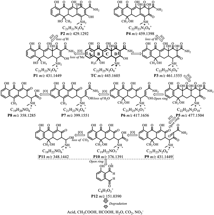
The absorption spectra of TC degradation by Fe-N-C-2 were analyzed using UV–vis spectroscopy (Fig. S18a in Supporting information). The results show that after the addition of H2O2, the intensity of the peaks at 357 nm and 276 nm gradually decreased over time, and the characteristic peaks disappeared, suggesting that the Fenton-like reaction destroyed the aromatic nucleus of TC and its intermediates [48]. The TOC degradation effect of Fe-N-C-2 degradation process was used to demonstrate the mineralization level of the process, and the TOC removal rate at 60 min was 16.39% (Fig. S18b in Supporting information). In order to further identify the intermediates and degradation pathways of TC by Fe-N-C/H2O2 system, liquid chromatography coupled to tandem mass spectrometry (LC-MS/MS) was used for the characterization in positive mode. A total of twelve intermediates were tentatively identified, with their structures and chemical formulas summarized in Table S8 (Supporting information) and the mass spectra shown in Fig. S19 (Supporting information). Fig. 5 demonstrates the proposed TC degradation pathways based on the intermediates. In pathway Ⅰ, the N,N-dimethylamine of TC undergoes oxidative rupture to yield P1 (m/z 431), which is followed by oxidative dehydrogenation to produce P2 (m/z 429). In pathway Ⅱ, the "D" ring of TC is first oxidatively hydroxylated to P3 (m/z 461), which is further oxidatively dehydrogenated to P4 (m/z 459) or hydroxylated to P5 (m/z 477). P6 (m/z 417) is formed from the oxidative ring opening reaction, and then be dehydrated to yield P7 (m/z 399). P7 is further undergoes to form P8 (m/z 358) by oxidative removal of the side chain. In pathway Ⅲ, P9 (m/z 431) is transformed from P5 through oxidation of the "D" ring. Then, P10 (m/z 376) is generated from P9 after the attack of the side chain, and subsequently decarboxylated to undergo oxidative hydroxylation to form P11 (m/z 348). Further transformation of these intermediates in the Fe-N-C/H2O2 system generates P12 (m/z 151), which in turn forms smaller molecular species and eventually H2O, CO2, NO3− and NH4+. The intermediates were not fully mineralized within 60 min, which explains the residual TOC after the treatment.
Finally, pharmaceutical simulated wastewater with high concentrations of organic matter and various ionic interferences was used to investigate the application of Fe-N-C catalyst (Text S4, Table S9, Figs. S20 and S21 in Supporting information). The results reveal that the removal rate of TC and TOC reached 70.49% and 35.97% at 540 min, suggesting the strong interference resistance of Fe-N-C/H2O2 system.
In summary, we studied the effects of Fe-N coordination number on the catalyst structure, as well as the degradation performance and possible mechanism of Fe-N-C samples. The EXAFS analysis revealed that the first shell Fe-N coordination numbers of Fe-N-C-1, Fe-N-C-2 and Fe-N-C-3 were determined as 3.3, 3.8 and 4.1, respectively. During the H2O2 activated process, the results showed that the degradation efficiency at coordination number 4 was better than 3. The EPR analysis demonstrated that •O2− played a key role as major reactive species in the catalytic reaction. By comparing the XPS energy spectra of Fe-N-C-2 before and after use, it was inferred that the Fe linked to pyridine nitrogen might be the catalytic active site of this material. This work presents the correlation between the Fe-N coordination number and catalytic activity, and demonstrates the possible mechanism and degradation pathways of these catalysts, establishing a foundation for the design and application of Fe-N-C catalysts.
The authors declare that they have no known competing financial interests or personal relationships that could have appeared to influence the work reported in this paper.
This work was supported by the National Key Research and Development Program of China (No. 2019YFC1906401-03) and the Harbin Institute of Technology National Engineering Research Center of Urban Water Resources Co., Ltd. (No. GJS-YF-LX202207280002).
Supplementary material associated with this article can be found, in the online version, at doi:
X.K. Gao, Z. Zhao. High mobility organic semiconductors for field-effect transistors[J]. Sci. China Chem., 2015,58:947-968. doi: 10.1007/s11426-015-5399-5
Y.G. Wen, Y.Q. Liu, Y.L. Guo, G. Yu, W.P. Hu. Experimental techniques for the fabrication and characterization of organic thin films for field-effect transistors[J]. Chem. Rev., 2011,111:3358-3406. doi: 10.1021/cr1001904
S.H. Kim, K. Hong, W. Xie. Electrolyte-gated transistors for organic and printed electronics[J]. Adv. Mater., 2013,25:1822-1846. doi: 10.1002/adma.v25.13
B. Singh, N.S. Sariciftci, J.G. Grote, F.K. Hopkins. Bio-organic-semiconductor-fieldeffect-transistor based on deoxyribonucleic acid gate dielectric[J]. J. Appl. Phys., 2006,100024514. doi: 10.1063/1.2220488
A. Petritz, A. Wolfberger, A. Fian. Cellulose as biodegradable high-k dielectric layer in organic complementary inverters[J]. Appl. Phys. Lett., 2013,103153303. doi: 10.1063/1.4824701
A. Dezieck, O. Acton, K. Leong. Threshold voltage control in organic thin film transistors with dielectric layer modified by a genetically engineered polypeptide[J]. Appl. Phys. Lett., 2010,97013307. doi: 10.1063/1.3459978
M. Irimia-Vladu, E.D. Głowacki, G. Voss, S. Bauer, N.S. Sariciftci. Green and biodegradable electronics[J]. Mater. Today, 2012,15:340-346. doi: 10.1016/S1369-7021(12)70139-6
M. Magliulo, K. Manoli, E. Macchia, G. Palazzo, L. Torsi. Tailoring functional interlayers in organic field-effect transistor biosensors[J]. Adv. Mater., 2015,27:7528-7551. doi: 10.1002/adma.v27.46
R.E. Marsh, R.B. Corey, L. Pauling. An investigation of the structure of silk fibroin[J]. Biochim. Biophys. Acta, 1955,16:1-34. doi: 10.1016/0006-3002(55)90178-5
R. Capelli, J.J. Amsden, G. Generali. Integration of silk protein in organic and light-emitting transistors[J]. Org. Electron., 2011,12:1146-1151. doi: 10.1016/j.orgel.2011.04.005
C.H. Wang, C.Y. Hsieh, J.C. Hwang. Flexible organic thin-film transistors with silk fibroin as the gate dielectric[J]. Adv. Mater., 2011,23:1630-1634. doi: 10.1002/adma.201004071
L.L. Shi, X.J. Xu, M.C. Ma, L.D. Li. High-performance, low-operating voltage, and solution-processable organic field-effect transistor with silk fibroin as the gate dielectric[J]. Appl. Phys. Lett., 2014,104023302. doi: 10.1063/1.4862198
X.L. Li, W. Shi, X.G. Yu, J.S. Yu. Performance improvement of organic field-effect transistor based nitrogen dioxide gas sensor using biocompatible PMMA/silk fibroin bilayer dielectric[J]. J. Mater. Sci. Mater. Electron., 2015,26:7948-7954. doi: 10.1007/s10854-015-3448-7
J.W. Chang, C.G. Wang, C.Y. Huang. Chicken albumen dielectrics in organic field-effect transistors[J]. Adv. Mater., 2011,23:4077-4081. doi: 10.1002/adma.v23.35
J.E. Nielsen, G. Vriend. Optimizing the hydrogen-bond network in Poisson-Boltzmann equation-based pKa calculations[J]. Proteins: Struct. Funct. Genet., 2001,43:403-412. doi: 10.1002/(ISSN)1097-0134
C.Y. Lee, J.C. Hwang, Y.L. Chueh. Hydrated bovine serum albumin as the gate dielectric material for organic field-effect transistors[J]. Org. Electron., 2013,14:2645-2651. doi: 10.1016/j.orgel.2013.07.004
C.Y. Hsieh, J.C. Hwang, T.H. Chang. Enhanced mobility of organic thin film transistors by water absorption of collagen hydrolysate gate dielectric[J]. Appl. Phys. Lett., 2013,103023303. doi: 10.1063/1.4813075
P. Guerrero, P.M. Stefani, R.A. Ruseckaite, K. de la Caba. Functional properties of films based on soy protein isolate and gelatin processed by compression molding[J]. J. Food Eng., 2011,105:65-72. doi: 10.1016/j.jfoodeng.2011.02.003
L.K. Mao, J.Y. Gan, J.C. Hwang, T.H. Chang, Y.L. Chueh. The role of water in the device performance of n-type PTCDI-C8 organic field-effect transistors with solution-based gelatin dielectric[J]. Org. Electron., 2014,15:920-925. doi: 10.1016/j.orgel.2014.01.023
M. González, L.A. Bagatolli, I. Echabe. Interaction of biotin with streptavidin[J]. J. Biol. Chem., 1997,272:11288-11294. doi: 10.1074/jbc.272.17.11288
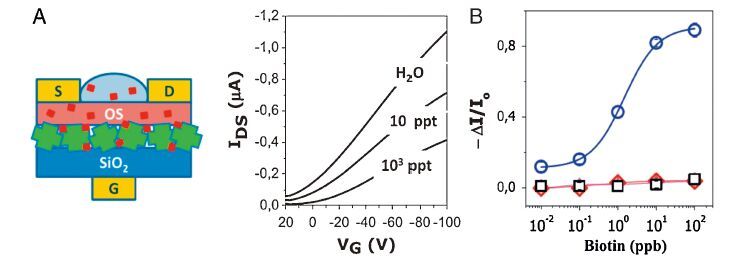
M.D. Angione, S. Cotrone, M. Magliulo. Interfacial electronic effects in functional biolayers integrated into organic field-effect transistors[J]. Proc. Natl. Acad. Sci. USA, 2012,109:6429-6434. doi: 10.1073/pnas.1200549109
M. Magliulo, A. Mallardi, R. Gristina. Part per trillion label-free electronic bioanalytical detection[J]. Anal. Chem., 2013,85:3849-3857. doi: 10.1021/ac302702n
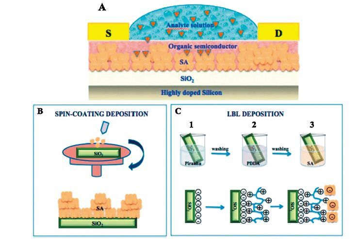
M. Magliulo, D. Altamura, C. Di Franco. Structural and morphological study of a poly(3-hexylthiophene)/streptavidin multilayer structure serving as active layer in ultra-sensitive OFET biosensors[J]. J. Phys. Chem. C, 2014,118:15853-15862. doi: 10.1021/jp504652u
X. Lin, J. Xie, L. Zhu. Hybrid ferritin nanoparticles as activatable probes for tumor imaging[J]. Angew. Chem. Int. Ed., 2011,50:1569-1572. doi: 10.1002/anie.201006757
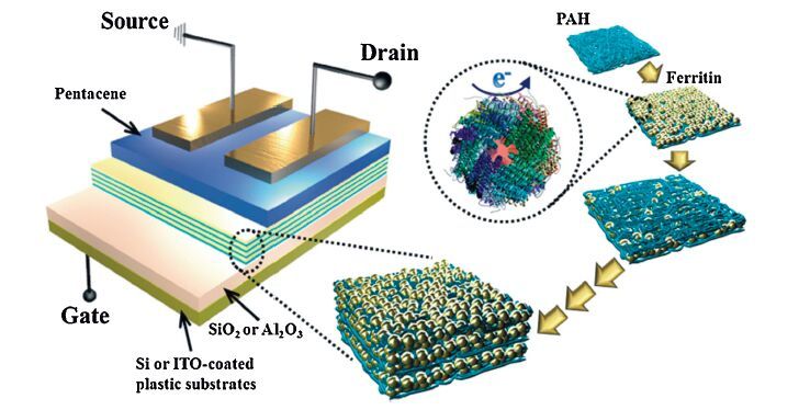
B.J. Kim, Y. Ko, J.H. Cho, J. Cho. Organic field-effect transistor memory devices using discrete ferritin nanoparticle-based gate dielectrics[J]. Small, 2013,9:3784-3791. doi: 10.1002/smll.v9.22
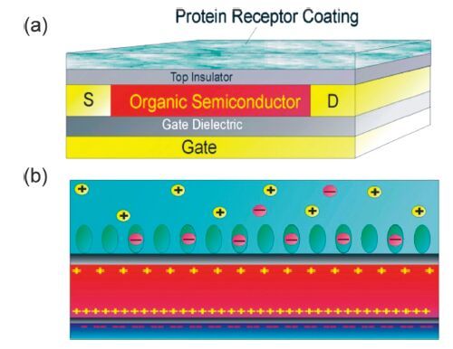
F. Maddalena, M.J. Kuiper, B. Poolman. Organic field-effect transistor-based biosensors functionalized with protein receptors[J]. J. Appl. Phys., 2010,108124501. doi: 10.1063/1.3518681
T.P.J. Knowles, M.J. Buehler. Nanomechanics of functional and pathological amyloid materials[J]. Nat. Nanotechnol., 2011,6:469-479. doi: 10.1038/nnano.2011.102
R. Paparcone, S. Keten, M.J. Buehler. Atomistic simulation of nanomechanical properties of Alzheimer's Aβ(1-40) amyloid fibrils under compressive and tensile loading[J]. J. Biomech., 2010,43:1196-1201. doi: 10.1016/j.jbiomech.2009.11.026
X.Y. Wang, Y.F. Li, C. Zhong. Amyloid-directed assembly of nanostructures and functional devices for bionanoelectronics[J]. J. Mater. Chem. B, 2015,3:4953-4958. doi: 10.1039/C5TB00374A
X.K. Gao, Z. Zhao. High mobility organic semiconductors for field-effect transistors[J]. Sci. China Chem., 2015,58:947-968. doi: 10.1007/s11426-015-5399-5
Y.G. Wen, Y.Q. Liu, Y.L. Guo, G. Yu, W.P. Hu. Experimental techniques for the fabrication and characterization of organic thin films for field-effect transistors[J]. Chem. Rev., 2011,111:3358-3406. doi: 10.1021/cr1001904
S.H. Kim, K. Hong, W. Xie. Electrolyte-gated transistors for organic and printed electronics[J]. Adv. Mater., 2013,25:1822-1846. doi: 10.1002/adma.v25.13
B. Singh, N.S. Sariciftci, J.G. Grote, F.K. Hopkins. Bio-organic-semiconductor-fieldeffect-transistor based on deoxyribonucleic acid gate dielectric[J]. J. Appl. Phys., 2006,100024514. doi: 10.1063/1.2220488
A. Petritz, A. Wolfberger, A. Fian. Cellulose as biodegradable high-k dielectric layer in organic complementary inverters[J]. Appl. Phys. Lett., 2013,103153303. doi: 10.1063/1.4824701
A. Dezieck, O. Acton, K. Leong. Threshold voltage control in organic thin film transistors with dielectric layer modified by a genetically engineered polypeptide[J]. Appl. Phys. Lett., 2010,97013307. doi: 10.1063/1.3459978
M. Irimia-Vladu, E.D. Głowacki, G. Voss, S. Bauer, N.S. Sariciftci. Green and biodegradable electronics[J]. Mater. Today, 2012,15:340-346. doi: 10.1016/S1369-7021(12)70139-6
M. Magliulo, K. Manoli, E. Macchia, G. Palazzo, L. Torsi. Tailoring functional interlayers in organic field-effect transistor biosensors[J]. Adv. Mater., 2015,27:7528-7551. doi: 10.1002/adma.v27.46
R.E. Marsh, R.B. Corey, L. Pauling. An investigation of the structure of silk fibroin[J]. Biochim. Biophys. Acta, 1955,16:1-34. doi: 10.1016/0006-3002(55)90178-5
R. Capelli, J.J. Amsden, G. Generali. Integration of silk protein in organic and light-emitting transistors[J]. Org. Electron., 2011,12:1146-1151. doi: 10.1016/j.orgel.2011.04.005
C.H. Wang, C.Y. Hsieh, J.C. Hwang. Flexible organic thin-film transistors with silk fibroin as the gate dielectric[J]. Adv. Mater., 2011,23:1630-1634. doi: 10.1002/adma.201004071
L.L. Shi, X.J. Xu, M.C. Ma, L.D. Li. High-performance, low-operating voltage, and solution-processable organic field-effect transistor with silk fibroin as the gate dielectric[J]. Appl. Phys. Lett., 2014,104023302. doi: 10.1063/1.4862198
X.L. Li, W. Shi, X.G. Yu, J.S. Yu. Performance improvement of organic field-effect transistor based nitrogen dioxide gas sensor using biocompatible PMMA/silk fibroin bilayer dielectric[J]. J. Mater. Sci. Mater. Electron., 2015,26:7948-7954. doi: 10.1007/s10854-015-3448-7
J.W. Chang, C.G. Wang, C.Y. Huang. Chicken albumen dielectrics in organic field-effect transistors[J]. Adv. Mater., 2011,23:4077-4081. doi: 10.1002/adma.v23.35
J.E. Nielsen, G. Vriend. Optimizing the hydrogen-bond network in Poisson-Boltzmann equation-based pKa calculations[J]. Proteins: Struct. Funct. Genet., 2001,43:403-412. doi: 10.1002/(ISSN)1097-0134
C.Y. Lee, J.C. Hwang, Y.L. Chueh. Hydrated bovine serum albumin as the gate dielectric material for organic field-effect transistors[J]. Org. Electron., 2013,14:2645-2651. doi: 10.1016/j.orgel.2013.07.004
C.Y. Hsieh, J.C. Hwang, T.H. Chang. Enhanced mobility of organic thin film transistors by water absorption of collagen hydrolysate gate dielectric[J]. Appl. Phys. Lett., 2013,103023303. doi: 10.1063/1.4813075
P. Guerrero, P.M. Stefani, R.A. Ruseckaite, K. de la Caba. Functional properties of films based on soy protein isolate and gelatin processed by compression molding[J]. J. Food Eng., 2011,105:65-72. doi: 10.1016/j.jfoodeng.2011.02.003
L.K. Mao, J.Y. Gan, J.C. Hwang, T.H. Chang, Y.L. Chueh. The role of water in the device performance of n-type PTCDI-C8 organic field-effect transistors with solution-based gelatin dielectric[J]. Org. Electron., 2014,15:920-925. doi: 10.1016/j.orgel.2014.01.023
M. González, L.A. Bagatolli, I. Echabe. Interaction of biotin with streptavidin[J]. J. Biol. Chem., 1997,272:11288-11294. doi: 10.1074/jbc.272.17.11288
M.D. Angione, S. Cotrone, M. Magliulo. Interfacial electronic effects in functional biolayers integrated into organic field-effect transistors[J]. Proc. Natl. Acad. Sci. USA, 2012,109:6429-6434. doi: 10.1073/pnas.1200549109
M. Magliulo, A. Mallardi, R. Gristina. Part per trillion label-free electronic bioanalytical detection[J]. Anal. Chem., 2013,85:3849-3857. doi: 10.1021/ac302702n
M. Magliulo, D. Altamura, C. Di Franco. Structural and morphological study of a poly(3-hexylthiophene)/streptavidin multilayer structure serving as active layer in ultra-sensitive OFET biosensors[J]. J. Phys. Chem. C, 2014,118:15853-15862. doi: 10.1021/jp504652u
X. Lin, J. Xie, L. Zhu. Hybrid ferritin nanoparticles as activatable probes for tumor imaging[J]. Angew. Chem. Int. Ed., 2011,50:1569-1572. doi: 10.1002/anie.201006757
B.J. Kim, Y. Ko, J.H. Cho, J. Cho. Organic field-effect transistor memory devices using discrete ferritin nanoparticle-based gate dielectrics[J]. Small, 2013,9:3784-3791. doi: 10.1002/smll.v9.22
F. Maddalena, M.J. Kuiper, B. Poolman. Organic field-effect transistor-based biosensors functionalized with protein receptors[J]. J. Appl. Phys., 2010,108124501. doi: 10.1063/1.3518681
T.P.J. Knowles, M.J. Buehler. Nanomechanics of functional and pathological amyloid materials[J]. Nat. Nanotechnol., 2011,6:469-479. doi: 10.1038/nnano.2011.102
R. Paparcone, S. Keten, M.J. Buehler. Atomistic simulation of nanomechanical properties of Alzheimer's Aβ(1-40) amyloid fibrils under compressive and tensile loading[J]. J. Biomech., 2010,43:1196-1201. doi: 10.1016/j.jbiomech.2009.11.026
X.Y. Wang, Y.F. Li, C. Zhong. Amyloid-directed assembly of nanostructures and functional devices for bionanoelectronics[J]. J. Mater. Chem. B, 2015,3:4953-4958. doi: 10.1039/C5TB00374A

Min Chen , Boyu Peng , Xuyun Guo , Ye Zhu , Hanying Li . Polyethylene interfacial dielectric layer for organic semiconductor single crystal based field-effect transistors. Chinese Chemical Letters, 2024, 35(4): 109051-. doi: 10.1016/j.cclet.2023.109051
Shaohua Zhang , Liyao Liu , Yingqiao Ma , Chong-an Di . Advances in theoretical calculations of organic thermoelectric materials. Chinese Chemical Letters, 2024, 35(8): 109749-. doi: 10.1016/j.cclet.2024.109749
Zhongchao Zhou , Jian Song , Yinghao Xie , Yuqian Ma , Hong Hu , Hui Li , Lei Zhang , Charles H. Lawrie . DFT calculation for organic semiconductor-based gas sensors: Sensing mechanism, dynamic response and sensing materials. Chinese Chemical Letters, 2025, 36(6): 110906-. doi: 10.1016/j.cclet.2025.110906
Xinyi Hong , Tailing Xue , Zhou Xu , Enrong Xie , Mingkai Wu , Qingqing Wang , Lina Wu . Non-Site-Specific Fluorescent Labeling of Proteins as a Chemical Biology Experiment. University Chemistry, 2024, 39(4): 351-360. doi: 10.3866/PKU.DXHX202310010
Zhaohong Chen , Mengzhen Li , Jinfei Lan , Shengqian Hu , Xiaogang Chen . Organic ferroelastic enantiomers with high Tc and large dielectric switching ratio triggered by order-disorder and displacive phase transition. Chinese Chemical Letters, 2024, 35(10): 109548-. doi: 10.1016/j.cclet.2024.109548
Genlin Sun , Yachun Luo , Zhihong Yan , Hongdeng Qiu , Weiyang Tang . Chiral metal-organic frameworks-based materials for chromatographic enantioseparation. Chinese Chemical Letters, 2024, 35(12): 109787-. doi: 10.1016/j.cclet.2024.109787
Zeyu Jiang , Yadi Wang , Changwei Chen , Chi He . Progress and challenge of functional single-atom catalysts for the catalytic oxidation of volatile organic compounds. Chinese Chemical Letters, 2024, 35(9): 109400-. doi: 10.1016/j.cclet.2023.109400
Xu Huang , Kai-Yin Wu , Chao Su , Lei Yang , Bei-Bei Xiao . Metal-organic framework Cu-BTC for overall water splitting: A density functional theory study. Chinese Chemical Letters, 2025, 36(4): 109720-. doi: 10.1016/j.cclet.2024.109720
Jiayin Zhou , Depeng Liu , Longqiang Li , Min Qi , Guangqiang Yin , Tao Chen . Responsive organic room-temperature phosphorescence materials for spatial-time-resolved anti-counterfeiting. Chinese Chemical Letters, 2024, 35(11): 109929-. doi: 10.1016/j.cclet.2024.109929
Haodong Wang , Xiaoxu Lai , Chi Chen , Pei Shi , Houzhao Wan , Hao Wang , Xingguang Chen , Dan Sun . Novel 2D bifunctional layered rare-earth hydroxides@GO catalyst as a functional interlayer for improved liquid-solid conversion of polysulfides in lithium-sulfur batteries. Chinese Chemical Letters, 2024, 35(5): 108473-. doi: 10.1016/j.cclet.2023.108473
Pu Zhang , Xiang Mao , Xuehua Dong , Ling Huang , Liling Cao , Daojiang Gao , Guohong Zou . Two UV organic-inorganic hybrid antimony-based materials with superior optical performance derived from cation-anion synergetic interactions. Chinese Chemical Letters, 2024, 35(9): 109235-. doi: 10.1016/j.cclet.2023.109235
Xinyu Ren , Hong Liu , Jingang Wang , Jiayuan Yu . Electrospinning-derived functional carbon-based materials for energy conversion and storage. Chinese Chemical Letters, 2024, 35(6): 109282-. doi: 10.1016/j.cclet.2023.109282
Wenbi Wu , Yinchu Dong , Haofan Liu , Xuebing Jiang , Li Li , Yi Zhang , Maling Gou . Modification of plasma protein for bioprinting via photopolymerization. Chinese Chemical Letters, 2024, 35(8): 109260-. doi: 10.1016/j.cclet.2023.109260
Jaeyong Ahn , Zhenping Li , Zhiwei Wang , Ke Gao , Huagui Zhuo , Wanuk Choi , Gang Chang , Xiaobo Shang , Joon Hak Oh . Surface doping effect on the optoelectronic performance of 2D organic crystals based on cyano-substituted perylene diimides. Chinese Chemical Letters, 2024, 35(9): 109777-. doi: 10.1016/j.cclet.2024.109777
Yixin Zhang , Ting Wang , Jixiang Zhang , Pengyu Lu , Neng Shi , Liqiang Zhang , Weiran Zhu , Nongyue He . Formation mechanism for stable system of nanoparticle/protein corona and phospholipid membrane. Chinese Chemical Letters, 2024, 35(4): 108619-. doi: 10.1016/j.cclet.2023.108619
Mingqi Wang , Shixin Fa , Jiate Yu , Guoxian Zhang , Yi Yan , Qing Liu , Qiuyu Zhang . Light-controlled protein imprinted nanospheres with variable recognition specificity. Chinese Chemical Letters, 2025, 36(2): 110124-. doi: 10.1016/j.cclet.2024.110124
Chengcheng Xie , Chengyi Xiao , Hongshuo Niu , Guitao Feng , Weiwei Li . Mesoporous organic solar cells. Chinese Chemical Letters, 2024, 35(11): 109849-. doi: 10.1016/j.cclet.2024.109849
Ying Li , Long-Jie Wang , Yong-Kang Zhou , Jun Liang , Bin Xiao , Ji-Shen Zheng . An improved installation of 2-hydroxy-4-methoxybenzyl (iHmb) method for chemical protein synthesis. Chinese Chemical Letters, 2024, 35(5): 109033-. doi: 10.1016/j.cclet.2023.109033
Si Ha , Jiacheng Zhu , Hua Xiang , Guoshun Luo . Hydrophobic tag tethering degrader as a promising paradigm of protein degradation: Past, present and future perspectives. Chinese Chemical Letters, 2024, 35(8): 109192-. doi: 10.1016/j.cclet.2023.109192
Wenhao Wang , Siyuan Peng , Zhengwei Huang , Xin Pan . Tuning amino/hydroxyl ratios of nanovesicles to manipulate protein corona-mediated in vivo fate. Chinese Chemical Letters, 2024, 35(11): 110134-. doi: 10.1016/j.cclet.2024.110134
 DownLoad:
 DownLoad: