Citation:
Qin Wenjing, Xu Chenchen, Zhao Yanfei, Yu Changmin, Shen Sheng, Li Lin, Huang Wei. Recent progress in small molecule fluorescent probes for nitroreductase[J]. Chinese Chemical Letters,
2018, 29(10): 1451-1455.
doi:
10.1016/j.cclet.2018.04.007

Recent progress in small molecule fluorescent probes for nitroreductase
English
Recent progress in small molecule fluorescent probes for nitroreductase
Abstract:
Nitroreductase (NTR) is a member of flavin-containing enzymes that exists widely in bacteria. Hypoxia, which is a characteristic of locally advanced solid tumors, resulting from an imbalance between oxygen consumption and supply, can result in NTR overexpression. Using either nicotinamide adenine dinucleotide (NADH) or nicotinamide adenine dinucleotide phosphate (NADPH) as a source of reducing equivalents, NTR can catalyze the reduction of nitroaromatic compounds to the corresponding amines. Based on this reduction mechanism, NTR can be applied not only in the bioremediation and degradation of organic nitrogen compounds, but also in the development of NTR-targeted fluorescent probes to detect the hypoxic status of cancer cells. This review aims to provide a summary of the progress in fluorescent probes for NTR in recent years and elucidate the main fluorescent mechanisms that have been applied to design probes.
-
Key words:
- Probe
- / Fluorescence
- / Nitroreductase
- / Small molecule probes
-
1. Introduction
Nitroreductases (NTRs) belong to the family of flavin-containing enzymes [1], which can reduce the nitroaromatic compounds to corresponding nitrite, hydroxylamine or amino derivatives with reduced NADH as an electron donor [2, 3]. These flavin-associated enzymes can be divided into two types [4-6]. Type Ⅰ NTRs are oxygen-insensitive, i.e., capable of reduction in the presence of molecular oxygen, whereas type Ⅱ NTRs are oxygen-sensitive and only function under extreme hypoxia environment [7]. The mechanistic basis of this distinction is that the type Ⅱ enzymes reduce nitro groups via successive single-electron transfers, which yield nitro radical anion intermediates that are rapidly backoxidized by molecular oxygen to their original forms, with the concomitant production of superoxide anion in a futile redox cycle [8]. In contrast, type Ⅰ NTRs proceed via concerted two-electron transfers, which allow them to achieve their reduced end-product (s) independent of the oxygen status of their environment [9].
It has been proved that the activity of NTR is related to hypoxia status in the cells [10]. Hypoxia is a common feature of solid tumors, which can cause an increase in reductive stress. Under hypoxic conditions, overexpression of NTRs has been observed [11, 12]. As a result, the level of O2 in the tumors can be reflected by the activity of NTR [13]. In addition, NTR has a promising application in the field of clinical diagnosis and drug screening [14-17].
Until now, various conventional methods have been reported to determine the activity of NTR, such as Clark electrode [18], nuclear magnetic resonance (NMR) [19], and electron paramagnetic resonance (EPR) [20]. However, conventional methods for NTR detection usually require expensive/sophisticated instruments and are ether time-consuming or troublesome sample pretreatment. In addition, the above methods show some limitations due to incapacity of real-time monitoring and low resolution. Among various detection approaches, fluorescence detection is advantageous as providing high sensitivity and simplicity of operation, as well as real-time imaging and noninvasive characteristics, which has become a powerful analytical tool to visualize the fluctuation and distribution of biomolecules [21-23]. In recent years, quite a number of small molecule fluorescent probes specific for NTR have been reported. Gratifyingly, some mechanisms and principles relating to the NTR-catalyzed reduction reaction have been proposed by these probes.
To present the recent progress in fluorescence detection and imaging of NTR, in this review, we summarize an overview of small molecule fluorescent probes targeting to NTR for monitoring hypoxia status in the biological systems. This review will classify and summary the fluorescent probes for NTR based on three basic types of fluorescence mechanisms: photo-induced electron transfer (PET), domino reaction and intramolecular charge transfer (ICT). In addition, some probes based on other mechanisms will be presented as well.
2. Fluorescent probes for NTR based on photo-induced electron transfer (PET) mechanism
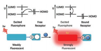
PET is an excited state electron transfer process by which an excited electron is transferred from donor to acceptor. The typical structure of PET probe is composed of three parts, including the fluorophore, the receptor and the spacer. The fluorophore is the part that can absorb light and emit fluorescence, while the receptor is to selectively binding to the donor and transmit the message to the fluorophore. The two parts is linked by the spacer [24]. One of the typical principles of PET sensors is shown in Fig. 1, in which the receptor is an electron donor and the fluorophore plays the role of an electron acceptor. Upon excitation of the fluorophore, an electron of the highest occupied molecular orbital (HOMO) is promoted to the lowest unoccupied molecular orbital (LUMO), which subsequently enables PET from the HOMO of the receptor to that of the fluorophore, resulting in fluorescence quenching. However, after the locking of free receptor, the energy of relevant HOMO becomes lower than that of the fluorophore. Therefore, PET process is inhibited and fluorescence quenching is suppressed. This process is called a-PET. Another PET path is d-PET, in which the fluorophore is the electron donor. When the donor is excited, the similar electron promotion will happen, making fluorescence to be quenched [24].
Figure 1

Based on the PET mechanism, Xu et al. [25] has designed a probe based on coumarin derivatives. In this probe, they introduced the strong electron-withdrawing nitro group in the 3-position of the coumarin as the recognition site for NTR. Upon addition of NTR, the PET pathway between nitro group and coumarin is suppressed, resulting in enhanced fluorescent emission at around 511 nm. Importantly, this probe was successfully applied to detect the endogenous NTR in the tumor tissue of mice with the penetration depth up to 90 μm.
Shao and coworkers [26] developed a "Turn-On" fluorescent probe by incorporating the same moiety 5-nitrofuran into BODIPY dye. Due to the merits of high fluorescence quantum yield, high extinction coefficient, excellent photochemical stability and low biological toxicity [27-30], BODIPY dyes have been widely used as chemosensors and labeling reagents. As expected, this probe showed weak fluorescence due to the PET process. After reacting with NTR, the fluorescence of this probe at 520 nm had 20-fold enhancement with a low detection limit of 9.6 ng/mL. Furthermore, the probe could be applied for monitoring the hypoxic status of A549 tumor cells as well as for real-time quantitative determination of NTR produced by Escherichia coli.
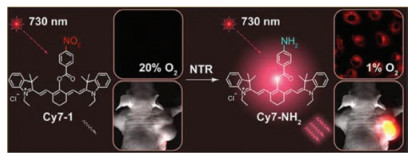
In order to monitor in vivo activity of NTR, Li et al. [31] developed a near-infrared (NIR) fluorescent detection probe (Cy7-1) for NTR based on cyanine dyes with fluorescence reporting structure and nitro aromatic groups as NTR moiety (Fig. 2). Confocal fluorescence imaging of hypoxic A549 cells has confirmed the NTR detection ability of Cy7-1 at the cellular level. Importantly, Cy7-1 can detect tumor hypoxia in a murine hypoxic tumor model, showing a rapid and significant enhancement of fluorescence characteristics suitable for in vivo fluorescence bioimaging of NTR.
Figure 2

3. Fluorescent probes for NTR based on domino reaction
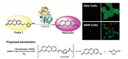
Actually, some NTR probes are often designed and developed by utilizing the domino reaction induced by reduction of the nitro group on the fluorophore to hydroxylamine or ammonia. Electron rearrangement and cleavage of carbon-oxygen bonds occur when the enzyme reduces the nitro group in the probe, releasing the fluorophore and causing a change in the optical signal. For example, Ma's group recently [11] reported a new spectroscopic "Off-On" probe, which was designed by connecting 5-nitrofuran as recognizing moiety to resorufin through an ether bond. As shown in Fig. 3, the probe 1 is first quenched by alkylation of the 7-hydroxy group. When this probe reacts with NTR in the presence of NADH, the reduction of the nitro moiety happens, followed by the 1, 6-rearrangement-elimination reaction and thereby the release of resorufin. Moreover, the probe can be used to monitor the hypoxic status of tumor cells via the detection of endogenous NTR. By replacing 5-nitrofuran with 5-nitrothiophene coupled with resorufin, the same group designed another NTR probe based on domino reaction [32].
Figure 3

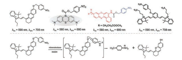
By tethering the p-nitrobenzyl moiety to different fluorophores, a number of probes with domino reaction have been reported as shown in Fig. 4 [33-36]. In the absence of NTR, those probes showed weak fluorescence. Upon addition of NTR, the p-nitrobenzyl moieties of these probes were reduced to form the p-aminobenzyl derivative, which is unstable and spontaneously collapses to release the initial fluorophores. As a result, the fluorescence was restored and the intensity of emission enhanced dramatically.
Figure 4

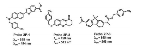
The development of two-photon (2P) fluorescence microscopy provides key advantages over conventional one-photon (1P) fluorescence imaging techniques, including increased penetration depth, lower tissue autofluorescence and self-absorption, and reduced photo-damage and photo-bleaching, and therefore is particularly useful for small molecule fluorescent probes designing in imaging deep tissues and animals [37-42]. Recently, several 2P fluorescent probes have been developed for NTR detection based on domino reaction. For example, Zhang et al. [43] developed a 2P fluorescent probe 2P-1 (Fig. 5) for NTR detection as well as the hypoxic status imaging in tumor cells and bioimaging in tissues. The probe was designed by introducing a p-nitrobenzyl group as NTR reaction moiety, which afford a very weak fluorescence background because of the quenching effect of the p-nitrobenzyl group through a PET process. The probe exhibited both 1P and 2P excited remarkable fluorescence enhancement (~70-fold) for NTR with a detection limit of 20 ng/mL. Liu et al. [44] designed and synthesized a novel 2P fluorescence "Turn-On" NTR probe 2P-2 (Fig. 5), based on a new platform GCTPOC. The mechanism in the detection of NTR is similar to the probe 2P-1 mentioned above. Importantly, a penetration depth of 70 μm by using 2P fluorescence microscopy was achieved in the tumor tissues. In 2017, a novel probe 2P-3 by using p-nitrobenzyl carbamate group as a recognition domain for NTR was reported by Zhai's group [45]. This probe showed a high sensitivity with a detection limit of 23.67 ng/mL, high selectivity, low cytotoxicity and good photostability. The probe was successfully proved for the detection of hypoxic status in cancer cells and tumor tissues with an imaging depth of up to 200 μm with 2P fluorescence microscopy, which implies that the probe is organism-permeable, and that the zebrafish embryos have a detectable amount of endogenous NTR.
Figure 5

4. Fluorescent probes for NTR based on intramolecular charge transfer (ICT) mechanism
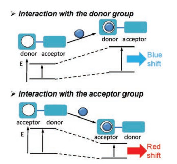
A fluorophore contains an electron-donating group conjugated to an electron-withdrawing group; in this case, it easily undergoes intramolecular charge transfer (ICT) from the donor to the acceptor upon excitation by light. As a result, the emission peak of the fluorescence spectra will be changed, namely as Stokes' shift. As shown in Fig. 6, when a group in the fluorophore acting as an electron donor interacts with the analyst, the latter reduces the electron-donating character of the group, resulting in a blue shift of the absorption spectrum together with a decrease of the extinction coefficient. On the contrary, if the analyst interacting with the acceptor group, it can enhance the electron-withdrawing character of this group and subsequent lead to a red shift and an increase of the extinction coefficient [24, 46]. Based on ICT mechanism, a number of ratiometric small molecule fluorescent probes have been designed for biomolecule detection, including NTR.
Figure 6

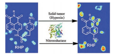
For example, Xiao et al. [10], for the first time, has reported a selective ratiometric fluorescent probe for NTR detection. As shown in Fig. 7, the electron-withdrawing carbamate group weakens the ICT effect and results in a fluorescent emission wavelength blue shift to 475 nm. Upon reduction, the amino group in the probe could be released and the fluorescence emission was restored at around 550 nm. The probe was also successfully applied in cell imaging of NTR.
Figure 7

In addition, Xu, et al. [47] synthesized the NIR probe semi-CyHP for NTR and hypoxia detection relies on tethering the p-nitrophenyl group to the indole moiety via an alkenyl linkage. The nitro group inhibits the ICT effect and results in a weak fluorescence emission. Upon reduction, the amino group reconstructed the electronic push-pull system and the fluorescence emission was restored, which was in accordance with an 85-fold enhancement of fluorescence intensity and could be used for cell imaging applications with minimal endogenous interference.
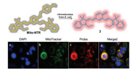
However, most of the current probes can only be used for the detection and imaging of NTR in the cytoplasm. In fact, mitochondria are the main source of nitroradicals in mammalian cells, it is essential to develop small molecule fluorescent probes to monitor mitochondria-specific NTR and elucidate its functions and dynamics. Until now, there are a few fluorescent probes concerning NTR activity in mitochondria. For example, Chevalier et al. [48] proposed mitochondrial probe Mito-NTR by using a profluorescent NIR dye. This probe was characterized for selective NIR imaging of NTR in the mitochondria, which is the first probe for the mitochondrial NTR activity detection (Fig. 8).
Figure 8

Next, Jiang et al. [49] developed a novel fluorescent benzoindocyanine probe (BICP) for mitochondrial NTR activity monitoring and imaging via extending a benzo-indole moiety into a benzoindocyanine based fluorophore (BICF) with a strong ICT effect. Live cell imaging of HeLa and A549 demonstrates that the developed BICP was able to realize sensitive and selective mitochondrial NTR activity probing with low background. The same mechanism was also used in Landry's group by incorporating nitrobenzyl groups to fluorescein [50]. In this study, both of the degree of hypoxia and NTR levels have been proved to be largely invariant in the tumor tissue whether 7 or 35 days post implantation.
5. Fluorescent probes for NTR based on aggregation-induced emission (AIE)
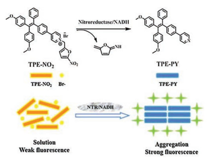
AIE fluorophores have high emission efficiency when their molecules are aggregated [51]. Due to this unique property, The AIE fluorophores have been exploited to serve as novel optical materials and probes for biosensing and imaging applications [52-55]. For example, You et al. [56] has recently reported a new fluorescent "Turn-On" probe for NTR detection by taking advantage of the AIE. TPE-NO2 can detect NTR at concentrations as low as 5 ng/mL in aqueous solution and monitor the endogenous NTR levels in tumor cells (Fig. 9).
Figure 9

Another example of small molecule fluorescent probe based on AIE was designed by Lin's group [57]. They used the tetraphenylethene as the fluorophore, and the nitro group as the NTR recognition site. As expected, this probe showed AIE characteristics, high sensitivity and selectivity, and low cytotoxicity. Importantly, the probe was successfully applied to monitor endogenous NTR in living HeLa cells.
6. Conclusions
By now, great progress has been made in small molecule fluorescent probes for NTRs. Furthermore, the development of fluorescent NTRs probes could facilitate exploring the level of hypoxia in the living system. It is believed that the ideal fluorescent probes with high performances must have the following characteristics: high selectivity, sensitivity, photostability and cell permeability. The following factors should be taken into account as well. First, the probes should have the high solubility in purely aqueous solution. Second, NIR or two-photon fluorescent probes with deeper tissue penetration, less photodamage and lower auto-fluorescence should be developed. In addition, exploring the fluorescence sensing mechanism is crucial to develop high performance fluorescent probes. In summary, the ongoing research in developing fluorescent probes for NTRs will provide an effective tool for investigating the biological function of NTR and the relationship between hypoxia and human disease.
Acknowledgments
This work was financially supported by the National Natural Science Foundation of China (Nos. 81672508 and 61505076), Jiangsu Provincial Foundation for Distinguished Young Scholars (No. BK20170041), Jiangsu Key Research and Development Program (No. BE2015699), Natural Science Foundation of Guangdong Province (No. 2017A030313299) and State Key Laboratory of Pulp and Paper Engineering (No. 201706).
-
-
[1]
A. Mukherjee, S.E. Rokita, J. Am. Chem. Soc. 137(2015) 15342-15345. doi: 10.1021/jacs.5b07540
-
[2]
B.D. Palmer, P.V. Zijl, W.A. Denny, W.R. Wilson, J. Med. Chem. 38(1995) 1229-1241. doi: 10.1021/jm00007a019
-
[3]
D.W. Bryant, D.R. McCalla, M. Leeksma, et al., Can. J. Microbiol. 27(1981) 81-86. doi: 10.1139/m81-013
-
[4]
P.R. Race, A.L. Lovering, R.M. Green, A. Ossor, et al., J. Biol. Chem. 280(2005) 13256-13264. doi: 10.1074/jbc.M409652200
-
[5]
M.M. Shan, J.C. Spain, Biochem. Biophys. Res. Commun. 220(1996) 563-568. doi: 10.1006/bbrc.1996.0443
-
[6]
B. Bhushan, A. Halasz, J. Hawari, Biochem. Biophys. Res. Commun. 322(2004) 271-276. doi: 10.1016/j.bbrc.2004.07.115
-
[7]
E.M. Williams, R.F. Little, A.M. Mowday, M.H. Rich, et al., Biochem. J. 471(2015) 131-153. doi: 10.1042/BJ20150650
-
[8]
F.J. Peterson, R.P. Mason, J. Hovsepian, J.L. Holtzman, J. Biol. Chem. 254(1979) 4009-4014. http://www.ncbi.nlm.nih.gov/pubmed/374406
-
[9]
C. Bryant, M. DeLuca, J. Biol. Chem. 266(1991) 4119-4125. http://www.ncbi.nlm.nih.gov/pubmed/1999405/
-
[10]
L. Cui, Y. Zhong, W. Zhu, et al., Org. Lett. 13(2013) 928-931. http://europepmc.org/abstract/MED/21268631
-
[11]
Z. Li, X. Li, X. Gao, et al., Anal. Chem. 85(2013) 3926-3932. doi: 10.1021/ac400750r
-
[12]
J.M. Brown, W.R. Wilson, Nat. Rev. Can. 4(2004) 437-447. doi: 10.1038/nrc1367
-
[13]
Y. Chen, L. Hu, Med. Res. Rev. 29(2009) 29-64. doi: 10.1002/med.v29:1
-
[14]
C. Berne, L. Betancor, H.R. Luckarift, J.C. Spain, Biomacromolecules 7(2006) 2631-2636. doi: 10.1021/bm060166d
-
[15]
W.A. Denny, Curr. Pharm. Des. 8(2002) 1349-1361. doi: 10.2174/1381612023394584
-
[16]
P.F. Searle, M.J. Chen, L. Hu, et al., Clin. Exp. Pharmacol. Physiol. 31(2004) 811-816. doi: 10.1111/cep.2004.31.issue-11
-
[17]
G. Xu, H.L. McLeod, Clin. Cancer Res. 7(2001) 3314-3324. http://www.ncbi.nlm.nih.gov/pubmed/11705842
-
[18]
H. Schagger, G. von Jagnw, Anal. Biochem. 166(1987) 368-379. doi: 10.1016/0003-2697(87)90587-2
-
[19]
R.J. Long, K.K. Papas, A. Sambanis, I. Constantinidis, J. Magn. Reson. 146(2000) 49-57. doi: 10.1006/jmre.2000.2112
-
[20]
M.J. Povich, Anal. Chem. 47(1975) 346-347. doi: 10.1021/ac60352a039
-
[21]
X. Li, X. Gao, W. Shi, H. Ma, Chem. Rev. 114(2014) 590-659. doi: 10.1021/cr300508p
-
[22]
Y. Yang, Q. Zhao, W. Feng, F. Li, Chem. Rev. 113(2013) 192-270. doi: 10.1021/cr2004103
-
[23]
C. Yu, X. Li, F. Zeng, F. Zheng, S. Wu, Chem. Commun. 49(2013) 403-405. doi: 10.1039/C2CC37329G
-
[24]
B. Valeur, I. Leray, Coord. Chem. Rev. 205(2000) 3-40. doi: 10.1016/S0010-8545(00)00246-0
-
[25]
A. Xu, Y. Tang, Y. Ma, et al., Sens. Actuators B:Chem. 252(2017) 927-933. doi: 10.1016/j.snb.2017.06.043
-
[26]
J. Xu, S. Sun, Q. Li, Y. Yue, Y. Li, S. Shao, Analyst 14(2015) 574-581. https://www.ncbi.nlm.nih.gov/pubmed/25422882
-
[27]
A. Loudet, K. Burgess, Chem. Rev. 107(2007) 4891-4932. doi: 10.1021/cr078381n
-
[28]
G. Ulrich, R. Ziessel, A. Harriman, Angew. Chem. Int. Ed. 47(2008) 1184-1201. doi: 10.1002/anie.200702070
-
[29]
S. Zhu, J. Zhang, G. Vegesna, et al., Org. Lett. 13(2010) 438-441. http://www.ncbi.nlm.nih.gov/pubmed/21175151/
-
[30]
Y. Li, Y. Sun, J. Li, et al., J. Am. Chem. Soc. 137(2015) 6407-6416. doi: 10.1021/jacs.5b04097
-
[31]
L. Li, J. Han, B. Nguyen, K. Burgess, J. Org. Chem. 73(2008) 1963-1970. doi: 10.1021/jo702463f
-
[32]
Z. Li, X. Gao, Y. Zhang, et al., Chem. Commun. 49(2013) 5859-5861. doi: 10.1039/c3cc42610f
-
[33]
T. Guo, L. Cui, J. Shen, et al., Chem. Commun. 49(2013) 10820-10822. doi: 10.1039/c3cc45367g
-
[34]
Y. Shi, S. Zhang, X. Zhang, Analyst 138(2013) 1952-1955. doi: 10.1039/c3an36807f
-
[35]
Z. Li, X. He, Z. Wang, et al., Biosens. Bioelectron. 63(2015) 112-116. doi: 10.1016/j.bios.2014.07.024
-
[36]
J. Bae, L. McNamara, M. Nael, et al., Chem. Commun. 51(2015) 12787-12790. doi: 10.1039/C5CC03824C
-
[37]
L. Qian, L. Li, S. Yao, Acc. Chem. Res. 49(2016) 626-634. doi: 10.1021/acs.accounts.5b00512
-
[38]
M. Pawlicki, H.A. Collins, R.G. Denning, H.L. Anderson, Angew. Chem. Int. Ed. 48(2009) 3244-3266. doi: 10.1002/anie.v48:18
-
[39]
H.M. Kim, B.R. Cho, Chem. Commun. (2009) 153-164. http://www.ncbi.nlm.nih.gov/pubmed/19099055
-
[40]
L. Li, X. Shen, Q. Xu, S. Yao, Angew. Chem. Int. Ed. 125(2013) 442-446. doi: 10.1002/ange.201205940
-
[41]
L. Li, C. Zhang, G. Chen, et al., Nat. Commun. 5(2014) 3267-3275. doi: 10.1038/ncomms4267
-
[42]
L. Li, J. Ge, H. Wu, Q. Xu, S. Yao, J. Am. Chem. Soc. 134(2012) 12157-12167. doi: 10.1021/ja3036256
-
[43]
J. Zhang, H. Liu, X. Hu, et al., Anal. Chem. 87(2015) 11832-11839. doi: 10.1021/acs.analchem.5b03336
-
[44]
Z. Liu, Y. Tang, A. Xu, W. Lin, Biosens. Bioelectron. 89(2017) 853-858. doi: 10.1016/j.bios.2016.09.107
-
[45]
B. Zhai, W. Hu, J. Sun, et al., Analyst 142(2017) 1545-1553. doi: 10.1039/C7AN00058H
-
[46]
M. Liu, H. Tan, Z. Liu, W. Wang, W. Zeng, Chin. J. Org. Chem. 33(2013) 1655-1667. doi: 10.6023/cjoc201301015
-
[47]
J. Yuan, Y. Xu, N. Zhou, et al., RSC Adv. 4(2014) 56207-56210. doi: 10.1039/C4RA10044A
-
[48]
A. Chevalier, Y. Zhang, O.M. Khdour, J.B. Kaye, S.M. Hecht, J. Am. Chem. Soc. 138(2016) 12009-12012. doi: 10.1021/jacs.6b06229
-
[49]
B. Huang, W. Chen, Y. Kuang, et al., Org. Biomol. Chem. 15(2017) 4383-4389. doi: 10.1039/C7OB00781G
-
[50]
S. Luo, R. Zou, J. Wu, M.P. Landry, ACS Sens. 2(2017) 1139-1145. doi: 10.1021/acssensors.7b00171
-
[51]
J. Xie, J. Lam, L. Cheng, et al., Chem. Commun. (2001) 1740-1741.
-
[52]
R.T.K. Kwok, C.W.T. Leung, J.W.Y. Lam, B.Z. Tang, Chem. Soc. Rev. 44(2015) 4228-4238. doi: 10.1039/C4CS00325J
-
[53]
C. Yu, Y. Wu, F. Zeng, et al., Biomacromolecules 14(2013) 4507-4514. doi: 10.1021/bm401548u
-
[54]
Y. Wu, S. Huang, F. Zeng, et al., Chem. Commun. 51(2015) 12791-12794. doi: 10.1039/C5CC04771D
-
[55]
H. Xie, F. Zeng, C. Yu, S. Wu, Polym. Chem. 4(2013) 5416-5424. doi: 10.1039/c3py00586k
-
[56]
X. You, L. Li, X. Li, et al., Chem. Asian J. 11(2016) 2918-2923. doi: 10.1002/asia.201600945
-
[57]
G. Xu, Y. Tang, Y. Ma, A. Xu, W. Lin, Spectrochim. Acta A:Mol. Biomol. Spectrosc. 188(2018) 197-201. doi: 10.1016/j.saa.2017.06.065
-
[1]
-
-

 DownLoad:

 下载:
下载:


 扫一扫看文章
计量
- PDF下载量: 1
- 文章访问数: 1120
- HTML全文浏览量: 51
 下载:
下载:
