Citation:
Zhao Chun-Ling, Hua Mei, Yang Can-Yu, Yang Yun-Hui. A novel aptasensor based on 3D-inorganic hybrid composite as immobilized substrate for sensitive detection of platelet-derived growth factor[J]. Chinese Chemical Letters,
2017, 28(7): 1417-1423.
doi:
10.1016/j.cclet.2017.02.010
A novel aptasensor based on 3D-inorganic hybrid composite as immobilized substrate for sensitive detection of platelet-derived growth factor
Received Date:
09 November 2016 Accepted Date:
06 February 2017 Revised Date:
14 January 2017 Available Online:
22 July 2017
Abstract:
A novel electrochemical detection approach for platelet-derived growth factor (PDGF) via "sandwich" structure is reported in this paper. 3D-4MgCO3·Mg(OH)2·4H2O-Au NPs inorganic hybrid composite was utilized as immobilized substrate for sensitive PDGF detection and Pt-Au bimetallic nanoparticles were labelled on PDGF aptamer to indirectly detect PDGF for the first time. The proposed aptasensor exhibited a high catalytic efficiency towards reduction of H2O2, hence the sensitive detection of PDGF was achieved. Results showed that the aptasensor exhibited excellent linear response to PDGF, in the range of 0.1 pg/mL-10 ng/mL (4 fmol/L-400 pmol/L), with detection limit of 0.03 pg/mL (1.2 fmol/L).
With increasing application of proteomic strategies for cancer diagnosis and disease studies, the detection of cancer-related proteins attracts increasing attention in the biomedical fields [1]. There has been increasing interest recently in protein sensing based on SAMs of aptamers [2, 3]. Aptamers generated by an in vitro selection process, termed as systematic evolution of ligands by exponential enrichment (SELEX) [4, 5], are short, single-stranded oligonucleotides (RNA or DNA) that can bind to various targets, including proteins, cells, small molecules and so on [6-10]. In comparison to antibodies, aptamers possess certain advantages, including their relatively simple and inexpensive synthesis through automation, tolerance to internal labeling, and long storage times without altering or losing their biological properties [11].
Synthesis of nanoparticles with desired size/morphology has enormous importance, especially in the compelling field of nanotechnology [12]. Gold nanoparticles (Au NPs) have attracted considerable interest in analytical and biomedical fields [13, 14]. The rapid and simple chemical synthesis, narrow size distribution, and good biocompatibility, have enabled Au NPs to be extensively used for the preparation of biosensors and signal amplification [15]. Au NPs are often combined with other biocompatible materials such as polymer matrix, inorganic micro/nanomaterials. Nanocomposite powders can be promising antimicrobial agents and fluorescent material for biodetection due to their optical and bioactive properties [16], among them, calcium carbonate (CaCO3) could be deemed as good candidate due to their porous surface structure, good biocompatibility and large surface area [17, 18]. Song et al. has synthesized porous hydromagnesite (4MgCO3·Mg(OH)2·4H2O) microspheres with rosette-like morphology by a facile pathway [19]. It also has large surface area and porous surface structure. It can be predict that 4MgCO3·Mg(OH)2·4H2O also has good biocompatibility because calcium and magnesium have very similar features.
To explore the distinct advantages of current inorganic hybrid composites is necessary. Investigations of bimetallic nanoparticles are also gaining broad attention very recently, due to their size and shape-dependent physical, chemical and electrochemical proper-ties. When Pt combines with another metal in a bimetallic composition, it has often shown higher catalytic activity, compared with the monometallic one [20, 21]. Pt-based bimetallic electro-catalysts have therefore been widely used in sensing and biosensing applications.
PDGF is a protein that regulates cell growth and division. However, a survey of literature shows there have been only a few reports on sensing for this important protein [22]. Lai and coworkers described an electrochemical aptasensor for the detection of PDGF in blood serum based on conformational switching of an immobilized aptamer on adaptively binding to the target. The detection limit was 1 nmol/L directly in undiluted, unmodified blood serum and at 50 pmol/L (1.25 ng/mL) in serum-diluted 2-fold with aqueous buffer [23]. Yu's group demonstrated sensitive electrochemical strategies for the detection of PDGF on the basis of rolling circle amplification followed by enzymatic silver deposition and proximity-dependent surface hybridization [24, 25]. It was demonstrated that the rolling circle amplification method was highly sensitive and specific with a wide detection range of 4 orders of magnitude and a detection limit as low as 10 fmol/L. Wang et al. also reported a research about PDGF detection combining aptamer with gold nanoparticles for electrochemical signal amplification. This PDGF detection approach obtains a low detection limit of 1 ×10-14 mol/L for purified samples, 1 ×10-12 mol/L for contaminated-ridden samples or undiluted blood serum [26]. Deng et al. have constructed a label-free electrochemical aptamer-based sensor (aptasensor) for detection of plateletderived growth factor (PDGF) based on the direct electrochemistry of glucose oxidase (GOD). The aptasensor showed excellent electrochemical response and the peak current decreased linearly with increasing logarithm of PDGF concentration from 0.005 nmol/ L to 60 nmol/L, with a relatively low detection limit of 1.7 pmol/L [27]. Bai et al. reported a sandwich-type electrochemical aptasensor for simultaneous sensitive detection of platelet-derived growth factor (PDGF) and fabrication of thrombin [28]. Chai et al. developed a novel electrochemiluminescence (ECL) aptasensor for platelet-derived growth factor B chain (PDGF-BB) assay, by assembling N-(aminobutyl)-N-ethylisoluminol functionalized gold nanoparticles (ABEI-Au NPs), with aptamers as nanoprobes [29]. Wang's group reported a novel fluorescent detection for PDGF-BB based on dsDNA-templated copper nanoparticles [30]. Recently, a novel VS2–graphene (VS2–GR) composite is firstly prepared by a facile one-step hydrothermal method to construct a sensitive, universal and label-free electrochemical aptamer-based sensor (aptasensor) for the detection of the platelet-derived growth factor BB (PDGF-BB) coupled with Exo Ⅲ-aided autocatalytic signal amplification [31]. All of these reports showed excellent linear response and high sensitivity for the aptasensors, but the methods were expensive and complicated. Development of a new detection method with simplicity, low cost, high sensitivity and stability is therefore necessary.
Based on the reasons mentioned above, we present a simple preparation and characterization of porous 3D-4MgCO3·Mg (OH)2·4H2O-Au NPs inorganic hybrid composite for the first time in this work. 3D-4MgCO3·Mg(OH)24H2O has a high mechanical strength and large surface area which can load abundant Au NPs, also can provide a friendly environment for aptamer to retain their activity. Pt-Au alloy nanoparticles were also synthesized by a facile pathway. In the presence of chitosan (CHIT), 3D-4MgCO3·Mg (OH)2·4H2O-Au NPs were modified on the surface of glassy carbon electrode. The thiolated PDGF aptamer Ⅰ was then immobilized on the electrode surface via self-assembled technology. In addition, PDGF aptamer Ⅱ was labeled with Pt-Au bimetallic nanoparticles. Since PDGF has two binding sites, one site binds to the aptamer immobilized on modified GCE and another site binds to the aptamer Ⅱ which has been labeled with Pt-Au NPs. The PDGF aptamer Ⅱ/PDGF/PDGF aptamer Ⅰ "sandwich" structure was finally formed. Because the Pt-Au bimetallic nanoparticles have great electrocatalytic activity for reduction of H2O2, quantitative determination of PDGF could be achieved by detecting the reduction current of H2O2. This method is novel, sensitive, stable and practicable.
2.
Results and discussion
2.1
Morphological characterization of 3D-4MgCO3·Mg(OH)2·4H2O and 3D-4MgCO3·Mg(OH)2·4H2O-Au NPs inorganic hybrid composite
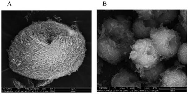
SEM is a powerful tool to characterize the morphology of different composites. Fig. 1 shows the SEM images of the porous hydromagnesite microspheres (3D-4MgCO3·Mg(OH)2·4H2O) microparticles before (Fig. 1A) and after loading of Au NPs (Fig. 1B). It was obvious to see that the prepared 3D-4MgCO3·Mg(OH)2·4H2O microparticles possessed porous rosette or three-dimension nest structure, large surface area and a uniformly multilayered structure, which could highly strengthen the amount and stability of the immobilized Au-NPs. After the adsorption of Au NPs, 4MgCO3·Mg(OH)2·4H2O microparticles retained the original three-dimension nest structure but became relatively smooth (Fig. 1B).
图 1
图 1
SEM images of 3D-4MgCO3·Mg(OH)2·4H2O (A) and 3D-4MgCO3·Mg(OH)2·4H2O-AuNPs (B).
Figure 1.
SEM images of 3D-4MgCO3·Mg(OH)2·4H2O (A) and 3D-4MgCO3·Mg(OH)2·4H2O-AuNPs (B).
2.2
XRD characterization of 3D-4MgCO3·Mg(OH)2·4H2O and 3D-4MgCO3·Mg(OH)2·4H2O-Au NPs inorganic hybrid composite
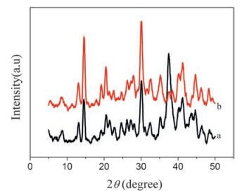
The XRD pattern of the spherical product (4MgCO3·Mg (OH)2·4H2O) is shown in Fig. 2(a). All the peaks in this figure can be indexed to the monoclinic crystalline phase of 4MgCO3·Mg (OH)2·4H2O in agreement with reference data (PD F 25-0513) [19]. Compared with Fig. 2(a), the XRD pattern of 3D-4MgCO3·Mg(OH)2·4H2O-Au NPs inorganic hybrid composite only has slightly change at 37 ° (Fig. 2(b)).
图 2
图 2
XRD pattern of 3D-4MgCO3·Mg(OH)2·4H2O (a) and 3D-4MgCO3.Mg (OH)2 4H O-Au NPs inorganic hybrid composite (b).
Figure 2.
XRD pattern of 3D-4MgCO3·Mg(OH)2·4H2O (a) and 3D-4MgCO3.Mg (OH)2 4H O-Au NPs inorganic hybrid composite (b).
2.3
Characterization of Pt-Au bimetallic nanoparticles
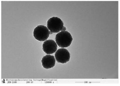
The as-synthesized Pt-Au bimetallic nanoparticles were characterized by TEM as shown in Fig. 3. The Pt-Au bimetallic nanoparticles showed red bayberry structure and a uniform size of approximately 290nm, which could provide a high loading amount for aptamer.
图 3
图 3
TEM images of Pt-Au bimetallic nanoparticles.
Figure 3.
TEM images of Pt-Au bimetallic nanoparticles.
2.4
Impedance characterization of various modified electrodes
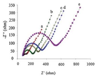
EIS could provide information on the impedance changes at various modified electrode surface. Fig. 4 shows the nyquist plots obtained on bare GCE, 3D-4MgCO3·Mg(OH)2·4H2O-Au NPs/CHIT/ GCE, aptamer Ⅰ/3D-4MgCO3·Mg(OH)2·4H2O-Au NPs/CHIT/GCE, PDGF/aptamer Ⅰ/3D-4MgCO3·Mg(OH)2·4H2O-Au NPs/CHIT/GCE, aptamer Ⅱ/PDGF/aptamer Ⅰ/3D-4MgCO3·Mg(OH)2·4H2O-Au NPs/ CHIT/GCE in a solution of 0.1mol/L KCl, containing 5.0mmol/L K3 Fe(CN)6 and K4Fe(CN)6. The impedance plot of the bare GCE exhibited a relative lowimpedance value and was almost a straight line, which was the diffusion-controlled process corresponding to Warburg impedance. After the bare GCE was modified by 3D-4MgCO3·Mg(OH)2·4H2O-Au NPs/CHIT, it showed a higher interfacial electron transfer resistance (Ret), indicating that modified 3D-4MgCO3·Mg(OH)2·4H2O-Au NPs/CHIT could obstruct the electron transfer. Impedance value went on increasing after the thiolated aptamers were self-assembled onto 3D-4MgCO3·Mg(OH)2·4H2OAu NPs/CHIT/GCE, indicating that the aptamer had been immobilized onto the electrode surface and obstructed electron transfer. After aptamer I/3D-4MgCO3·Mg(OH)2·4H2O-Au NPs/CHIT/GCE was incubated with PDGF and aptamer Ⅱ respectively, the Ret increased obviously, indicating that the PDGF had combined with aptamer Ⅰ, followed by reacting with aptamer Ⅱ to form a sandwich structure.
图 4
图 4
Nyquist plots of impedance spectra obtained in 5 mmol/L [Fe(CN)6]3-/4-, (a) bare GCE, (b)3D-4MgCO3·Mg(OH)2·4H2O-Au NPs/CHIT/GCE, (c) aptamer Ⅰ/3D-4MgCO3·Mg(OH)2·4H2O-Au NPs/CHIT/GCE, (d)PDGF/aptamer Ⅰ/3D-4MgCO3.Mg(OH)2.4H2O-Au NPs/CHIT/GCE, (e) aptamer Ⅱ/PDGF/aptamer Ⅰ/3D-4MgCO3.Mg (OH)2.4H2O-Au NPs/CHIT/GCE.
Figure 4.
Nyquist plots of impedance spectra obtained in 5 mmol/L [Fe(CN)6]3-/4-, (a) bare GCE, (b)3D-4MgCO3·Mg(OH)2·4H2O-Au NPs/CHIT/GCE, (c) aptamer Ⅰ/3D-4MgCO3·Mg(OH)2·4H2O-Au NPs/CHIT/GCE, (d)PDGF/aptamer Ⅰ/3D-4MgCO3.Mg(OH)2.4H2O-Au NPs/CHIT/GCE, (e) aptamer Ⅱ/PDGF/aptamer Ⅰ/3D-4MgCO3.Mg (OH)2.4H2O-Au NPs/CHIT/GCE.
The experimental conditions, such as the incubation time of PDGF, concentration of 3D-4MgCO3·Mg(OH)2·4H2O-Au NPs, PDGF aptamer Ⅱ labeled with Pt-Au alloy and H2O2 were also investigated. Different incubation times of PDGF might cause an obvious difference in the response peak current, which presents the optimal reaction time between the aptamer and its target. Thus, a series of times were investigated to find the optimum incubation time using the same analyte concentrations in the study(Fig.S1 inSupporting information).It took some timetoform steady complexes after the target analytes were incubated on the surface of the aptasensor. Results showed that the optimum incubation time for PDGF was 75min. The current response increased and then reached a plateau with the increase of the concentrations of 3D-4MgCO3·Mg(OH)2·4H2O-Au NPs/CHIT because the loading amount of thiolated PDGF aptamer Ⅰ increased (Fig. S2 in Supporting information). Therefore, 2.0mg/mL of 3D-4MgCO3·Mg(OH)2·4H2O-Au NPs/CHIT was adopted as optimum concentrations. It can be seen from Fig.S3 (Supporting information) that the current response increased as the concentration of PDGF aptamer Ⅱ labelled with Pt-Au alloy increases and reached a maximum at 0.8 μmol/L. The reason why current decreased after that may be owe to the electron transfer obstruction of too much aptamer. Thus, 0.8 μmol/L was selected as optimum concentration of PDGF aptamer Ⅱ labeled with Pt-Au alloy. The current response increased withthe increase of theconcentrationsof H2O2 (Fig. S4 in Supporting information). However, the concentration of H2O2 in excess of 1.0mol/L is unnecessary. Therefore, 1.0mol/L of H2O2 was adopted as optimum concentrations added in 10mL of PBS (100 mL).
2.6
Study on different catalytic efficiency of 3D-4MgCO3·Mg (OH)2·4H2O-AuNPs inorganic hybrid composite and Pt-Au bimetallic nanoparticles
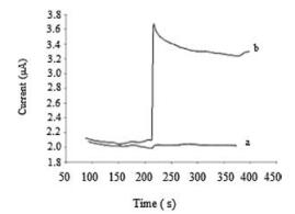
In order to prove whether the catalytic reduction of H2O2 was mainly dependent on Pt-Au bimetallic nanoparticles, a controlled experiment was carried out. The result is shown in Fig. 5. Curve a was obtained at the 3D-4MgCO3·Mg(OH)2·4H2O-AuNPs inorganic hybrid composite-modified electrode for the catalytic reduction of H2O2, while curve b was obtained at the Pt-Au bimetallic nanoparticles Isabelle PDGF aptamer Ⅱ/PDGF/PDGF aptamer Ⅰ "sandwich" structure-modified electrode for the catalytic reduction of H2O2. It can be seen that the catalytic efficiency of the 3D-4MgCO3·Mg(OH)2·4H2O-AuNPs inorganic hybrid composite was much lower than that of Pt-Au bimetallic nanoparticles Isabelle PDGF aptamer Ⅱ/PDGF/PDGF aptamer Ⅰ "sandwich" structuremodified electrode. It demonstrates that the 3D-4MgCO3·Mg (OH)2·4H2O-Au NPs inorganic hybrid composite was just used as immobilized substrate in this research. The result also indicates that the Pt-Au bimetallic nanoparticles had excellent catalytic efficiency towards reduction of H2O2.
图 5
图 5
The different catalytic efficiency of 3D-4MgCO3·Mg(OH)2·4H2O-AuNPs inorganic hybrid (a)and Pt-Au bimetallic nanoparticles (b) to the reduction of H2O2 at -0.1 V.
Figure 5.
The different catalytic efficiency of 3D-4MgCO3·Mg(OH)2·4H2O-AuNPs inorganic hybrid (a)and Pt-Au bimetallic nanoparticles (b) to the reduction of H2O2 at -0.1 V.
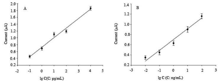
Since the Pt-Au bimetallic nanoparticles had high catalytic efficiency towards the reduction of H2O2 in this work, an aptasensor, using the Pt-Au bimetallic nanoparticles as labels, was constructed for the detection of PDGF via the "sandwich" structure. Under the optimized experimental conditions, the proposed aptasensor had a linear relationship between the current response towards the concentration of PDGF. The calibration curve is shown in Fig. 6A. The catalytic current increased linearly with the concentration of PDGF within the range of 0.1 pg/mL–10 ng/mL (4 fmol/L–400 pmol/L), with a relative coefficient of 0.9826. The detection limit of the aptasensor was 0.03 pg/mL(1.2 fmol/L) at 3s. As a contrast, Fig. 6B shows the calibration curve of the aptasensor based on Au NPs without 4MgCO3·Mg(OH)2·4H2O. It's detection limit is 10 pg/mL, which is 333 times higher than that of the aptasensor based on 3D-4MgCO3·Mg(OH)2·4H2O-Au NPs. This result indicates that 4MgCO3·Mg(OH)2·4H2O can load more Au NPs and enhance the sensitivity. A comparison of different electrochemical aptasensors for the determination of PDGF is shown in Table 1, demonstrating the detection limit of this aptasensor was much lower than that of other electrochemical aptasensors, which can be attributed to the high catalytic efficiency of Pt-Au bimetallic nanoparticles using as label and the 3D structure of 4MgCO3·Mg (OH)2·4H2O-Au NPs inorganic hybrid because it can load a large amount of PDGF aptamer.
图 6
图 6
The calibration curve of the aptasensor based on 3D-4MgCO3·Mg(OH)2·4H2O-AuNPs inorganic hybrid (A) and only Au NPs (B). Each data point is the average of three independent assays.
Figure 6.
The calibration curve of the aptasensor based on 3D-4MgCO3·Mg(OH)2·4H2O-AuNPs inorganic hybrid (A) and only Au NPs (B). Each data point is the average of three independent assays.
2.8
Reproducibility, selectivity and stability of the prepared aptasensor
To evaluate the reproducibility of the aptasensor, a series of measurements were prepared for the detection of 1 ng/mL PDGF. The relative standard deviation (RSD) was 4.8% for five parallel measurements, indicating that reproducibility of the proposed aptasensor was acceptable.
To investigate the selectivity of the aptasensor, the electrochemical aptasensor was incubated with 30 ng/mL ascorbic acid, uric acid, glycine, and glucose, respectively. Results are shown in Table 2, indicating that the selectivity of the aptasensor was quite good.
表 2
表 2
Possible interferences tested with aptasensor.
Table 2.
Possible interferences tested with aptasensor.
The stability of the aptasensor was also examined. The aptasensor was incubated with 1.0 ng/mL PDGF and then measured with cyclic voltammetry. After 100 consequent cyclic voltammetry, the current decreased to 93% of its initial value. The slow decrease in the current response may be attributed to the unique nest structure and excellent biocompatibility of the 3D-4MgCO3·Mg (OH)2·4H2O–Au NPs inorganic hybrid composite.
2.9
The practicability of electrochemical aptasensor in real blood serum
In order to confirm the practicability of the aptasensor in real diluted serum samples, serum sample was diluted and mixed with various concentrations of the PDGF. The recovery is elaborated in Table 3. The results are satisfactory.
In this study, a novel electrochemical aptasensor, based on 3D-4MgCO3·Mg(OH)2·4H2O–Au NPs inorganic hybrid composite as immobilized substrate and Pt-Au bimetallic nanoparticles as labels, has been proposed and tested for detection of PDGF. The Pt-Au bimetallic nanoparticles showed high catalytic efficiency towards the reduction of H2O2. The aptasensor displayed a linear response for detecting PDGF, within a wide PDGF concentration range (0.1 pg/mL–10 ng/mL), low detection limit, good reproducibility, selectivity, and acceptable stability. Compared with aptasensors only based on Au NPs andother types of amperometric aptasensors, the synthesized aptasensor reported in this study exhibited much more excellent properties, which may provide many potential applications in clinical diagnosis.
4.
Experimental
4.1
Materials and instruments
PDGF aptamer Ⅰ 5'-SH(C6) ACA GGC TAC GGC ACG TAG AGC ATC ACC ATG ATC CTG-3', PDGF aptamer Ⅱ 5'-SH(C6) CAG GCT ACG GCA CGT AGA GCA TCA CCA TGA TCC TG-30 and PDGF analyte were both synthesized by Takara biotechnology (Dalian, China).
2-Mercaptoethanol (MCE, HSCH2CH2OH) was obtained from Lanji Biological Limited Corporation (Shanghai, China). Chitosan (MW ~ 1 ×10⁶; 75%–80% deacetylation), deoxyadenosine triphosphate (dATP), sodium dodecyl sulfonate (SDS), polyvinylpyrrolidone (PVP) and tris-(2-carbox-yethyl) phosphine hydrochlorideride (TCEP), which was used to cut disulfide bonds to get free-SH groups, were both purchased from Sigma. Tetrachloroauric acid (HAuCl4) and H2PtCl6·6H2O were obtained from Kunming Borui Chemical Limited Corporation. All other chemicals were of analytical-reagent grade and double distilled water was used throughout. The assay solution was 10 mmol/L phosphate buffer solution (PBS pH 7.4). Serum specimens were obtained from infirmary of Yunnan Normal University and stored at 4 ℃.
Cyclic voltammetry (CV), electrochemical impedance spectroscopy (EIS), and amperometric i-t curve (i-t) were performed with a CHI 660D electrochemistry workstation (Shanghai CHI Apparatus Corporation, China). A standard three electrode system, using saturated calomel electrode (SCE) as the reference electrode, and a platinum wire electrode as the counter electrode, the working electrode, was the "sandwich" structure modified glassy carbon electrode. The scanning electron microscope (SEM) image was obtained using XL30ESEM-TMP microscope (Philips, Holland). The transmission electron microscopy (TEM) images were recorded on a JEM-2100F transmission electron microscope (JEOL, Japan), operating at 200 kV.
4.2
Methods
4.2.1
Synthesis of porous 3D-4MgCO3·Mg(OH)2·4H2O-Au NPs inorganic hybrid composite
The hydromagnesite microspheres (4MgCO3·Mg(OH)2·4H2O) were prepared according to the literature [19]. 160 mL of MgCl2 solution (0.5 mol/L) was placed in a beaker at 293.2 K. Subsequently, 160 mL of NaCO3 solution (0.5 mol/L) was rapidly added into the vigorously stirred reactor within 4 s. The mixture was further stirred for about 15 min with same stirring rate and strength. The temperature was maintained at 293.2 K for 6 h under static conditions. The obtained white precipitate was collected and filtered off, washed with ethanol for three times, and then placed in an oven at 353.2 K for 5 h to obtain MgCO3·3H2O precursor.
The synthesized MgCO3·3H2O (1.0 g) was then added into deionized water which had been already heated to 353.2 K under stirring, and agitation was stopped rapidly. The sample was dispersed at the bottom of the beaker at 352.2 K for 3 h. The obtained precipitate was collected and filtered off, washed with ethanol for three times and dried in an oven at 353.2 K overnight to give the porous hydromagnesite microspheres (4MgCO3·Mg (OH)2·4H2O).
Au NPs were prepared by the conventional citrate reduction of HAuCl4 in aqueous solution according to the literature [34]. In brief, 100 mL of HAuCl4 solution (1 mmol/L) was heated to boiling with vigorous stirring. 10 mL of 38.8 mmol/L sodium citrate solution was then added and the stirring was continued for another 10 min. The color changed from yellow to deep red, indicating the formation of gold nanoparticles.
The preparation of porous 3D-4MgCO3·Mg(OH)2·4H2O-Au NPs inorganic hybrid composite was performed as follows:10 mg of 4MgCO3·Mg(OH)2·4H2O was dispersed in 10 mL of Au colloid solution and sonicated for 2 min. The obtained light purple product was 3D-4MgCO3·Mg(OH)2·4H2O-Au NPs inorganic hybrid composite. After centrifugation, the hybrid composite was washed with double distilled water and dried [19].
4.2.2
Synthesis of Pt-Au bimetallic nanoparticles
Pt-Au bimetallic nanoparticles were synthesized by solvent reduction [35-37]. 1.11 g of PVP and 0.13 g H2PtCl6.6H2O were dissolved in a mixture solution containing 130 mL of methanol and 150 mL of deionized water. Subsequently, 20 mL of 0.1 mol/L NaOH dissolved in methanol was added. The resulting mixture was refluxed in a 70 ℃ oil bath, with stirring for 3 h. The obtained solution was of Pt seeds.
5 mL of 0.0254 mol/L HAuCl4·3H2O and 10 mL of 0.1 mol/L sodium citrate were added into 150 mL of as–prepared Pt seeds above. The reaction solution was refluxed with an oil bath at 70 ℃ under stirring for 1 hour. The obtained Pt-Au bimetallic nanoparticles were centrifuged at 16, 000 rpm for 30 min. The solution was concentrated for five times and then washed with methanol and deionized water respectively. Finally, Pt-Au bimetallic nanoparticles were re-dispersed in deionized water for following experiment.
4.2.3
Preparation of Pt-Au Bimetallic Nanoparticles Tagged Aptamer
30 mL of 235μmol/L dATP was firstly added into 1 mL of five time's concentrated Pt-Au bimetallic nanoparticles solution and incubated at room temperature for about 15–20 min with stirring. 5 μL of 1% SDS was then slowly introduced into the mixture solution and kept steady for 5 min. This was repeated for three times and incubated at room temperature for 10 min with stirring. 50 μL of 2 mol/L NaCl was then slowly added into the mixture solution (2 μL added, mixed and kept at 3 minutes interval each time). This was followed by addition of 10 μL PDGF aptamer Ⅱ (100 μmol/L), which was mixed and incubated at room temperature for 3 h under stirring. The resulting product was then centrifuged at 12, 000 rpm for 20 min. The supernatant was abandoned after centrifugation and precipitate dispersed in 1 μL of PBS buffer(10 mmol/L), mixed and centrifuged for 15 min at 12, 000 rpm for three times. The obtained product was finally dispersed into 1 μL of eluent buffer, containing 20 mmol/L Na3PO4·12H2O, 10% sucrose, 5% BSA, 0.25% Tween 20 and stored at 4 ℃.
4.2.4
Fabrication of "sandwich" sensing system
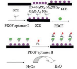
Glassy carbon electrode (GCE 3 mm diameter) was used as base electrode for the fabrication of the aptasensor. Before modification, glassy carbon electrode was first polished with emery paper and alumina slurry, followed by thorough rinsing with double distilled water. After successive sonication in 1:1 nitric acid, absolute alcohol, and deionized water, the electrode was rinsed with deionized water and allowed to dry at room temperature. 10 μL of 3D-4MgCO3·Mg(OH)2·4H2O–Au NPs/CHIT solution, which was prepared with 3D-4MgCO3·Mg(OH)2·4H2O–Au NPs and 0.5% CHIT (1:1), was then pipetted onto the surface of GCE and kept overnight. 10 μL of 0.2 μmol/L thiolated PDGF aptamer Ⅰ, containing 1 mmol/L EDTA, 10 mmol/L TCEP and 0.1 mol/L NaCl, was then placed onto the modified electrode above and kept overnight again. The modified electrode was rinsed with abundant PBS (10 mmol/L NaH2PO4, 10 mmol/L Na2HPO4 and 0.1 mol/L NaCl, pH 7.4), and subsequently blocked with 10 μL of 1 mmol/L MCE, for 1 h, in order to decrease nonspecifically attached aptamer. The prepared PDGF aptasensor was rinsed with PBS (pH 7.4). Before further modification of the aptamer-modified electrode surface, it should be first treated with PBS, to keep the aptamer stable. The sensor was then allowed to incubate at 37 ℃ for 1 h in the presence of 10 μL of different concentrations of PDGF. After being washed with PBS buffer, the aptamer Ⅱ, Isabelle with Pt-Au-NPs, was introduced onto the electrode and incubated for another 1 h. The "sandwich" sensing system was then prepared. The stepwise procedure for the electrochemical aptamer biosensor is shown in Fig. 7. As comparison, aptasensor based on Au NPs was constructed according to above method using Au colloid solution instead of 3D-4MgCO3·Mg(OH)2·4H2O–Au NPs.
图 7
图 7
The stepwise procedure for synthesis of the electrochemical aptamer biosensor.
Figure 7.
The stepwise procedure for synthesis of the electrochemical aptamer biosensor.
4.2.5
Experimental measurements and signal detection
The PBS buffer (pH 7.4) was used for all the electrochemical measurements. CV experiments were carried out in quiescent solutions at a scan rate of 100 mV/s. Amperometric experiments for the aptasensor were recorded in a stirred system. After the background current was stabilized, 1 mol/L H2O2 was added into the buffer and the current change was recorded at -0.1 V. All fabrication steps were monitored with 5.0 mmol/L K3Fe(CN)6/K4Fe (CN)6 mixture solution containing 0.1 mol/L KCl.
Acknowledgment
This work was supported by the National Natural Science Foundation of China (Nos. 214650236, 21165023).
Sun Y.H., Cai S., Lau C.W., Zhu J.H., Lu J.Z.. Novel PDGF aptasensor based on gold nanoparticle triggered chemiluminescence[J]. Procedia Eng.,
2011, 25:
1565-1568.
doi: 10.1016/j.proeng.2011.12.387
[2]
Bunka D.H.J., Stockley P.G.. Aptamers come of age—at last[J]. Nat. Rev. Microbiol.,
2006, 4:
588-596.
doi: 10.1038/nrmicro1458
[3]
Willner I., Zayats M.. Electronic aptamer-based sensors[J]. Angew. Chem. Int. Ed.,
2007, 46:
6408-6418.
doi: 10.1002/(ISSN)1521-3773
[4]
Ellington A.D., Szostak J.W.. In vitro selection of RNA molecules that bind specific ligands[J]. Nature,
1990, 346:
818-822.
doi: 10.1038/346818a0
[5]
Tuerk C., Gold L.. Systematic evolution of ligands by exponential enrichment: RNA ligands to bacteriophage T4 DNA polymerase[J]. Science,
1990, 249:
505-510.
doi: 10.1126/science.2200121
[6]
Jayasena S.D.. Aptamers: an emerging class of molecules that rival antibodies in diagnostics[J]. Clin. Chem.,
1999, 45:
1628-1650.
[7]
Patel D.J., Suri A.K.. Structure, recognition and discrimination in RNA aptamer complexes with cofactors, amino acids, drugs and aminoglycoside antibiotics[J]. Rev. Mol. Biotechnol.,
2000, 74:
39-60.
doi: 10.1016/S1389-0352(99)00003-3
[8]
Clark S.L., Remcho V.T.. Aptamers as analytical reagents[J]. Electrophoresis,
2002, 23:
1335-1340.
doi: 10.1002/(ISSN)1522-2683
[9]
Luzi E., Minunni M., Tombelli S., Mascini M.. New trends in affinity sensing: aptamers for ligand binding[J]. TrAC Trends. Anal. Chem.,
2003, 22:
810-818.
doi: 10.1016/S0165-9936(03)01208-1
[10]
You K.M., Lee S.H., Im A., Lee S.B.. Aptamers as functional nucleic acids: in vitro selection and biotechnological applications[J]. Biotechnol. Bioprocess Eng.,
2003, 8:
64-75.
doi: 10.1007/BF02940259
[11]
Huang C.C., Huang Y.F., Cao Z.H., Tan W.H., Chang H.T.. Aptamer-modified gold nanoparticles for colorimetric determination of platelet-derived growth factors and their receptors[J]. Anal. Chem.,
2005, 77:
5735-5741.
doi: 10.1021/ac050957q
[12]
Huang B., Tong F.Y., Chen Y.T.. Fabrication and bioproperties of raspberrytype hybrid nanoparticles of Au-thioethyl pendant Ligand@Chitosan[J]. J. Biomed. Nanotechnol.,
2013, 9:
115-123.
doi: 10.1166/jbn.2013.1476
[13]
Leng C., Lai G.S., Yan F., Ju H.X.. Gold nanoparticle as an electrochemical label for inherently crosstalk-free multiplexed immunoassay on a disposable chip[J]. Anal. Chim. Acta,
2010, 666:
97-101.
doi: 10.1016/j.aca.2010.03.060
Tang D.P., Ren J.J.. In situ amplified electrochemical immunoassay for carcinoembryonic antigen using horseradish peroxidase-encapsulated nanogold hollow microspheres as labels[J]. Anal. Chem.,
2008, 80:
8064-8070.
doi: 10.1021/ac801091j
[16]
Wiglusz R.J., Kedziora A., Lukowiak A., Doroszkiewicz W., Strek W.. Hydroxyapatites and europium(Ⅲ) doped hydroxyapatites as a carrier of silver nanoparticles and their antimicrobial activity[J]. J. Biomed. Nanotechnol.,
2012, 8:
605-612.
doi: 10.1166/jbn.2012.1424
[17]
Li F., Feng Y., Wang Z.. Direct electrochemistry of horseradish peroxidase immobilized on the layered calcium carbonate?gold nanoparticles inorganic hybrid composite[J]. Biosens. Bioelectron.,
2010, 25:
2244-2248.
doi: 10.1016/j.bios.2010.03.006
[18]
Cai W.Y., Xu Q., Zhao X.N., Zhu J.J., Chen H.Y.. Porous gold-nanoparticle-CaCO3 hybrid material: preparation, characterization, and application for horseradish peroxidase assembly and direct electrochemistry[J]. Chem. Mater.,
2006, 18:
279-284.
doi: 10.1021/cm051442i
[19]
Song X.F., Yang C., Wang J., Sun S.Y., Yu J.G.. Synthesis of porous hydromagnesite microspheres with rosette-like morphology[J]. Chin. J. Inorg. Chem.,
2011, 27:
1008-1014.
[20]
Zhang H., Yin Y.J., Hu Y.J.. Pd@Pt core-shell nanostructures with controllable composition synthesized by a microwave method and their enhanced electrocatalytic activity toward oxygen reduction and methanol oxidation[J]. J. Phys. Chem. C,
2010, 114:
11861-11867.
[21]
Fu X.M., Liu Z.J., Cai S.X.. Electrochemical aptasensor for the detection of vascular endothelial growth factor (VEGF) based on DNA-templated Ag/Pt bimetallic nanoclusters[J]. Chin. Chem. Lett.,
2016, 27:
920-926.
doi: 10.1016/j.cclet.2016.04.014
Lai R.Y., Plaxco K.W., Heeger A.J.. Aptamer-based electrochemical detection of picomolar platelet-derived growth factor directly in blood serum[J]. Anal. Chem.,
2007, 79:
229-233.
doi: 10.1021/ac061592s
[24]
Zhou L., Ou L.J., Chu X., Shen G.L., Yu R.Q.. Aptamer-based rolling circle amplification: a platform for electrochemical detection of protein[J]. Anal. Chem.,
2007, 79:
7492-7500.
doi: 10.1021/ac071059s
[25]
Zhang Y.L., Huang Y., Jiang J.H., Shen G.L., Yu R.Q.. Electrochemical aptasensor based on proximity-dependent surface hybridization assay for single-step reusable, sensitive protein detection[J]. J. Am. Chem. Soc.,
2007, 129:
15448-15449.
doi: 10.1021/ja0773047
[26]
Wang J., Meng W.Y., Zheng X.F., Liu S.L., Li G.X.. Combination of aptamer with gold nanoparticles for electrochemical signal amplification: application to sensitive detection of platelet-derived growth factor[J]. Biosens. Bioelectron.,
2009, 24:
1598-1602.
doi: 10.1016/j.bios.2008.08.030
[27]
Deng K., Xiang Y., Zhang L.Q., Chen Q.H., Fu W.L.. An aptamer-based biosensing platform for highly sensitive detection of platelet-derived growth factor via enzyme-mediated direct electrochemistry[J]. Anal. Chim. Acta.,
2013, 759:
61-65.
doi: 10.1016/j.aca.2012.11.018
[28]
Bai L.J., Yuan R., Chai Y.Q.. Simultaneous electrochemical detection of multiple analytes based on dual signal amplification of single-walled carbon nanotubes and multi-labeled graphene sheets[J]. Biomaterials,
2012, 33:
1090-1096.
doi: 10.1016/j.biomaterials.2011.10.012
[29]
Chai Y., Tian D.Y., Gu J., Cui H.. A novel electrochemiluminescence aptasensor for protein based on a sensitive N-(aminobutyl)-N-ethylisoluminol-functionalized gold nanoprobe[J]. Analyst.,
2011, 136:
3244-3251.
doi: 10.1039/c1an15298j
[30]
Yang X.H., Sun S., Liu P.. A novel fluorescent detection for PDGF-BB based on dsDNA-templated copper nanoparticles[J]. Chin. Chem. Lett.,
2014, 25:
9-14.
doi: 10.1016/j.cclet.2013.10.032
[31]
Huang K.J., Liu Y.J., Zhai Q.F.. Ultrasensitive biosensing platform based on layered vanadium disulfide-graphene composites coupling with tetrahedronstructured DNA probes and exonuclease Ⅲ assisted signal amplification[J]. J. Mater. Chem. B,
2015, 3:
8180-8187.
doi: 10.1039/C5TB01239B
[32]
Huang K.J., Shuai H.L., Zhang J.Z.. Ultrasensitive sensing platform for plateletderived growth factor BB detection based on layered molybdenum selenide-graphene composites and Exonuclease Ⅲ assisted signal amplification[J]. Biosens Bioelectron.,
2016, 77:
69-75.
doi: 10.1016/j.bios.2015.09.026
[33]
Fang L.X., Huang K.J., Liu Y.. Novel electrochemical dual-aptamer-based sandwich biosensor using molybdenum disulfide/carbon aerogel composites and Au nanoparticles for signal amplification[J]. Biosens. Bioelectron.,
2015, 71:
171-178.
doi: 10.1016/j.bios.2015.04.031
[34]
Doron A., Katz E., Willner I.. Organization of Au colloids as monolayer films onto ITO glass surfaces: application of the metal colloid films as base interfaces to construct redox-active monolayers[J]. Langmuir,
1995, 11:
1313-1317.
doi: 10.1021/la00004a044
[35]
He W., Liu J.Y., Qiao Y.J.. Simple preparation of Pd-Pt nanoalloy catalysts for methanol-tolerant oxygen reduction[J]. J. Power Sources,
2010, 195:
1046-1050.
doi: 10.1016/j.jpowsour.2009.09.006
[36]
Xu J.B., Zhao T.S., Liang Z.X.. Carbon supported platinum?gold alloy catalyst for direct formic acid fuel cells[J]. J. Power Sources,
2008, 185:
857-861.
doi: 10.1016/j.jpowsour.2008.09.039
[37]
Yi Q.F., Yu W.Q., Niu F.J.. Novel nanoporous binary Au-Ru electrocatalysts for glucose oxidation[J]. Electroanalsis,
2010, 22:
556-563.
doi: 10.1002/elan.v22:5
Figure 1
SEM images of 3D-4MgCO3·Mg(OH)2·4H2O (A) and 3D-4MgCO3·Mg(OH)2·4H2O-AuNPs (B).
Figure 5
The different catalytic efficiency of 3D-4MgCO3·Mg(OH)2·4H2O-AuNPs inorganic hybrid (a)and Pt-Au bimetallic nanoparticles (b) to the reduction of H2O2 at -0.1 V.
Figure 6
The calibration curve of the aptasensor based on 3D-4MgCO3·Mg(OH)2·4H2O-AuNPs inorganic hybrid (A) and only Au NPs (B). Each data point is the average of three independent assays.



 下载:
下载:




 下载:
下载:


 下载:
下载: