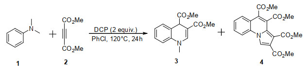
Scheme 1.
Syntheses of 1,4-dihydroquinoline (3) and pyrrolo[1,2-a]quinoline (4)
Synthesis, Crystal Structure and Biological Activity of Dimethyl 1-Methyl-1,4-dihydroquinoline-3,4-dicarboxylate and Tetramethyl Pyrrolo-[1,2-a]quinoline-2,3,4,5-tetracarboxylate
Xue-Mei XU , Zai-Gang LUO , Xin-Xin HAN , Qian-Nan LIU , Rui LI

1,4-Dihydroquinoline derivatives, one kind of nitrogen-containing heterocycles, are an important class of biologically active compounds, which are widely used in organic synthesis and pharmaceutical chemistry[1-3]. In addition, among various N-heterocycles, pyrrolo[1,2-a]-quinoline derivatives also have received much attention for their unique properties in functional materials[4] and biological activities[5] as well as in natural products[6]. Therefore, many kinds of well-established methods for the synthesis of these nitrogen-containing heterocycles are available in the literature in the last several decades[7-12]. Although these reported methods have made a significant contribution to the preparation of these N-heterocycles, the development of a simple, efficient, and environmentally friendly protocol for the synthesis of 1,4-hydroquinolines and pyrrolo[1,2-a]quinolines is still needed.
Over the last three decades, numerous small-molecule HIV-1 integrase inhibitors have been described[13, 14]. Recently, the formation of N-containing heterocycles has also been extensively investigated using N-methyl anilines as cheap, safe, and widely accessible starting materials[15-17]. In the present study, in order to gain new molecular entities with potential biological activities, we used 1,4-dihydroquinoline and pyrrolo[1,2-a]quinoline skeleton as the platforms to design two new class of integrase inhibitors. Herein, we report the synthesis of dimethyl 1-methyl-1,4-dihydroquinoline-3,4-dicarboxylate (3) and tetramethyl pyrrolo[1,2-a]quinoline-2,3,4,5-tetracarboxylate (4) using N, N-dimethyl aniline and dimethyl but-2-ynedioate as the starting materials in the presence of dicumyl peroxide (DCP) as the oxidant in one-pot, as shown in Scheme 1. The structures of them were confirmed via 1H NMR, 13C NMR and HRMS. Meanwhile, the crystal structures of compounds (3) and (4) were determined by X-ray single-crystal diffraction analysis. And the inhibition to the strand transfer process of HIV-1 integrase of the title compounds was also evaluated.
The melting point was measured on a SGW X-4 monocular microscope melting point apparatus with thermometer unadjusted. 1H NMR and 13C NMR spectra were acquired on a 500 MHz Bruker Avance spectrometer with CDCl3 as solvent. H RMS was obtained by ESI on a TOF mass analyzer. X-ray diffraction was performed using a Bruker Smart Apex CCD diffractometer.
Unless otherwise noted, all materials were obtained from commercial suppliers and purified by standard procedures. Column chromatography was performed with silica gel (200~300 mesh, Qingdao Haiyang Chemical Co., Ltd, China).
General procedure for the synthesis of 1,4-dihydroquinoline and pyrrolo[1,2-a]quinoline is described as follows: A Schlenk tube equipped with a magnetic stirring bar was charged with N, N-dimethyl aniline 1 (0.2 mmol), dimethyl but-2-ynedioate 2 (0.4 mmol), DCP (0.4 mmol) and PhCl (2 mL). Then the tube was sealed and the resulting mixture was heated to 120 ℃ for 24 h. After cooling, the solvent was diluted with water (5 mL) and extracted with dichloromethane (3 × 10 mL). The combined organic layers were dried over anhydrous Na2SO4 and concentrated by a rotary evaporator, and the residue was purified by column chromatography on silica gel (V(petroleum ether)/V(ethyl acetate) = 10:1~6:1, 0.6 > Rf > 0.3) to provide the desired products 3 and 4. The details of synthetic work were also reported by us, recently[18].
Compound 3: dimethyl 1-methyl-1,4-dihydroquinoline-3,4-dicarboxylate, white solid, m.p.: 124~125 ℃, 30 mg, 57% yield. 1H NMR (500 MHz, CDCl3, ppm) δ: 7.48 (s, 1H), 7.39 (d, J = 7.5 Hz, 1H), 7.23 (d, J = 7.5 Hz, 1H), 7.03 (t, J = 7.5 Hz, 1H), 6.86 (d, J = 8.0 Hz, 1H), 4.89 (s, 1H), 3.73 (s, 3H), 3.68 (s, 3H), 3.32 (s, 3H); 13C NMR (125 MHz, CDCl3, ppm) δ: 173.56, 167.78, 143.58, 138.09, 130.06, 128.69, 123.74, 120.66, 113.46, 96.76, 52.62, 51.41, 43.43. 39.41. HRMS (ESI) m/z: calcd. for C14H16NO4 [M+H]+: 262.1079, found 262.1076.
Compound 4: tetramethyl pyrrolo[1,2-a]quinoline-2,3,4,5-tetracarboxylate, yellow solid, m.p.: 179~181 ℃, 18 mg, 23% yield. 1H NMR (500MHz, CDCl3, ppm) δ: 8.43 (s, 1H), 7.97 (t, J = 7.5 Hz, 2H), 7.69~7.72 (m, 1H), 7.50 (t, J = 7.5 Hz, 1H), 3.98 (s, 3H), 3.93 (s, 3H), 3.92 (s, 3H), 3.91 (s, 3H); 13C NMR (125 MHz, CDCl3, ppm) δ: 166.32, 165.21, 165.13, 163.43, 132.21, 131.02, 127.86, 126.55, 126.20, 124.95, 123.21, 119.63, 119.31, 117.38, 114.85, 113.35, 52.97, 52.84, 52.35, 52.02; HRMS (ESI) m/z: calcd. for C20H18NO8 [M+H]+: 400.1032, found 400.1026.
X-ray crystallographic data of the colorless block crystal 3 and light-yellow crystal of 4 were collected and mounted on a glass fiber for measurement at room temperature, respectively. X-ray crystallographic data were collected at 289.12(10) K for 3 and 288.81(10) K for 4, respectively. All measurements of the title compounds were made on a Bruker Smart Apex CCD diffractometer equipped with graphite-monochromated MoKα radiation (λ = 1.54184 Å for 3 and 4). The structures of compounds 3 and 4 were solved by direct methods, and then the non-hydrogen atoms were refined anisotropically with SHELXS-97 by applying a full-matrix least-squares procedure based on F2 values after they were located from the trial structure[19]. Moreover, the hydrogen atom positions were fixed geometrically at calculated distances. At the same time, they were allowed to ride on the parent atoms. For compound 3: A total of 3939 reflections were selected in the range of 3.96≤θ≤65.99° (h: −8~8, k: −6~9, l: −26~20) by using a ψ-ω scan mode, of which 2236 were independent with R = 0.0515 and 1827 were observed with I > 2σ(I). The final refinement showed R = 0.0515, wR = 0.1394 with GOF = 1.044 (w = 1/[σ2(Fo2) + (0.0917P)2 + 0.1161P], where P = (Fo2 + 2Fc2)/3), (Δρ)max = 0.223, (Δρ)min = −0.281 e/Å3 and (∆/σ)max = 0.001. For compound 4: Out of the 5936 reflections collected in the range of 4.10≤θ≤66.01° (h: −8~5, k: −14~14, l: −23~24) by using a ψ-ω scan mode, 3216 were independent with R = 0.0612 and 2253 were observed with I > 2σ(I). The final refinement showed R = 0.0612, wR = 0.1548 with GOF = 1.055 (w = 1/[σ2(Fo2) + (0.0930P)2], where P = (Fo2 + 2Fc2)/3), (Δρ)max = 0.271, (Δρ)min = −0.344 e/Å3 and (∆/σ)max = 0.000. Data were collected by Rapid Auto program[20, 21]. The hydrogen atoms bound to carbon atoms were determined by accurate theoretical calculation. The representative bond lengths and bond angles for compounds 3 and 4 are illustrated in Tables 1 and 2, respectively.
 DownLoad:
CSV
DownLoad:
CSV
| Bond | Dist. | Angle | (º) | |
| N(1)–C(1) | 1.403(3) | C(9)–N(1)–C(1) | 119.18(15) | |
| N(1)–C(9) | 1.363(2) | N(1)–C(1)–C(6) | 119.44(17) | |
| N(1)–C(10) | 1.454(2) | C(2)–C(1)–N(1) | 121.04(17) | |
| C(6)–C(7) | 1.520(3) | C(1)–C(6)–C(7) | 120.33(17) | |
| C(7)–C(8) | 1.501(3) | C(5)–C(6)–C(7) | 120.89(17) | |
| C(7)–C(11) | 1.526(2) | C(6)–C(7)–C(11) | 108.88(14) | |
| C(8)–C(9) | 1.345(3) | C(8)–C(7)–C(6) | 110.25(14) | |
| C(8)–C(13) | 1.457(3) | C(9)–C(8)–C(7) | 20.09(17) |
DownLoad:
CSV
| Bond | Dist. | Angle | (º) | |
| O(1)-C(8) | 1.194(3) | C(1)–N(1)–C(11) | 122.4(2) | |
| O(2)–C(8) | 1.320(3) | C(14)–N(1)–C(1) | 128.3(2) | |
| O(7)–C(15) | 1.207(4) | C(14)–N(1)–C(11) | 109.1(2) | |
| O(8)–C(15) | 1.340(4) | N(1)–C(1)–C(6) | 118.1(2) | |
| O(8)–C(20) | 1.434(4) | C(2)–C(1)–N(1) | 121.1(2) | |
| N(1)–C(1) | 1.404(3) | C(1)–C(6)–C(7) | 119.9(2) | |
| N(1)–C(11) | 1.405(3) | C(9)–C(7)–C(6) | 120.4(2) | |
| N(1)–C(14) | 1.359(3) | N(1)–C(11)–C(9) | 118.3(2) | |
| C(8)–C(7) | 1.502(4) | C(12)–C(11)–N(1) | 107.3(2) | |
| C(9)–C(10) | 1.501(3) | C(11)–C(12)–C(13) | 107.3(2) | |
| C(12)–C(16) | 1.484(4) | C(14)–C(13)–C(12) | 107.9(2) | |
| C(15)–C(13) | 1.460(4) | N(1)–C(14)–C(13) | 108.4(2) |
The inhibition effects of compounds 3 and 4 were measured by HIV-1 integrase strand transfer activity assay, which was carried out as described previously with some minor modifications[22, 23]. Compounds diluted in DMSO were pre-incubated with 800 ng of integrase at 37.8 ℃ in the reaction buffer in the absence of Mn2+ for 10 min. Subsequently, 1.5 pmol of donor DNA and 9 pmol of target DNA were added and the reaction was initiated by the addition of 10 mmol/L Mn2+ into the final reaction volume. The reactions were carried out at 37.8 ℃ for 1 h and subsequent detection procedure was applied to detect the assay signals. Integrase inhibitor, baicalein, was used as the control compound (positive control), whereas no compound but only DMSO in the reaction mixture was set as the drug-free control (negative control). The inhibition effects of compounds 3 and 4 were calculated based on the positive and negative controls.
The structures for the target compounds 3 and 4 were confirmed by 1H NMR, 13C NMR and HRMS. For example, the 1H NMR spectrum of 1,4-dihydroquinoline 3 shows the signals of 7.48 and 4.89 ppm as singlet due to the =CH and CH in the N-containing six-membered ring, and the signals of CH3 at 3.73, 3.68 and 3.32 as singlet. The phenyl signals appeared at 7.39~6.86 ppm as three doublets and one triplet. The 1H NMR spectrum of pyrrolo[1,2-a]quinoline 4 also shows the signals of 8.43 ppm as singlet due to the =CH in the N-containing five-membered ring, and the signals of CH3 at 3.98, 3.93, 3.92 and 3.91 as singlet. The phenyl signals appeared at 7.97~7.50 ppm as two triplets and one multiplet. The 13C NMR signals of 1,4-dihydroquinoline 3 and pyrrolo[1,2-a]quinoline 4 possess the normal value. And the HRMS spectra of 3 and 4 show molecular ion peaks and [M+H]+ at m/z 262.1076 and 400.1026, respectively. Furthermore, the solid of compounds 3 and 4 was recrystallized from dichloromethane/ethanol to give colorless single crystal of 3 and light-yellow crystal of 4 suitable for single-crystal X-ray diffraction. The crystals of the title compounds are stable in air at room temperature.
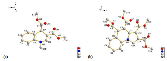
X-ray diffraction of the molecular structure of the bicyclic 1,4-dihydroquinoline 3 and polycyclic pyrrolo[1,2-a]quinoline 4 is demonstrated in Fig. 1. The bicyclic skeleton of 1,4-dihydroquinoline 3 is not coplanar (Fig. 1a). The torsion angles of C1–C6–C7–C8 and C9–N1–C1–C6 are 25.7(2)° and –12.4(2)°, respectively. The two ester groups connected with C7 and C8 are approximately perpendicular to each other, for the torsion angle of N1–C9–C8–C13 is –179.28(16)°, while the torsion angle of C9–C8–C7–C11 is 96.1(2)°. Obviously, the polycyclic skeleton of pyrrolo[1,2-a]quinoline 4 is coplanar (Fig. 1b), and the bond lengths and bond angles are within normal ranges. And the four bulky substituents, methoxy carbonyl groups connected with C7, C9, C12 and C13 of the molecular frame structure, are in a staggered reverse alignment manner. It means that the molecule has thermodynamically stable conformation. However, the intermolecular and intramolecular hydrogen bonds are not found in the two molecules.
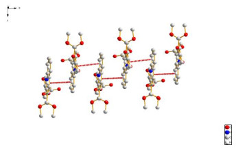
Moreover, the crystal packing of 4 illustrated in Fig. 2 reveals that the overall packing has reverse staggered parallel arrangement. Besides, one kind of π-π interaction between the two adjacent molecules at upper and lower levels can be observed, which formed a one-dimensional interaction model. As can be detected by the packing diagram with stratified arrangement, the distance between the upper and lower levels is 3.5177 Å.
Compounds 3, 4 and positive control compound baicalein were tested against purified integrase and the data are summarized in Table 3.
DownLoad:
CSV
| Compounds | Inhibition ratio (%)b |
| 3 | 48.52 |
| 4 | 38.98 |
| Baicalein | 100 |
| Control | 0 |
| a HIV-1 IN inhibitory activities were measured according to the procedure described in Refs. 22 and 23. b Inhibition of strand transfer at the concentration of 50 μM. |
|
As shown in Table 3, the value of inhibition ratio of compound 3 is 48.52% at the concentration of 50 μM, which means it has a moderate inhibitory activity towards HIV integrase; while compound 4 owns a lower inhibitory activity than compound 3 with the inhibition ratio of 38.98%, which maybe correlates with bulky steric hindrance of the four adjacent ester groups blocking the interaction of the molecule and HIV integrase. Further work based on these structures is in progress.
In summary, a facile method for the synthesis of 1,4-dihydroquinoline 3 and pyrrolo[1,2-a]quinoline 4 using N, N-dimethyl aniline and dimethyl but-2-ynedioate as the starting materials in one-pot process was reported, and their structures were confirmed by 1H NMR, 13C NMR and HRMS. X-ray diffraction of the molecular structure disclosed that the skeleton of 1,4-dihydroquinoline 3 is noncoplanar, while pyrrolo[1,2-a]-quinoline 4 owns a coplanar frame structure. One-dimensional interaction model of compound 4 was formed by one kind of π-π interaction between the two adjacent molecules at upper and lower levels. The HIV-1 integrase strand transfer activity assay results showed that compound 3 has more potent inhibitory activity than 4. The mechanism study and applications of these nitrogen-containing heterocycles are underway in our lab.
Bossert, F.; Meyer, H.; Wehinger, E. 4-Aryldihydropyridines, a new class of highly active calcium antagonists. Angew. Chem. Int. Ed. 1981, 20, 762–769. doi: 10.1002/anie.198107621
Mentese, M. Y.; Bayrak, H.; Uygun, Y.; Mermer, A.; Ulker, S.; Karaoglu, S. A.; Demirbas, N. Microwave assisted synthesis of some hybrid molecules derived from norfloxacin and investigation of their biological activities. Eur. J. Med. Chem. 2013, 67, 230–242. doi: 10.1016/j.ejmech.2013.06.045
Mistry, S. N.; Valant, C.; Sexton, P. M.; Capuano, B.; Christopoulos, A.; Scammells, P. J. Synthesis and pharmacological profiling of analogues of benzyl quinolone carboxylic acid (BQCA) as allosteric modulators of the M1 muscarinic receptor. J. Med. Chem. 2013, 56, 5151–5172. doi: 10.1021/jm400540b
Sonnenschein, H.; Hennrich, G.; Resch-Genger, U.; Schulz, B. Fluorescence and UV/Vis spectroscopic behaviour of novel biindolizines. Dyes Pigm. 2000, 46, 23–27. doi: 10.1016/S0143-7208(00)00032-2
Hazra, A.; Mondal, S.; Maity, A.; Naskar, S.; Saha, P.; Paira, R.; Sahu, K. B.; Paira, P.; Ghosh, S.; Sinha, C.; Samanta, A.; Banerjee, S.; Mondal, N. B. Amberlite-IRA-402 (OH) ion exchange resin mediated synthesis of indolizines, pyrrolo [1,2-a] quinolines and isoquinolines: antibacterial and antifungal evaluation of the products. Eur. J. Med. Chem. 2011, 46, 2132–2140. doi: 10.1016/j.ejmech.2011.02.066
Fan, H.; Peng, J. N.; Hamann, M. T.; Hu, J. F. Lamellarins and related pyrrole-derived alkaloids from marine organisms. Chem. Rev. 2008, 108, 264–287. doi: 10.1021/cr078199m
Yu, M.; Kim, S. G. Asymmetric organocatalytic michael addition/aza-cyclization coupled with sequential michael addition for synthesizing densely polycyclic-fused dihydroquinolines. Tetrahedron Lett. 2015, 56, 4159–4162. doi: 10.1016/j.tetlet.2015.04.112
Wu, X. J.; Xu, X. P.; Su, X. M.; Chen, G.; Zhang, Y.; Ji, S. J. A Novel, highly efficient, one-pot synthesis of 1,4-dihydroquinoline derivatives in the presence of a Pd(OAc)2/DABCO catalytic system. Eur. J. Org. Chem. 2009, 4963–4970.
Chu, X. Q.; Zi, Y.; Meng, H.; Xu, X. P.; Ji, S. J. Synthesis of 1,4-dihydroquinoline derivatives under transition-metal-free conditions and their diverse applications. Org. Biomol. Chem. 2014, 12, 4243–4251. doi: 10.1039/c4ob00475b
Wu, F. S.; Zhao, H. Y.; Xu, Y. L.; Hu, K.; Pan, Y. M.; Ma, X. L. Catalyst-free synthesis of pyrrolo[1,2-a]quinolines via dehydration/[3+2] cycloaddition directly from 2-methylquinolines, aldehydes, and alkynoates. J. Org. Chem. 2017, 82, 4289–4296. doi: 10.1021/acs.joc.7b00280
Yu, Y.; Liu, Y.; Liu, A. X.; Xie, H. X.; Li, H.; Wang, W. Ligand-free Cu-catalyzed [3+2] cyclization for the synthesis of pyrrolo[1,2-a]quinolines with ambient air as a terminal oxidant. Org. Biomol. Chem. 2016, 14, 7455–7458. doi: 10.1039/C6OB01316C
Wu, L.; Sun, J.; Yan, C. G. Efficient synthesis of pyrrolo[2,1-a]isoquinoline and pyrrolo[1,2-a]quinoline derivatives via one-pot two-step metal-catalyzed three-component reactions. Chin. J. Chem. 2012, 30, 590–596. doi: 10.1002/cjoc.201100238
Dayam, R.; Deng, J.; Neamati, N. HIV-1 integrase inhibitors: 2003–2004 update. Med. Res. Rev. 2006, 26, 271−309. doi: 10.1002/med.20054
Dayam, R.; Gundla, R.; Al-Mawsawi, L. Q.; Neamti, N. HIV-1 integrase inhibitors: 2005–2006 update. Med. Res. Rev. 2008, 28, 118−154. doi: 10.1002/med.20116
Li, C. J. Cross-dehydrogenative coupling (CDC): exploring C−C bond formations beyond functional group transformations. Acc. Chem. Res. 2009, 42, 335–344. doi: 10.1021/ar800164n
Pandey, G.; Sudha Rani, K.; Lakshamaiah, G. Direct carbon-carbon bond formation strategy at α-position of tertiary amines by photoinduced electron transfer (PET) processes. Tetrahedron Lett. 1992, 33, 5107–5110. doi: 10.1016/S0040-4039(00)61203-0
Zhou, H.; Lu, P.; Gu, X.; Li, P. Visible-light-mediated nucleophilic addition of an α-aminoalkyl radical to isocyanate or isothiocyanate. Org. Lett. 2013, 15, 5646−5649. doi: 10.1021/ol402573j
Luo, Z. G.; Han, X. X.; Liu, C. F.; Liu, Q. N.; Li, R.; Liu, P.; Xu, X. M. Catalyst-free synthesis of 1,4-dihydroquinolines and pyrrolo[1,2-a]quinolines via intermolecular [4+2]/[3+2] radical cyclization of N-methylanilines with alkynoates. Synthesis 2020, 52, 1067–1075. doi: 10.1055/s-0039-1691541
Bruker 2000, SMART (Version 5.0), SAINT-plus (Version 6), SHELXTL (Version 6.1), and SADABS (Version 2.03); Bruker AXS Inc. : Madison, WI.
Sheldrick, G. M. SHELXS-97, Program for X-ray Crystal Structure Solution. University of Göttingen, Germany 1997.
Sheldrick, G. M. SHELXL-97, Program for the Refinement of Crystal Structure. University of Göttingen: Göttingen, Germany 1997.
He, H. Q.; Ma, X. H.; Liu, B.; Chen, W. Z.; Wang, C. X.; Chen, S. H. A novel high-throughput format assay for HIV-1 integrase strand transfer reaction using magnetic beads. Acta Pharmacologica. Sinica 2008, 29, 397−404. doi: 10.1111/j.1745-7254.2008.00748.x
Luo, Z. G.; Zhao, Y.; Ma, C.; Cao, L.; Ai, S. H.; Hu, J. S.; Xu, X. M. Synthesis, crystal structure and anti-integrase activity of 25,27-bis [(Z)-4-(p-methoxyphenyl)-4-hydroxybut-3-en-2-one-1-methyl]-26,28-dihydroxycalix[4]arene. Chin. J. Struct. Chem. 2014, 33, 1117−1122.

Figure 1 (a) Coordination environment of compound 3, (b) Coordination environment of compound 4. The selected atoms are omitted for clarity
Table 1. Selected Bond Lengths (Å) and Bond Angles (°) of Compound 3
| Bond | Dist. | Angle | (º) | |
| N(1)–C(1) | 1.403(3) | C(9)–N(1)–C(1) | 119.18(15) | |
| N(1)–C(9) | 1.363(2) | N(1)–C(1)–C(6) | 119.44(17) | |
| N(1)–C(10) | 1.454(2) | C(2)–C(1)–N(1) | 121.04(17) | |
| C(6)–C(7) | 1.520(3) | C(1)–C(6)–C(7) | 120.33(17) | |
| C(7)–C(8) | 1.501(3) | C(5)–C(6)–C(7) | 120.89(17) | |
| C(7)–C(11) | 1.526(2) | C(6)–C(7)–C(11) | 108.88(14) | |
| C(8)–C(9) | 1.345(3) | C(8)–C(7)–C(6) | 110.25(14) | |
| C(8)–C(13) | 1.457(3) | C(9)–C(8)–C(7) | 20.09(17) |
 下载: 导出CSV
Table 2. Selected Bond Lengths (Å) and Bond Angles (°) of Compound 4
| Bond | Dist. | Angle | (º) | |
| O(1)-C(8) | 1.194(3) | C(1)–N(1)–C(11) | 122.4(2) | |
| O(2)–C(8) | 1.320(3) | C(14)–N(1)–C(1) | 128.3(2) | |
| O(7)–C(15) | 1.207(4) | C(14)–N(1)–C(11) | 109.1(2) | |
| O(8)–C(15) | 1.340(4) | N(1)–C(1)–C(6) | 118.1(2) | |
| O(8)–C(20) | 1.434(4) | C(2)–C(1)–N(1) | 121.1(2) | |
| N(1)–C(1) | 1.404(3) | C(1)–C(6)–C(7) | 119.9(2) | |
| N(1)–C(11) | 1.405(3) | C(9)–C(7)–C(6) | 120.4(2) | |
| N(1)–C(14) | 1.359(3) | N(1)–C(11)–C(9) | 118.3(2) | |
| C(8)–C(7) | 1.502(4) | C(12)–C(11)–N(1) | 107.3(2) | |
| C(9)–C(10) | 1.501(3) | C(11)–C(12)–C(13) | 107.3(2) | |
| C(12)–C(16) | 1.484(4) | C(14)–C(13)–C(12) | 107.9(2) | |
| C(15)–C(13) | 1.460(4) | N(1)–C(14)–C(13) | 108.4(2) |
下载: 导出CSV
Table 3. Inhibition of HIV-1 Integrase Strand Transfer Catalytic Activitiesa
| Compounds | Inhibition ratio (%)b |
| 3 | 48.52 |
| 4 | 38.98 |
| Baicalein | 100 |
| Control | 0 |
| a HIV-1 IN inhibitory activities were measured according to the procedure described in Refs. 22 and 23. b Inhibition of strand transfer at the concentration of 50 μM. |
|
下载: 导出CSV

 扫一扫看文章

扫一扫关注我们


 下载:
下载: