Table 1.
Singlet Oxygen Quenching Rates of 28 Carotenoids (KQΔ, 109M-1S-1)
Citation:
Kai ZHAO, Wei JIANG, Chong MENG. Key Parameters Analysis and Regulation of Singlet Oxygen Quenching Rate of Carotenoids[J]. Chinese Journal of Structural Chemistry,
2020, 39(7): 1226-1234.
doi:
10.14102/j.cnki.0254–5861.2011–2568

Key Parameters Analysis and Regulation of Singlet Oxygen Quenching Rate of Carotenoids
English
Key Parameters Analysis and Regulation of Singlet Oxygen Quenching Rate of Carotenoids
Abstract:
28 kinds of carotenoids are studied to reveal the key parameters and regulation on the singlet oxygen quenching rate. First, the quantum chemistry parameters of carotenoids calculated by Gaussian software combined with substitution parameters were used to construct the quantitative structure-activity relationship model (QSAR) of the singlet oxygen quenching rate of carotenoids. The key parameters affecting the antioxidant activity of carotenoids are revealed, and the data predicted via the QSAR model were provided for subsequent research. Then, a three-dimensional (3D) pharmacophore model was used to regulate and modify the antioxidant activity of carotenoids. The correlation coefficients of the modeling group (R2) and verification group (Rpre2) of the established QSAR model were 0.945 and 0.916, respectively, which can be used for the analysis of antioxidant activity of carotenoids; the antioxidant activity of carotenoids can be significantly regulated by the number of conjugated C=C bonds, the energy difference between frontier molecular orbitals and the partial Mulliken charge in C1 and the π…π* excitation energy E(s); the antioxidant activity of carotenoids can be effectively regulated by the hydrogen bond acceptor pharmacophores on both sides of the conjugated C=C bonds and the hydrophobic groups on the conjugated C=C bond; the hydrophobic substituents attached to conjugated C=C bonds can effectively improve the singlet oxygen quenching rate of carotenoids.
-
1. INTRODUCTION
Carotenoids, a class of compounds consisting of eight isoprenoid units, are ubiquitous in green plants and are one of the main antioxidants in leafy vegetables[1]. As a major class of natural antioxidants, carotenoids are widely used in the medicine, food, sports supplements and in other industries[2, 3]. Studies have shown that the intake of carotenoid-rich foods can prevent serious chronic diseases and improve immunity for humans[4]. For example, lycopene has excellent effects of antioxidation, anti-fatigue, and enhancing athletic ability. Lycopene products have been introduced as sports supplements for sports practice[5]. Astaxanthin has certain effects on antioxidation, cardiovascular inflammation, gastric ulcers and blood pressure recovery in mice[6]. β-Carotene can not only be used to treat vitamin A deficiency but also to reduce the incidence of heart disease and cancer[7].
Oxygen free radicals can react with important macromolecules such as DNA, proteins, carbohydrates and lipids in the body, damage their functions, and cause aging and a series of diseases (cancer, cardiovascular diseases, etc.)[8, 9]. However, carotenoids can reduce these risks in two ways: reacting with free radicals to form harmless products; and scavenging free radicals by destroying free radical chains[10, 11]. Different carotenoids have different elimination (quenching) effects on oxygen free radicals. Antioxidant activity is generally measured by the singlet oxygen quenching capacity (rate) and peroxide radiation capturing capacity. Previous studies have shown that the singlet oxygen quenching ability of carotenoids is mainly affected by the number of unsaturated double bonds and the characteristics of terminal groups[12]. Lycopene can prevent lipid peroxidation and protect biofilms from damage by oxygen free radicals by quenching singlet oxygen[13, 14].
Because of the large number of carotenoid homologs and the poor stability of some homologs in the environment, it is time-consuming, laborious and unrealistic to carry out antioxidant experiments on each homolog. The lack of basic data is not conducive to systematic summarization and study of the antioxidant properties of carotenoids. Quantitative structure-activity relationship modeling (QSAR) provides a method for predicting and analyzing the properties of undetected substances using existing experimental data[15, 17].
The lack of experimental data limits the application of QSAR modeling in carotenoids. Sun et al. established a QSAR model of cell gap junction function of carotenoids, which showed that the cell gap junction function was related to affinity reaction[18]. Soffers established a QSAR model of carotenoid antioxidant activity containing nine conjugated C=C bonds, which showed that the radical scavenging action was correlated with ionization potentials[19]. However, there were some problems in previous QSAR models[19, 21]: the selected parameters were not intuitive and not conducive to summarizing the rules; more intuitive and specific substituent parameters were not involved; and the established QSAR models were only used to reveal individual key parameters, not for further research.
Based on the above problems, the purpose of this work is to establish a QSAR model of the singlet oxygen quenching rate of carotenoids by means of quantum chemical and substituent parameters, to elaborate key parameters affecting the antioxidant activity of carotenoids, and to provide predicted data for subsequent modifying research. Then, a typical carotenoid was effectively modified using 3D pharmacophore modeling to improve the singlet oxygen quenching rate.
2. METHODS
2.1 Experimental data
Dietmar et al. selected 28 carotenoids (Table 1)[12] to measure the singlet oxygen quenching rate (KQΔ) at 37 ℃ in mixed solution (C2H5OH, CHCl3, and D2O, 50:50:1) as an index of antioxidant activity. The higher singlet oxygen quenching rate indicates higher antioxidant activity.
Table 1

 DownLoad:
CSV
DownLoad:
CSV
No. Carotenoids KQΔ No. Carotenoids KQΔ L1 
0.2 L15 
12.0 L2 
0.5 L16 
12.6 L3 
0.1 L17# 
10.2 L4 
3.0 L18 
9.0 L5# 
1.6 L19 
11.1 L6 
8.1 L20# 
12.7 L7 
10.2 L21 
12.1 L8# 
11.1 L22 
13.3 L9 
12.7 L23 
13.0 L10 
11.0 L24 
12.9 L11 
11.7 L25# 
13.8 L12# 
8.4 L26# 
12.3 L13 
9.0 L27 
8.8 L14 
12.4 L28 
8.4 2.2 Calculation and modelling methods
GaussView software was used to draw the initial molecular structures of 28 kinds of carotenoids and to set optimization tasks. The optimal structures and quantum chemical parameters of carotenoids were calculated via Gaussian 09 software[22] with the B3LYP/6-31+(d) level by density functional theory (DFT)[23].
Quantum chemical descriptors were obtained from the output files of Gaussian software, including the charge parameters, polarization parameters, and energy parameters: energy of the lowest unoccupied molecular orbital (ELUMO, eV), energy of the highest occupied molecular orbital (EHOMO, eV), ELUMO-EHOMO (∆E, eV), the dipole moment (μ, Debye), partial Mulliken charges in typical atoms (qC1, qC2, qC3, qC4, qC5, qC6, qC7, qC8, e), total energy (TE, eV), the mean polarizability (α, 10-30 esu), the anisotropy polarizability (∆α, 10-30 esu), the approximate polarizability (αxx, αyy, αzz, αxy, αxz, αyz) and its ten components (βxxx, βxxy, βxyy, βyyy, βxxz, βxyz, βyyz, βxzz, βyzz, βzzz), the π…π* excitation energy (E(s), 103 cm-1), and molar volume (V, cm3/mol). Structural parameters include the number of conjugated C=C bonds (NC=C), groups (NCH3), hydroxyl groups (NOH), O atoms (NO), C atoms (NC), and H atoms (NH). C(1)–C(4) and C(5)–C(8) are the number of the four left-most and four right-most C atoms in the conjugated C=C bonds, respectively (as shown in L1 in Fig. S1 in SI).
The total experimental data (n = 28) were divided into a modeling set (n = 21) and a testing set (n = 7), relying on the interval-sampling method[24]. The experimental data were ordered according to the magnitude of KQΔ and those with the order number of 4n (n = 1, 2, 3, 4, 5, 6, and 7) were put in the modeling group. 7 selected samples in the verification group, labeled with a "#", are listed Table 1. The quantum chemical parameters combined with the artificially designed structural parameters were selected as independent variables to establish the QSAR model of the KQΔ values of carotenoids using multiple linear regression method via SPSS software. During the process, the variable screening principle of stepwise analysis was selected to generate the model; collinearity diagnostics (variance inflation factor) among independent variables and residual analysis (Durbin-Watson) were also considered to ensure the validity of the model.
2.3 Model evaluation method
The performance of the established QSAR model needs to be evaluated from three perspectives: fitness, robustness, and predictability[25]. In the present work, the conventional square of the correlation coefficient (R2), Fisher test values (F), and the standard deviation (SD) were selected to test the fitness. For robustness, the correlation coefficients during leave-one-out cross-validation (q2), prediction error sum of squares (PRESS) and Y-randomization test were used. The conventional square produced by the verification group (R2 pre), Tropsha parameters (k and k') and Roy′ Rm2 metrics were used to reflect the predictive ability. Tropsha et al. considered a QSAR model to have good predictive ability if the following conditions were satisfied[26]:
$ R_{\mathrm{pre}}^2>0.6$ $0.85 \leqslant k \leqslant 1.15 \text { or } 0.85 \leqslant k^{\prime} \leqslant 1.15$ The detailed definitions of R2 pre, k and k' are presented below:
$ {R}_{\mathrm{p}\mathrm{r}\mathrm{e}}^{2}=1-\frac{{\sum }_{i=1}^{test}{\left({y}_{i}-{\widetilde{y}}_{i}\right)}^{2}}{{\sum }_{i=1}^{test}{\left({y}_{i}-{\overline{y}}_{tr}\right)}^{2}} $ $ k=\frac{\sum {y}_{i}{\widetilde{y}}_{i}}{\sum {\widetilde{y}}_{i}^{2}} , {k}^{\text{'}}=\frac{\sum {y}_{i}{\widetilde{y}}_{i}}{\sum {y}_{i}^{2}} $ where
$ {\widetilde{y}}_{i} $ are the predicted values using the established QSAR model,$ {y}_{i} $ are the experimental values, and$ {\overline{y}}_{tr} $ is the mean value of the dependent variables of the modeling group.The Rm2 metric is calculated based on the correlations between the experimental and predicted values with (R2) and without (R02) intercept for the least squares regression lines as shown in the following equation[27]:
$ {R}_{m}^{2}={R}^{2}(1-\left|\sqrt{{R}^{2}-{R}_{0}^{2}}\right|) $ The Rm2 value for a given model larger than 0.5 indicates good external predictability of the developed model[28].
2.4 3D pharmacophore modeling
The 3D pharmacophore model was constructed using the Discovery Studio 2.5 software package (DS) developed by Accelrys[29]. The given parameters are as follows: five pharmacophore features (hydrogen bond donor D, hydrophobic H, aromatic ring A, hydrogen bond acceptor HBa, hydrophobic aromatic Ha); a maximum of 255 diverse conformers were generated for each molecule; the energy threshold for generating diverse conformers was 10 eV; the "best" method was chosen for calculation; and the convergence index "configuration cost" was 17. When the configuration cost is less than 17, the established 3D pharmacophore model met the convergence requirement. The performance of the model was validated by configuration cost and total cost: the configuration cost value of the model should be less than 17; a significant pharmacophore model should have a value of total cost that is close to the fixed cost (the most ideal pharmacophore model) and far from the null cost (the worst pharmacophore model).
3. RESULTS AND DISCUSSION
3.1 QSAR modeling and analysis
The established QSAR model of the carotenoid quenching rate KQΔ for singlet oxygen is shown as follows. The results of QSAR modeling are summarized in Table 2.
$ \begin{aligned} & \mathrm{K}_{\mathrm{Q}}^{\Delta}=34+0.127 N_{\mathrm{C}=\mathrm{C}}-40.4 \Delta \mathrm{E}+ \\ & 2.54 q_{\mathrm{Cl}}-0.728 E(\mathrm{s}) \end{aligned} $ (1) Table 2
Table 2. Performance of the QSAR ModelDownLoad:
CSV
Performance Feature Value Criterion Fitting R2 0.956 > 0.90 F 108.88 / sig. 0.000 < 0.01 Robustness q2 0.905 > 0.5 PRESS 36.98 / Y-randomization test R2 0.084 < 0.80 Y-randomization test R2 CV 0.000 < 0.01 Predicted performance k 0.993 (0.85, 1.15) k' 0.998 (0.85, 1.15) Rpre2 0.919 > 0.6 Rm2 (modeling group) 0.756 > 0.5 Rm2 (verification group) 0.658 > 0.5 Rm2 (28 congeners) 0.738 > 0.5 From Table 2, it can be found that the QSAR model of the KQΔ values of carotenoids satisfies the performance requirements of fitting, robustness and predictability. All of these results indicate that the established QSAR model can be used to predict the unknown KQΔ values of carotenoids that are difficult to determine experimentally.
The quenching rates KQΔ of carotenoids were significantly positively correlated with the number of conjugated C=C bonds. The energy transfer of conjugated C=C bonds can change reactive oxygen species into stable oxygen molecules and avoid the attack of singlet oxygen on lipid double bonds. Therefore, the greater the number of conjugated C=C bonds, the more difficult the decomposition and transformation of the structure of carotenoids by singlet oxygen are to exert its quenching effect on singlet oxygen[30]. As shown in Table 1, the KQΔ values of carotenoids with fewer conjugated C=C bonds numbered L1, L2, L3, and L4 (KQΔ values of 0.2, 0.5, 0.1, and 3.0) are lower than those with more conjugated C=C bonds numbered L6, L25, L11 and L19 (KQΔ values of 8.1, 13.8, 11.7, and 11.1).
The quenching rates KQΔ of carotenoids were significantly negatively correlated with ∆E. A smaller ∆E means a smaller difference in molecular orbital energy between HOMO and LUMO and an easier electron transition by absorbing external energy, reflecting a higher chemical reaction activity. The bimolecular quenching of singlet oxygen can in principle happen either by chemical reaction forming oxidation products or by physical energy transfer[11]. The carotenoids with smaller ∆E are more likely to be stimulated by the singlet oxygen and participate in the reaction. Sun et al. also found that the energy difference between HOMO and LUMO and the ionization energy are related parameters affecting the antioxidant activity of carotenoids[20].
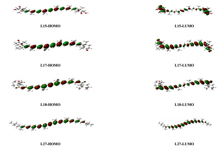
Taking the most common carotenoids (astaxanthin-L15, canthaxanthin-L17, capsorubin-L18, lycopene-L27) in the environment as an example, the HOMO and LUMO of them are shown in Fig. 1. Compared with the HOMO diagrams, the electron conjugation of LUMO diagrams decreases significantly (the electron cloud density distribution is uncoupled and dispersive) and the LUMO diagrams of different carotenoid molecules have more difference. The carotenoids (L15, L17) with smaller energy difference have greater difference between HOMO and LUMO, which strengthened the flow of electron cloud density and was beneficial to the redox reaction.
Figure 1
 Figure 1. Diagrams of HOMO and LUMO of several carotenoids
The partial Mulliken charge in typical atom C1 (qC1) was affected by the properties of substituted groups and can positively regulate the singlet oxygen quenching rates of carotenoids. For example, the KQΔ value of the carotenoid numbered L16 (12.6) is higher than those of carotenoids numbered L10 (11.0) and L15 (12.0). There are two more electronegative O atoms on the left side of the carotenoid numbered L16, while carotenoids numbered L10 and L15 have only one more electronegative O atom. Studies so far demonstrated that the quenching ability of carotenoids can be influenced by introducing oxo or hydroxyl substituents in the β-ionone end group[31-32].
The π…π* excitation energy E(s) can negatively regulate the singlet oxygen quenching rates of carotenoids. The triplet energy of the carotenoid is decisive for the efficiency of the energy transfer of single oxygen to the carotenoid because the triplet energy of the carotenoid is close to the energy of singlet oxygen[33].
$ \begin{aligned} & { }^1 \mathrm{O}_2\left({ }^1 \Delta_{\mathrm{g}}\right)+{ }^1 \mathrm{Car} \rightarrow{ }^3 \mathrm{O}_2\left({ }^1 \Delta \Sigma_{\mathrm{g}}^{-}\right)+{ }^3 \mathrm{Car}^* \\ & { }^3 \mathrm{Car}^* \rightarrow{ }^1 \mathrm{Car}+\Delta \end{aligned}$ Involved orbitals of carotenoids during electron transition are shown in Table 3. The electron transitions mainly occur from the orbitals of HOMO-1 and HOMO to LUMO and LUMO+1, obtained from the atomic composition analysis of frontier molecule orbitals. The electron density mainly transfers from the conjugated C=C bonds of HOMO-1/HOMO to the C1 side chain in LUMO/LUMO+1. In addition, the electron density redistributes on the conjugated C=C bonds through the transition of π→π* electrons, especially the reduction of charge density on the methyl attached to conjugated C=C bonds can promote the breaking of C-CH3 bond. It can also be seen that the partial Mulliken charges of C1 (qC1) significantly regulate the reactivity of carotenoids.
Table 3
Table 3. Orbital Components and Atoms Components of π…π* ExcitationDownLoad:
CSV
Carotenoids E(s) (103 cm-1) λmax (nm) Orbital components (%) Atoms components (%) L15 19.53 512 HOMO-1→LUMO
(48.6)
HOMO→LUMO+1
(24.4)C=C(82.5)
C-CH3(5.7)C1(8.3)
→
C=C(60.3)C1(4.2)
side-chains(27.6)L17 20.37 491 HOMO-1→LUMO
(51.5)
HOMO→LUMO+1
(31.8)C=C(81.2)
C-CH3(7.2)C1(5.7)
→
C=C(57.2)C1(3.2)
side-chains(30.4)L18 20.57 486 HOMO-1→LUMO
(52.3)
HOMO-1→LUMO+1
(24.1)C=C(84.7)
C-CH3(4.5)C1(5.2)
→
C=C(69.3)C1(4.8)
side-chains(19.8)L27 20.66 484 HOMO-1→LUMO
(49.6)
HOMO→LUMO+1
(29.8)C=C(87.4)
C-CH3(4.1)C1(3.8)
→
C=C(77.1)C1(3.7)
side-chains(10.4)3.2 Antioxidant modification of carotenoids based on 3D pharmacophore modeling
According to the modeling method in section 2.4, nine 3D pharmacophore models of the quenching rate KQΔwere formed, with the parameters listed in Table 4.
Table 4
Table 4. Parameters of the 3D Pharmacophore ModelDownLoad:
CSV
Index Model Total cost RMSE R2 Pharmacophorea KQΔ M1 98.122 0.092 0.874 HBaHBaHHH M2 99.629 0.096 0.742 HBaHBaHHH M3 100.139 0.101 0.621 HBaHBaHHH M4 100.304 0.102 0.525 HBaHBaHHH M5 100.314 0.104 0.416 HBaHBaHHH M6 100.553 0.109 0.335 HBaHBaHHH M7 100.671 0.111 0.190 HBaHHH M8 100.692 0.116 0.151 HBaHBaHHH M9 101.088 0.121 0.105 HBaHBaHHH Configuration cost 12.599 Fixed cost 91.087 Null cost 118.438 a HBa: hydrogen bond acceptor; H: hydrophobic The established 3D pharmacophore models of the singlet oxygen quenching rate of carotenoids mainly contained hydrogen bond acceptors (HBa) and hydrophobic (H) features. M1 was chosen as the optimal pharmacophore model: the RMSE value of M1 was the smallest (0.092); the fitting coefficient R2 was the largest (0.874); the total cost value of M1 (98.122) was the closest to the fixed cost value (91.087) and the most distant from the null cost value (118.438); and the convergence value of M1 was (12.599) less than 17.
The pharmacophore model M1 contains two hydrogen bond acceptors and three hydrophobic pharmacophores. Taking the carotenoid numbered L1 in Table 1 as an example, the binding mode between L1 and the pharmacophore model M1 is shown in Fig. 2. The hydrogen bond acceptors (light green label) cover both sides of the carotenoid. The oxygen atoms of carbonyl substituents have strong electronegativity and can bind to the surrounding singlet oxygen or other substances, thus causing singlet oxygen quenching. Hydrophobic pharmacophore characteristics (blue labeling) are mainly located on the conjugated C=C double bond, especially near the methyl region. Previous studies and the above established QSAR model mentioned that more conjugated C=C bonds on carotenoids are correlated with a higher singlet oxygen quenching rate of carotenoids; however, the influence of methyl substitution on the conjugated double bond was often ignored.
Figure 2
 Figure 2. Diagram of representative carotenoid L1 embedded within the pharmacophore model M1
To further verify the effect of methyl groups (or other hydrophobic substituents) attached to conjugated C=C bonds on the singlet oxygen quenching rate of carotenoids, molecular modification of L1 was carried out. The specific modification scheme was to replace a methyl in L1 with ethyl (labeled L1-1), propyl (labeled L1-2), vinyl (labeled L1-3), and chlorine (labeled L1-4). At the same time, hydrophilic hydroxyl radicals (labeled L1-5) were added for comparative analysis. The KQΔvalues of five modified congeners were predicted using the established QSAR model, which are 0.26, 0.48, 0.84, 0.44 and 0.16, respectively. The KQΔ values of the first four modified substances with stronger hydrophobicity were higher than that of the corresponding L1, and the KQΔ values of L1-5 were lower than L1. The same modification design and results of singlet oxygen quenching rate were also confirmed in L17 and L18 (Table 5). The above analysis results indicated that the hydrophobic substituents can effectively adjust the singlet oxygen quenching rate of carotenoids.
Table 5
Table 5. Modification Design Scheme and KQΔ Values of 3 CarotenoidsDownLoad:
CSV
Congeners Modification scheme KQΔ Congeners Modification scheme KQΔ L1 Methyl 0.20 L17 Methyl 10.20 L1-1 Ethyl 0.26 L17-1 Ethyl 10.64 L1-2 Propyl 0.48 L17-2 Propyl 10.92 L1-3 Vinyl 0.84 L17-3 Vinyl 11.34 L1-4 Chlorine 0.44 L17-4 Chlorine 10.83 L1-5 Hydroxyl 0.16 L17-5 Hydroxyl 9.77 L18 Methyl 9.00 L18-3 Vinyl 9.92 L18-1 Ethyl 9.35 L18-4 Chlorine 9.61 L18-2 Propyl 9.68 L18-5 Hydroxyl 8.67 4. CONCLUSION
The correlation coefficients of the modeling group (R2) and verification group (Rpre2) of the established QSAR model are 0.945 and 0.916, respectively. The established QSAR model satisfies the required performance parameters for fitting, robustness and predictability and can be used to predict and research the antioxidant activity of carotenoids against singlet oxygen.
The singlet oxygen quenching rates of carotenoids were positively correlated with the number of conjugated C=C bonds and the partial Mulliken charge in C1, and were negatively correlated with the frontier molecular orbital energy difference and the π…π* excitation energy.
The optimal 3D pharmacophore model of the singlet oxygen quenching rate of carotenoids primarily contained hydrogen bond acceptors and hydrophobic features. The antioxidant activity of carotenoids can be effectively regulated by the hydrogen bond acceptor pharmacophores on both sides of the conjugated C=C bond and the hydrophobic groups on the conjugated C=C bond. For typical carotenoids, the hydrophobic substituents attached to conjugated C=C bonds can effectively improv the singlet oxygen quenching rate of carotenoids.
-
-
[1]
Yungyuen, W.; Ma, G.; Zhang, L. C.; Yamawaki, K.; Takagi, T.; Kiriiwa, Y.; Ikoma, Y.; Matsumoto, H.; Yoshioka, T.; Nesumi, H. Regulation of carotenoid metabolism in response to different temperatures in citrus juice sacs in vitro. Sci. Hortic. 2017, 238, 384–390.
-
[2]
Asker, D. High throughput screening and profiling of high-value carotenoids from a wide diversity of bacteria in surface seawater. Food Chem. 2018, 261, 103–111. doi: 10.1016/j.foodchem.2018.03.109
-
[3]
Benmeziane, A.; Boulekbache-Makhlouf, L.; Mapelli-Brahm, P.; Khodja, N. K.; Remini, H.; Madani, K.; Meléndez-Martínez, A. J. Extraction of carotenoids from cantaloupe waste and determination of its mineral composition. Food Res. Int. 2018, 111, 391–398. doi: 10.1016/j.foodres.2018.05.044
-
[4]
Zeng, Y. C.; Mu, G. P.; Huang, S. F.; Zeng, X. H.; Cheng, H.; Li, Z. X. Effects of lycopene on number and function of human peripheral blood endothelial progenitor cells cultivated with high glucose. Nutr. Res. Pract. 2014, 8, 368–376. doi: 10.4162/nrp.2014.8.4.368
-
[5]
Rao, A. V.; Rao, L. G. Carotenoids and human health. Pharmacol. Res. 2007, 55, 207–216. doi: 10.1016/j.phrs.2007.01.012
-
[6]
Kamath, B. S.; Srikanta, B. M.; Dharmesh, S. M.; Sarada, R.; Ravishankar, G. A. Ulcer preventive and antioxidative properties of astaxanthin from Haematococcus pluvialis. Eur. J. Pharmacol. 2008, 590, 387–395. doi: 10.1016/j.ejphar.2008.06.042
-
[7]
Islam, S. N.; Nusrat, T.; Begum, P.; Ahsan, M. Carotenoids and β-carotene in orange fleshed sweet potato: a possible solution to vitamin A deficiency. Food Chem. 2016, 199, 628–631. doi: 10.1016/j.foodchem.2015.12.057
-
[8]
De Lucca, L.; Rodrigues, F.; Jantsch, L. B.; Kober, H.; Neme, W. S.; Gallarreta, F. M. P.; Gonçalves, T. L. Delta-aminolevulinate dehydratase activity and oxidative stress markers in preeclampsia. Biomed. Pharmacother. 2016, 84, 224–229. doi: 10.1016/j.biopha.2016.09.033
-
[9]
Yang, Z. H.; Luo, S.; Wei, Z. S.; Ye, T.; Spinney, R.; Chen, D.; Xiao, R. Rate constants of hydroxyl radical oxidation of polychlorinated biphenyls in the gas phase: a single-descriptor based QSAR and DFT study. Environ. Pollut. 2016, 211, 157–164. doi: 10.1016/j.envpol.2015.12.044
-
[10]
Amir, M.; Khan, A.; Mujeeb, M. Phytochemical analysis and in vitro antioxidant activity of zingiber officinale. Free Radical. Antioxi. 2011, 1, 75–81. doi: 10.5530/ax.2011.4.12
-
[11]
Foote, D. S.; Denny, R. W. Chemistry of singlet oxygen. Ⅶ. Quenching by. beta. -carotene. J. Am. Chem. Soc. 1968, 90, 6233–6235. doi: 10.1021/ja01024a061
-
[12]
Baltschun, D.; Beutner, S.; Briviba, K.; Martin, H. D.; Paust, J.; Peters, M.; Röver, S.; Sies, H.; Stahl, W.; Steigel, A.; Stenhorst, F. Singlet oxygen quenching abilities of carotenoids. Eur. J. Org. Chem. 1997, 9, 1887–1893.
-
[13]
Heymann, T.; Heinz, P.; Glomb, M. A. Lycopene inhibits the isomerization of β-carotene during quenching of singlet oxygen and free radicals. J. Agr. Food Chem. 2015, 63, 3279–3287 doi: 10.1021/acs.jafc.5b00377
-
[14]
Fukuzawa, K.; Inokami, Y.; Tokumura, A.; Terao, J.; Suzuki, A. Rate constants for quenching singlet oxygen and activities for inhibiting lipid peroxidation of carotenoids and α-tocopherol in liposomes. Lipids. 1998, 33, 751–756. doi: 10.1007/s11745-998-0266-y
-
[15]
Singh, A. K. Development of quantitative structure-activity relationship (QSAR) models for predicting risk of exposure from carcinogens in animals. Cancer Invest. 2001, 19, 611–620. doi: 10.1081/CNV-100104289
-
[16]
Chen, K. X.; Shen, Q. Q.; Shen, S. Y.; Zhou, X. T.; Li, Z. G.; Chen, Z. X. In-silico prediction of the sweetness of aspartame analogues from QSPR analysis. Chin. J. Struct. Chem. 2018, 37, 1689–1702.
-
[17]
Jin, H.; Hua, S. G.; Feng, M. B.; Chen, L. QSAR modeling of toxicity of quaternary ammonium compounds to Chlorella pyrenoidosa using 2D and 3D descriptors. Chin. J. Struct. Chem. 2015, 34, 1793–1802.
-
[18]
Sun, Y. J.; Wu, D.; Liu, D. H. Quantitative structure-activity relationship studies on the antioxidant activity and gap junctional communication of carotenoids. Chin. J. Struct. Chem. 2010, 29, 1362–1372.
-
[19]
Soffers, A. E.; Marjon, J. H.; Haandel, V. Antioxidant activities of carotenoids: quantitative relationships between theoretical calculations and experimental literature data. Free Radical Res. 1990, 30, 233–240.
-
[20]
Sun, Y. J.; Pang, J.; Ye, X. Q.; Lv, Y.; Li, J. Quantitative structure-activity relationship study on the antioxidant activity of carotenoids. Chin. J. Struct. Chem. 2009, 28, 163–170.
-
[21]
Woodall, A. A.; Ming Lee, S. W.; Weesie, R. J.; Jackson, M. J. Oxidation of carotenoids by free radicals: relationship between structure and reactivity. BBA-Gen. Subjects 1997, 1336, 33–42. doi: 10.1016/S0304-4165(97)00006-8
-
[22]
Frisch, M. J.; Trucks, G. W.; Schlegel, H. B.; Frisch, M. J.; Trucks, G. W.; Schlegel, H. B.; Scuseria, G. E.; Robb, M. A.; Cheeseman, J. R.; Scalmani, G.; Barone, V.; Mennucci, B.; Petersson, G. A.; Nakatsuji, H.; Caricato, M.; Li, X.; Hratchian, H. P.; Izmaylov, A. F.; Bloino, J.; Zheng, G.; Sonnenberg, J. L.; Hada, M.; Ehara, M.; Toyota, K.; Fukuda, R.; Hasegawa, J.; Ishida, M.; Nakajima, T.; Honda, Y.; Kitao, O.; Nakai, H.; Vreven, T.; Montgomery, J. A.; Peralta, J. E.; Ogliaro, F.; Bearpark, M.; Heyd, J. J.; Brothers, E.; Kudin, K. N.; Staroverov, V. N.; Kobayashi, R.; Normand, J.; Raghavachari, K.; Rendell, A.; Burant, J. C.; Iyengar, S. S.; Tomasi, J.; Cossi, M.; Rega, N.; Millam, J. M.; Klene, M.; Knox, J. E.; Cross, J. B.; Bakken, V.; Adamo, C.; Jaramillo, J.; Gomperts, R.; Stratmann, R. E.; Yazyev, O.; Austin, A. J.; Cammi, R.; Pomelli, C.; Ochterski, J. W.; Martin, R. L.; Morokuma, K.; Zakrzewski, V. G.; Voth, G. A.; Salvador, P.; Dannenberg, J. J.; Dapprich, S.; Daniels, A. D.; Farkas, O.; Foresman, J. B.; Ortiz, J. V.; Cioslowski, J.; Fox, D. J. Gaussian Inc., Pittsburgh PA 2009, Gaussian 09, Revision A. 02.
-
[23]
Hohenberg, P.; Kohn, W. Inhomogeneous electron gas. Phys. Rev. 1964, 136, B864–B871. doi: 10.1103/PhysRev.136.B864
-
[24]
Zhu, H.; Wang, M. C. A semi-stationary copula model approach for bivariate survival data with interval sampling. Int. J. Biostat. 2015, 11, 151–173.
-
[25]
Jiang, L.; Li, Y. How do the substituents affect and regulate the relative retention times of polychlorinated biphenyls during gas chromatography? J. Chemometr. 2015, 29, 606–614. doi: 10.1002/cem.2744
-
[26]
Tropsha, A.; Gramatica, P.; Gombar, V. K. The importance of being earnest: validation is the absolute essential for successful application and interpretation of QSPR models. Qsar Comb. Sci. 2003, 22, 69–77. doi: 10.1002/qsar.200390007
-
[27]
Ojha, P. K.; Mitra, I.; Das, R. N.; Roy, K. Further exploring rm2 metrics for validation of QSPR models. Chemom. Intell. Lab. Syst. 2011, 107, 194–205. doi: 10.1016/j.chemolab.2011.03.011
-
[28]
Arkan, E.; Shahlaei, M.; Pourhossein, A.; Fakhri, K.; Fassihi, A. Validated QSAR analysis of some diaryl substituted pyrazoles as CCR2 inhibitors by various linear and nonlinear multivariate chemometrics methods. Eur. J. Med. Chem. 2010, 45, 394–406.
-
[29]
Huang, H. J.; Lee, C. C.; Chen, C. Y. C. Pharmacological chaperone design for reducing risk factor of Parkinson's disease from traditional Chinese medicine. E. Evid. Based Complement Alternat Med. 2014, 2014, 830490–830490.
-
[30]
Conn, P. F.; Schalch, W.; Truscott, T. G. The singlet oxygen and carotenoid interaction. J. Photoch. Photobio. B 1991, 11, 41–47. doi: 10.1016/1011-1344(91)80266-K
-
[31]
Devasagayam, T. P. A.; Sundquist, A. R.; Di Mascio, P.; Kaiser S.; Sies H. Activity of thiols as singlet molecular oxygen quenchers. J. Photochem. Photobiol. B 1991, 9, 105–116. doi: 10.1016/1011-1344(91)80008-6
-
[32]
Truscott, T. G. The photophysics and photochemistry of the carotenoids. J. Photochem. Photobiol. B: Biol. 1990, 6, 359–371. doi: 10.1016/1011-1344(90)85110-I
-
[33]
Krisky, N. I. Carotenoid protection against oxidation. Pure Allp. Chem. 1979, 51, 649–660. doi: 10.1351/pac197951030649
-
[1]
-
Figure 2 Diagram of representative carotenoid L1 embedded within the pharmacophore model M1
Table 1. Singlet Oxygen Quenching Rates of 28 Carotenoids (KQΔ, 109M-1S-1)
No. Carotenoids KQΔ No. Carotenoids KQΔ L1 0.2 L15 12.0 L2 0.5 L16 12.6 L3 0.1 L17# 10.2 L4 3.0 L18 9.0 L5# 1.6 L19 11.1 L6 8.1 L20# 12.7 L7 10.2 L21 12.1 L8# 11.1 L22 13.3 L9 12.7 L23 13.0 L10 11.0 L24 12.9 L11 11.7 L25# 13.8 L12# 8.4 L26# 12.3 L13 9.0 L27 8.8 L14 12.4 L28 8.4  下载: 导出CSV
Table 2. Performance of the QSAR Model
Performance Feature Value Criterion Fitting R2 0.956 > 0.90 F 108.88 / sig. 0.000 < 0.01 Robustness q2 0.905 > 0.5 PRESS 36.98 / Y-randomization test R2 0.084 < 0.80 Y-randomization test R2 CV 0.000 < 0.01 Predicted performance k 0.993 (0.85, 1.15) k' 0.998 (0.85, 1.15) Rpre2 0.919 > 0.6 Rm2 (modeling group) 0.756 > 0.5 Rm2 (verification group) 0.658 > 0.5 Rm2 (28 congeners) 0.738 > 0.5
下载: 导出CSV
Table 3. Orbital Components and Atoms Components of π…π* Excitation
Carotenoids E(s) (103 cm-1) λmax (nm) Orbital components (%) Atoms components (%) L15 19.53 512 HOMO-1→LUMO
(48.6)
HOMO→LUMO+1
(24.4)C=C(82.5)
C-CH3(5.7)C1(8.3)
→
C=C(60.3)C1(4.2)
side-chains(27.6)L17 20.37 491 HOMO-1→LUMO
(51.5)
HOMO→LUMO+1
(31.8)C=C(81.2)
C-CH3(7.2)C1(5.7)
→
C=C(57.2)C1(3.2)
side-chains(30.4)L18 20.57 486 HOMO-1→LUMO
(52.3)
HOMO-1→LUMO+1
(24.1)C=C(84.7)
C-CH3(4.5)C1(5.2)
→
C=C(69.3)C1(4.8)
side-chains(19.8)L27 20.66 484 HOMO-1→LUMO
(49.6)
HOMO→LUMO+1
(29.8)C=C(87.4)
C-CH3(4.1)C1(3.8)
→
C=C(77.1)C1(3.7)
side-chains(10.4)
下载: 导出CSV
Table 4. Parameters of the 3D Pharmacophore Model
Index Model Total cost RMSE R2 Pharmacophorea KQΔ M1 98.122 0.092 0.874 HBaHBaHHH M2 99.629 0.096 0.742 HBaHBaHHH M3 100.139 0.101 0.621 HBaHBaHHH M4 100.304 0.102 0.525 HBaHBaHHH M5 100.314 0.104 0.416 HBaHBaHHH M6 100.553 0.109 0.335 HBaHBaHHH M7 100.671 0.111 0.190 HBaHHH M8 100.692 0.116 0.151 HBaHBaHHH M9 101.088 0.121 0.105 HBaHBaHHH Configuration cost 12.599 Fixed cost 91.087 Null cost 118.438 a HBa: hydrogen bond acceptor; H: hydrophobic
下载: 导出CSV
Table 5. Modification Design Scheme and KQΔ Values of 3 Carotenoids
Congeners Modification scheme KQΔ Congeners Modification scheme KQΔ L1 Methyl 0.20 L17 Methyl 10.20 L1-1 Ethyl 0.26 L17-1 Ethyl 10.64 L1-2 Propyl 0.48 L17-2 Propyl 10.92 L1-3 Vinyl 0.84 L17-3 Vinyl 11.34 L1-4 Chlorine 0.44 L17-4 Chlorine 10.83 L1-5 Hydroxyl 0.16 L17-5 Hydroxyl 9.77 L18 Methyl 9.00 L18-3 Vinyl 9.92 L18-1 Ethyl 9.35 L18-4 Chlorine 9.61 L18-2 Propyl 9.68 L18-5 Hydroxyl 8.67
下载: 导出CSV
-


 扫一扫看文章
计量
- PDF下载量: 2
- 文章访问数: 685
- HTML全文浏览量: 3
 下载:
下载:
